Ef
fi
cacy of HPV-16 E7 Based Vaccine in a TC-1
Tumoric Animal Model of Cervical Cancer
Maryam Fazeli, M.Sc.1, Hoorieh Soleimanjahi, Ph.D.1*, Amir Ghaemi, Ph.D.1,
Mahdieh Farzanepour, M.Sc.1, Amir Amanzadeh, Ph.D.2, Seyed Reza Hashemi, M.D.3
1. Department of Virology, Faculty of Medical Sciences, Tarbiat Modares University, Tehran, Iran 2. National Cell Bank of Iran, Pasteur Institute of Iran, Pasteur Avenue, Tehran, Iran 3. Shahid Modares Hospital, Shahid Beheshti University of Medical Sciences, Tehran, Iran
* Corresponding Address: P.O. Box: 14115-111, Department of Virology, Faculty of Medical Sciences, Tarbiat Modares University, Tehran, Iran
Email: [email protected]
Received: 4/Jan/2010, Accepted: 17/Jun/2010 Abstract
Objective: The human papillomavirus as an etiological agent of cervical cancer does not grow adequately in tissue culture systems. The tumor cell line TC-1 continuously ex-presses the E6 and E7 oncogenic proteins of HPV, and is considered a suitable tool in laboratory investigations and vaccine researches against cervical cancer.
Materials and Methods: The TC-1 cell line was grown in RPMI 1650 supplemented with 10% FBS, glutamine and antibiotics, and was used for tumor development in mice. Six to seven week-old tumor bearing C57BL/6 mice were divided into 3 groups consisting of 7 mice per group. The first group received pcDNA-E7, the second group received pcDNA3, and the third group received phosphate buffered saline (PBS). The treated animals were monitored for their tumor size progression and survival. At last, the tumoric tissues from autopsied animals were fixed and examined with Mayer's hematoxylin and eosin (H&E). All experiments were done in accordance with guidelines of the Laboratory Animal Ethical Commission of Tarbiat Modares University. Data analysis was performed using the one-way ANOVA followed by Tukey's test in both experimental and control groups. A p-value <0.05 was considered significant.
Results: There were significant decreases in tumor growth; there were also improve-ments in survival among mice in the treated groups (p<0.041). H&E stained sections from untreated mice were studied independently in a blinded fashion by two observers and showed malignant neoplasms composed of severely pleomorphic tumor cells with nuclear enlargement, high nuclear-cytoplasmic (N/C) ratios, and prominent nucleoli in solid and fascicular patterns of growth. High mitotic activity with extensive necrosis was also noted in both test and control groups.
Conclusion: The TC-1 lung metastatic model can be used to test the efficacy of various E7-based therapeutic cancer vaccine strategies for cervical cancer and the prevention of HPV-related neoplasia.
Keywords: TC-1, C57BL Mice, DNA Vaccine
Cell Journal(Yakhteh), Vol 12, No 4, Winter 2011, Pages: 483-488
Introduction
Human papillomavirus (HPV) infections have been implicated in the development of epithelial malig-nancies, especially cervical cancer, and are consid-ered the most frequent cause of sexually transmit-ted infections (STIs) (1-3). Each year, genital HPV infection affects 440 million people worldwide; it causes about 200,000 deaths, 80% of which occur in developing countries (3, 4). The high risk HPV types 16 and 18 have particularly been constantly implicated in causing cervical cancer (3, 5). Since cervical cancer related case-control stud-ies and clinical trials are not ethically permit-ted, a tumor model in C57BL/6 mice which
In this study, TC-1 cells were used to develop a tumor model in C57BL/6 mice for investigation of tumor production and evaluation of tumor features. This model can be used to evaluate the effectiveness of the HPV vaccine.
It is shown that HPV vaccinations potentially can protect against cervical cancer, indicating that immunoreactivity of HPV-16 E7 based vaccine against HPV plays a key role in cancer preven-tion and therapy (10).
Materials and Methods
Cell line
The TC-1 cells were purchased from a cell bank (Pasteur Institute of Iran). They were cultured in RPMI 1640 (Gibco) supplemented with 10% heat-inactivated fetal calf serum, insulin, growth factor, 2 mM L-glutamine, 1 mM pyruvate, 0.1mM minimal essential medium with nones-sential amino acids, 100U penicillin/ml and 100
μg streptomycin/ml and was incubated at 37°C in
5% CO2 (8).
Construction of DNA expression vector
As shown previously, the HPV16-E7 gene was isolated using PCR, cloned into pTZ57R/T-E7 and sequenced. The target gene was sub-cloned into the unique EcoRI and XbaI cloning sites of the pcDNA3 expression vector (Invitrogen,
Canada). The competent DH5α strain of E. Coli
cells were transformed using a confirmed re-combinant pcDNA3-E7 vector in Luria-Bertani medium (Fig 1).
The presence of the HPV16-E7 gene in the con-structed vector (pcDNA3-E7) was determined us-ing restriction enzyme analysis (11). Large-scale plasmid preparation was performed according to the standard polyethylene glycol (PEG) precipi-tation method (12).
Mice
Six to seven week-old female C57BL/6 mice were obtained from the Pasteur Institute of Iran. Given free access to food and water, the mice were housed as of one week before the experi-ment, and were maintained in a good standard condition. All experiments were done in accord-ance with the Animal Care and Use Protocol of Tarbiat Modares University.
Optimization of tumor formation
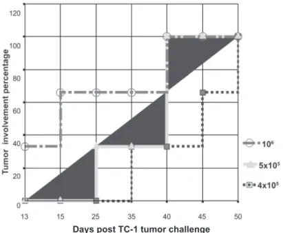
Inoculation of HPV-16 E6 and E7 expressing TC-1 tumor cells caused rapid tumor forma-tion. TC-1 cells were subcutaneously injected into the left flanks of the C57BL/6 mice at var-ious dosages (4×10⁵, 5×10⁵, 106 cells/mouse),
suspended in 100μl phosphate buffered saline
(PBS).
Monitoring of tumor model
The C57BL/6 mice were challenged by subcutane-ous injections of TC-1 cells (5×10⁵) suspended in 100 μl PBS to their left flanks. After 2 weeks, the resulting tumors were obvious and palpable in the mice. Tumor sizes (in millimeters) were reported as the average of all measured dimensions. Each tumor's smallest diameter (a) and biggest (b) diam-eters were measured and its volume was calculated using the formula: V= (a2b)/2 (13).
Immunization of mice
Two weeks after the initial inoculation of the mice with TC 1 cells, the animals were grouped into 3 cages and each group was in-tramuscularly vaccinated 3 times in two-week
intervals with 100μl PBS (negative control),
100μg pcDNA3 (negative control), and 100μg
pcDNA3-E7 (test). They were then monitored every other day for survival as well as infection symptoms, and their tumor diameter sizes were measured and their means were calculated and recorded.
Histological study
The mice were euthanized and histologically studied. Autopsied tissues from control and test animals were fixed in 10% phosphate-buffered formalin. They were imbedded in paraffin and were sectioned at 4-6 μm; they were then stained with Mayer’s hematoxylin and eosin (H&E). The slides were mounted and microscopically evalu-ated (×40) (Fig 2).
Statistical analysis
To compare results between the different groups, the univariate analysis of variance (ANOVA) was performed using the SPSS 11.0 statistical soft-ware. Differences were considered statistically significant when p-value <0.05.
Results
Rapid tumor formation
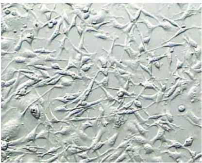
Fig 2 shows the confluent monolayer of TC-1 cells. These adherent cells have a fibroblastic appearance and continuously express E6 and E7 oncogenic pro-teins of HPV. As shown in fig.3, rapid tumor growth appeared in mice inoculated with 5×10⁵ TC-1 cells.
Cancer prevention and therapy in the immu-nized mice
The mice were inoculated as described in ma-terial and methods and monitored for evidence of tumor growth by palpation and inspection of two of their perpendicular tumor diam-eters every other day. Data analysis was per-formed using the one-way ANOVA followed by Tukeys’ test in the experimental and con-trol groups. A p-value <0.05 was considered significant. There were significant decreases in tumors sizes and improvements in survival among the treated mice (p<0.041). The mean tumor volumes and tumor sizes were not sig-nificantly different between the two control groups (p=0.9), whereas significant differenc-es were observed in the DNA vaccinated group (p=0.00) (Tables 1, 2).
Table 1: Comparison of tumor size between test and control groups. The difference between the sizes in pcDNA3 and pcDNA3/E7 shows a meaningful reduction and is statisti-cally significant.
P value Groups
0.00
PBS & pcDNA3-E7
0.041
pcDNA3 & pcDNA3-E7
0.90
PBS & pcDNA3
P-values were calculated using univariate analysis of variance. The E7 groups had significant differences of p= 0.04 and 0.00 in comparison to the pcDNA3 and PBS control groups.
A neoformed tumor lesion is composed of undifferen-tiated polyhedral, round and plump spindle cells with high N/C ratios, eosinophilic cytoplasms with indis-tinct borders, highly pleomorphic nuclei with coarse granular chromatin, and many giant cells with multiple nuclei. Furthermore, such a lesion displays a high mi-totic rate with atypical features, and is packed together by a cellular sheath with large necrotic areas.
It also invades its peripheral skeletal muscle fibers.
In our specimens, none of these special cytoplas-mic and nuclear features of a viral infection, which are common in the highly undifferentiated HPV-induced tumor lesions, were identified (Fig 3).
Fig 4: A high grade intraepithelial neoplastic lesion. This figure shows cells of tumoric tissue cells after injection with TC-1 cells injection (×40). TC-1 tumoric model showing many tumoric giant cells with multiple nuclei; no differen-tiation was detectable on the surface.
Table 2: Measurement of tumor size in mice from the PBS, pcDNA3 and pcDNA3- E7 groups on different days
pcDNA3/E7 mean ± SD pcDNA3
mean ± SD PBS
mean ± SD Days p.i
305 ± 5.5 600 ± 24.3
607 ± 25.7
34
1860.2 ± 3.4 4469 ± 21.1
4123.5 ± 20.4
55
1492.3 ± 1.5 5482.1 ± 20
4998 ± 8.2
66
Days post TC-1 tumor challenge
Fig 3: Tumor growth kinetic of TC-1. The cells were subcutaneously injected into C57BL/6 mice subcutaneously with various doses (4×10⁵, 5×10⁵, 106 cells/mouse). The mice were then
Discussion
HPV is an epithelium tropic DNA virus and is re-sponsible for one of the most widespread sexually transmitted infections (4).
High-risk HPV infections are the major cause of cervical cancer with a high mortality rate in wom-en. (10, 14).
Tumor models in C57BL/6 mice have been used in cervical cancer studies since clinical trials are not ethically allowed. Therefore, TC-1 cells are produced for experimental purposes be-cause they continuously express the E6 and E7 oncoproteins (15). These cells have gained an appropriate importance status for vaccine re-searches against cervical carcinoma.
Administration of HPV vaccines plays a cen-tral role in preventing human papilloma virus (HPV) infections in people. The majority of ani-mal studies using viral vectors for the HPV16 E7 DNA vaccine have focused on prophylaxis to generate immune responses which could re-ject a subsequently inre-jected tumor challenge. The only available vaccine approved by the U.S. food and drug administration (FDA) for human use is Gardasil, a quadrivalent recombinant vac-cine manufactured and marketed by Merck (16). Several studies have shown that such purified recombinant HPV proteins and HPV DNA vac-cines have been successfully used to generate cell-mediated immune responses in experimen-tal animal models (17, 18).
We conducted therapeutic vaccination trials in mice with already established tumors. Thru a number of immune measure assessments, Meshkat et al. showed that immunization of C57BL/6 mice induced efficient immune responses and generat-ed HPV16 E7-specific cytotoxic T lymphocytes (CTLs) (11). In this study we used the same con-struct and therapeutic strategies which are capa-ble of generating an antitumor response in tumor bearing mice.
In this study, tissues were perused independently and showed malignant neoplasms composed of severely pleomorphic tumor cells with nuclear enlargement, high N/C ratios and prominent nu-cleoli in solid and fascicular patterns of growth. High mitotic activity with extensive necrosis was also noted in both test and control groups. It seems that immunization with this construct alone could not improve the mitotic activity. Therefore, the linkage of different genes or gene fragments to a DNA vaccine represents a pro-spective approach for increasing the potency of DNA vaccines.
With continued attempts in the development of
HPV therapeutic vaccines, HPV therapeutic DNA vaccines will appear as a significant ap-proach which can be combined with existing forms of therapy such as chemo and radiation therapies. Therapeutic strategies such as a DNA vaccine capable of generating an antitumor re-sponse, should be explored as a way to treat in-fected patients.
Conclusion
Currently, there is a major thrust to develop vac-cines in an effort to prevent infectious diseases. In order to test the efficacy of these vaccines, suitable animal models are needed. Normally, ab-solute protection against disease is the ultimate goal, but in cases such as cervical cancer, it may take years for HPV-associated lesions to appear; thus, protection against the initial infection would be a faster way to determine efficacy. In conclu-sion, the results of this study might improve our knowledge in an attempt to treat the TC-1 tumor-cell challenged animals as the first step in a suc-cessful preventive vaccine development.
Acknowledgments
This study was supported by Deputy of Research in Tarbiat Modares University.
There is no conflict of interest in this article.
References
Smith HO, Tiffany MF, Qualls CR, Key CR. The rising 1.
incidence of adenocarcinoma relative to squamous cell carcinoma of the uterine cervix in the United States--a 24-year population-based study. Gynecol Oncol. 2000; 78(2): 97-105.
Mohar A, Frias-Mendivil M. Epidemiology of cervical 2.
cancer. Cancer Invest. 2000; 18(6): 584-90.
Bosch FX, Manos MM, Munoz N, Sherman M, Jansen 3.
AM, Peto J, et al. Prevalence of human papillomavirus in cervical cancer: a worldwide perspective. Interna-tional biological study on cervical cancer (IBSCC) Study Group. J Natl Cancer Inst. 1995; 87(11): 796-802. La Torre G, de Waure C, Chiaradia G, Mannocci A, Ric-4.
ciardi W. HPV vaccine efficacy in preventing persistent cervical HPV infection: a systematic review and meta-analysis. Vaccine. 2007; 25(50): 8352-8358.
Niakan M, Eftekhar Z, Jamali Zavareh M,Golalipour F, 5.
Faghihzadeh S, Jalali MR. Human Papillomavirus geno-type as a major determinant of the course of cervical cancer. Yakhteh Medical Journal. 2004; 5(20):154-157. Wu TC, Hsieh ST, Purow BW, Kurman RJ. Demonstra-6.
tion of human papillomavirus (HPV) genomic amplifica-tion and viral-like particles from CaSki cell line in SCID mice. J Virol Methods. 1997; 65(2): 287-298.
Heard I, Tassie JM, Schmitz V, Mandelbrot L, Kazatchk-7.
ine MD, Orth G. Increased risk of cervical disease among human immunodeficiency virus-infected women with se-vere immunosuppression and high human papillomavi-rus load(1). Obstet Gynecol. 2000; 96(3): 403-409. Chen CH, Ji H, Suh KW, Choti MA, Pardoll DM, Wu TC. 8.
E7-ex-pressing murine tumor metastases in the liver and lungs. Gene Ther. 1999; 6(12): 1972-1981.
Lin KY, Guarnieri FG, Staveley-O'Carroll KF, Levitsky HI, 9.
August JT, Pardoll DM, et al. Treatment of established tu-mors with a novel vaccine that enhances major histocom-patibility class II presentation of tumor antigen. Cancer Res. 1996; 56(1): 21-26.
Harper DM, Franco EL, Wheeler C, Ferris DG, Jenkins D, 10.
Schuind A, et al. Efficacy of a bivalent L1 virus-like parti-cle vaccine in prevention of infection with human papillo-mavirus types 16 and 18 in young women: a randomised controlled trial. Lancet. 2004; 364(9447): 1757-1765. Meshkat Z, Soleimanjahi H, Mahmoudi M, Hassan ZM, 11.
Mirshahabi H, Meshkat M, et al. CTL responses to a DNA vaccine encoding E7 gene of human papillomavirus type 16 from an Iranian isolate. Iran J Immunol. 2008; 5(2): 82-91.
Sambrook J, Russel D. Molocular Cloning. 2001; AI.19-12.
AI.24
Li Y, Subjeck J, Yang G, Repasky E, Wang XY. Genera-13.
tion of anti-tumor immunity using mammalian heat shock protein 70 DNA vaccines for cancer immunotherapy. Vac-cine. 2006; 24(25): 5360-5370.
zur Hausen H. Papillomavirus infections--a major cause 14.
of human cancers. Biochim Biophys Acta. 1996; 1288(2): F55-78.
Han L, Wang W, Fang Y, Feng Z, Liao S, Li W, et al. 15.
Soluble B and T lymphocyte attenuator possesses anti-tumor effects and facilitates heat shock protein 70 vac-cine-triggered antitumor immunity against a murine TC-1 cervical cancer model in vivo. J Immunol. 2009; 183(12): 7842-7850.
HPV Vaccine Information For Young Women. Centers for 16.
Disease Control and Prevention (CDC). Available from: URL://www.cdc.gov/std/hpv/STDFact-HPV-vaccine-young-women.htm. Retrieved 13 Feb 2010.
Brinkman JA, Xu X, Kast WM. The efficacy of a DNA vac-17.
cine containing inserted and replicated regions of the E7 gene for treatment of HPV-16 induced tumors. Vaccine. 2007; 25(17): 3437-3444.
Feltkamp MC, Smits HL, Vierboom MP, Minnaar RP, de 18.