1. Department of Pediatrics, Hospital Prof. Doutor Fernando Fonseca E.P.E., Amadora, Portugal
2. Department of Pediatrics, Hospital Dona Estefânia – CHLC – EPE 3. Instituto Português de Reumatologia (IPR), Lisbon, Portugal
Juvenile Systemic Lupus Erythematosus in Portugal:
clinical and immunological patterns of disease
expression in a cohort of 56 patients
ACTA REUMATOL PORT. 2013;38:274-285
AbstrAct
Objective: To define the pattern of disease expression and to gain better understanding in patients with juve-nile onset systemic lupus erythematosus (SLE) in Por-tugal.
Methods: The features of unselected patients with sys-temic lupus erythematosus who had disease onset be-fore the age of 18 years were retrospectively analysed in three Portuguese centres with Pediatric Rheumato-logy Clinic over a 24-year period (1987-2011). Demo-graphic, clinical and laboratory manifestations, thera-py and outcome were assessed.
Results: A cohort of 56 patients with a mean age at di-sease onset of 12.6±4.04 years (mean±1SD) (range, 1.0--17.0 years) and a mean period of follow-up of 5.5±5.4 years. Forty six (82.1%) patients were female. The most common disease manifestations were musculoskeletal (87.5%), mucocutaneous (80.3%) and haematological abnormalities (75%). Lupus nephritis was diagnosed in 46.4% of patients and consisted of glomerular ne -phritis in all cases. Neuropsychiatric manifestations oc-curred in 21.4% but severe central nervous system complications were uncommon, as brain infarcts and organic brain syndrome in 4 (7.1%) patients. Antinu-clear antibodies and anti-double stranded DNA were positive in most patients in (98.2% and 71.4% respec-tively), as well as low C3 and/or C4 were observed fre-quently (85.7%). Generally, most patients had a good response to therapy as demonstrated by a significant decreasing of SLEDAI score from disease presentation to the last evaluation. The SLEDAI at diagnosis, the
ma-Marta Cabral1, Carlos Escobar1, Marta Conde2, Margarida Ramos2, José A. Melo Gomes3
ximum SLEDAI and the incidence of complications were significantly higher in patients with neurolupus and/or lupus nephritis. Therapy included oral steroids (87.5%), hydroxychloroquine (85.7%), azathioprine (55.4%), IV cyclophosphamide (28.6%) along with other drugs. Six (10.7%) patients were treated with ri-tuximab. Long-term remission was achieved in 32%, disease was active in 68%, adverse reactions to thera-py occurred in 53.6% and complications/severe mani-festations in 23.2%. Two patients died, being active di-sease and severe infection the causes of death.
Conclusions: This study suggests that in our patients the clinical and laboratory features observed were si-milar to juvenile systemic lupus erythematosus patients from other series. Clinical outcome was favourable in the present study. Complications from therapy were frequent. Objective: To define the pattern of disease ex-pression and to gain better understanding in patients with juvenile onset systemic lupus erythematosus (SLE) in Portugal.
Methods: The features of unselected patients with sys-temic lupus erythematosus who had disease onset be-fore the age of 18 years were retrospectively analysed in three Portuguese centres with Pediatric Rheumato-logy Clinic over a 24-year period (1987-2011). Demo-graphic, clinical and laboratory manifestations, thera-py and outcome were assessed.
Results: A cohort of 56 patients with a mean age at di-sease onset of 12.6±4.04 years (mean±1SD) (range, 1.0--17.0 years) and a mean period of follow-up of 5.5±5.4 years. Forty six (82.1%) patients were female. The most common disease manifestations were musculoskeletal (87.5%), mucocutaneous (80.3%) and haematological abnormalities (75%). Lupus nephritis was diagnosed in 46.4% of patients and consisted of glomerular ne -phritis in all cases. Neuropsychiatric manifestations occur red in 21.4% but severe central nervous system
complications were uncommon, as brain infarcts and organic brain syndrome in 4 (7.1%) patients. Antinu-clear antibodies and anti-double stranded DNA were positive in most patients in (98.2% and 71.4% res-pectively), as well as low C3 and/or C4 were observed frequently (85.7%). Generally, most patients had a good response to therapy as demonstrated by a signi-ficant decreasing of SLEDAI score from disease presentation to the last evaluation. The SLEDAI at diagno -sis, the maximum SLEDAI and the incidence of com-plications were significantly higher in patients with neurolupus and/or lupus nephritis. Therapy included oral steroids (87.5%), hydroxychloroquine (85.7%), azathioprine (55.4%), IV cyclophosphamide (28.6%) along with other drugs. Six (10.7%) patients were trea-ted with rituximab. Long-term remission was achie-ved in 32%, disease was active in 68%, adverse reac-tions to therapy occurred in 53.6% and complica-tions/severe manifestations in 23.2%. Two patients died, being active disease and severe infection the cau-ses of death.
Conclusions: This study suggests that in our patients the clinical and laboratory features observed were si-milar to juvenile systemic lupus erythematosus pa-tients from other series. Clinical outcome was favou-rable in the present study. Complications from thera-py were frequent.
Keywords: Juvenile; Systemic Lupus Erythematosus.
introduction
Juvenile systemic lupus erythematosus (jSLE) is a chro-nic multisystem autoimmune disease of unpredicted course and prognosis1,2. It manifests with a wide
spec-trum of clinical and immunological abnormalities, which range from skin rashes and oral ulcers to life-threatening neurological, hematological and renal in-volvement.2,3,4Although SLE is most commonly
diag-nosed in women during the second to fourth decades of life, disease onset can occur at any age. It is rare in childhood, but in 15 to 20% of all SLE patients the diagnosis is made for the first time before 16 years old3,5 and, to our best knowledge, there is only one
stu-dy (published in 1994) that aims to demonstrate de-mographic, clinical and laboratory characteristics and outcome of SLE in Portuguese pediatric patients6.
Several investigators have reported that age at on-set has a modifying effect on disease expression7. This
is important because examination of more homoge-neous subsets, such as childhood onset patients, may allow an earlier diagnosis, better treatment, and more accurate prognosis. It has been noted that certain fea-tures of SLE usually associated with severity, such as nephritis or central nervous system dysfunction, are more common in patients with childhood onset SLE (cSLE)7.
The incidence of SLE varies according to each po-pulation’s characteristics, such as patients’ age, gender, ethnicity, and the period of time studied. Epidemiolo-gic studies suggest that SLE occurrence differs among different countries, and even among different areas of the same country8,9. These differences are also observed
among population groups of the same race living in dif-ferent parts of the world, suggesting that besides gene-tic susceptibility, geographic and environmental fac-tors are probably implicated in development of this connective tissue disease9,10.
In this study, to better define the pattern of disease expression in jSLE patients, we have retrospectively analyzed the clinical and immunological features, treat-ments, complications and outcome of 56 patients in whom the first manifestations appeared in childhood or adolescence.
PAtients And Methods
The present study reports a cohort of 56 SLE patients with disease onset before the age of 18 year-old, that were followed consecutively either as inpatients or out-patients over a 24-year period (from January 1987 un-til November 2011) in three different referral centers, with different epidemiological characteristics and cul-tures of its patients: 1) a private Rheumatology center and a reference for pediatric rheumatologic patients from different regions of the country, 2) a tertiary pe-diatric hospital in the centre of Lisbon and 3) an hos-pital in the suburban area of Lisbon, with an impor-tant prevalence of immigrant patients (Amadora).
We considered cSLE when age onset was below 10 year-old.
All patients fulfilled at least four of the American College of Rheumatology criteria for the classification of SLE11,12.
We performed a retrospective review of the records from patients diagnosed as jSLE. The data were retrieved on a predesigned protocol form. Information ga -thered included patients’ age at onset, gender,
ethnici-ty, disease duration (calculated from symptoms onset to the end of the study or patients’ death), follow up duration, different clinical features at presentation and follow up, complications, therapy and outcome. La-boratory data collected included hematological, renal and immunological parameters consisting of comple-te blood counts (leucopenia was considered for whicomple-te blood cell count (WBC) <4,000/mm3, anemia for he-moglobin (Hb) < 11g/dL, lymphopenia for lymphocy-tes <1,500/mm3, thrombocytopenia for platelets <150 × 103/mm3), serum creatinine, 24-hours urine pro-tein excretion, antinuclear antibodies (ANA), double stranded (ds) DNA antibodies, SSA/Ro, -SSB/La, anti–Sm, ribonucleoprotein (RNP), anti-cardiolipin (aCL), anti-b2glycoprotein I (anti-b2GPI) (IgG and IgM) antibodies and lupus anticoagulant (LAC), the lowest levels of complement components 3 and 4 (C3 and C4) (low C3:<90mg/dl; low C4:<20mg/dl) and the highest erythrocyte sedimenta-tion rate (ESR) value (elevated ESR: >20mm at 1 hour). Hemolytic anemia was considered when direct Coombs’ test was positive.
Lupus nephritis or nephropathy was defined by the presence of any of the following indicators: proteinu-ria >0.5g/day, persistent cellular cast and/or hematu-ria, decreased glomerular filtration rate, abnormalities on the renal biopsy and end-stage renal disease (ESRD) treated by dialysis or transplant.
Myositis was considered if confirmation of muscle inflammation by muscular enzymes elevation and magnetic resonance.
Pericarditis was considered in the presence of peri-cardial pain and/or effusion and/or compatible elec-trocardiogram abnormalities.
Neuropsychiatric symptoms were considered when the patient with the established diagnosis of active jSLE presented the following manifestations: psychiatric di-sorders (anxiety and/or depression, psychosis, deli-rium, paranoid features or hallucination, confirmed by psychiatrist evaluation), moderate to severe heada-che, ataxia, peripheral neuropathy, myopathy, chorea or seizure not attributed to another causes, cerebro-vascular accident (confirmed by computerized tomo-graphy or magnetic resonance imaging) or other type of cognitive disorder. Organic brain syndrome was considered in the presence of altered mental function with impaired orientation, memory or other cognitive function or decreased psychomotor activity.
Data related to different treatment options, hospi-talization and disease outcome were also gathered. The
outcome measures were: 1) remission (defined as long-term (more than 6 months) absence of disease activi-ty manifestations, including clinical features and la-boratory indices), either on treatment or off treatment; 2) active disease on treatment and its degree of activi-ty according to the last SLEDAI score (mild if ≤3, mo-derate if >3 and ≤12 and severe if >12); 3) complica-tions (secondary to the underlying disease or as thera-py adverse effects) and 4) death. Diagnosis of anti-phospholipid syndrome had to meet diagnostic criteria13. Only severe infection was considered as
com-plication/severe manifestation and it was defined as any infection that resulted in hospitalization, in delay in an existing hospitalization and/or was life threate-ning and/or caused death. The clinical activity was as-sessed by the Systemic Lupus Erythematosus Disease Activity Index (SLEDAI) score at the time of initial pre-sentation, during and after treatment14,15. The
maxi-mum SLEDAI during follow-up was calculated retros-pectively for all patients from medical record. We in-cluded the latest SLEDAI, documented in medical re-cord, for those who defaulted or died before concluding the study in November 2011.
Statistical analysis of the data was performed using
SPSS® 19.0 (SPSS Inc., Chicago, Illnois, EUA).
Conti-nuous data were expressed as mean T ± standard de-viation (SD) or median and range or interquartile ran-ge, and categorical variables as percentages. For con-tinuous variables, mean values were compared using T-student test or Mann-Whitney test depending on normality distribution of results. Categorical variables were analyzed using Chi-square test or Fisher exact test, as needed.
results
Fifty-six patients, 91.1% Caucasian, 82.1% female with a ratio female/male of 4.6/1 and a mean (±SD) age at diagnosis of 12.6±4.04 years (range, 1.0-17.0 years), were enrolled. Mean (±SD) period of follow-up was 5.5±5.4 years (range, 0-288 months). Patient charac-teristics are shown in Table I. The delay on diagnosis was on average 12.4±19.0 months (extremes of 0.5 and 84 months). 23.2% had relatives with autoimmu-ne diseases, four (7.1%) with SLE, and there was a cou-ple of monozygotic twin sisters concordant for jSLE. 26.8% had some comorbidities; from them we high-light: sickle cell disease, Down syndrome, asthma, Gra-ves disease, hepatic hemangioma, vitiligo and
pituita-ry hypoplasia.
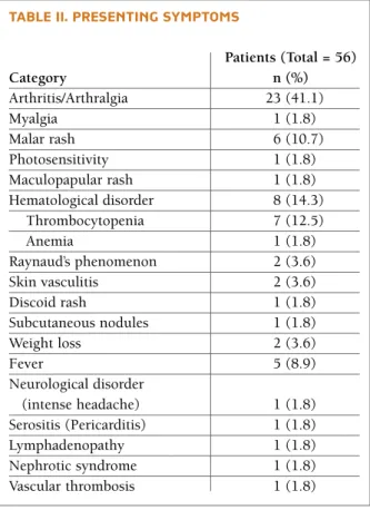
The presenting symptoms at diagnosis are summa-rized in Table II. Musculoskeletal, mucocutaneous and hematological involvement were the major clinical ma-nifestations. The cumulative frequencies of systemic involvement are presented in Table III, being articular involvement the most prevalent (85.7%). General symptoms (fever and/or asthenia and/or weigh lost) were present in 42 (75%) patients. Malar rash and Ray-naud’s phenomenon were the commonest mucocuta-neous features. On the other hand, discoid lupus oc-curred in only one patient.
Hematologic abnormalities were found in 75% pa-tients. Lymphopenia was the commonest and it was identified in 80.8% patients. Anemia at any time of the disease occurred in 48.2% patients, but only 25% had confirmed hemolytic anemia. Thrombocytopenia was seen in 37.5%, and occurred around the time of diag-nosis in nearly half of the patients; however, in six of them thrombocytopenia predated the definite diagno-sis of jSLE by months and even years (mean 33,5±28.5 months; extremes of 5 months and 7 years).
Twenty-six (46.4%) had renal involvement and con-sisted of glomerular nephritis in all cases. Twenty of them (76.9%) underwent renal biopsy. Biopsies were assigned to WHO classes as follows: minimal change (class I) in 1 (5%) patient; mesangial glomerulone -phritis (class II) in 5 (25%); focal segmental prolifera-tive glomerulonephritis (class III) in 4 (20%), diffuse proliferative glomerulonephritis (class IV) in 9 (45%) and membranous glomerulonephritis in 1 (5%). No isolated interstitial nephritis was seen.
Pericarditis was present in 25% and it was
recur-rent in two patients (one patient had four differecur-rent epi-sodes).
Neuropsychiatric systemic lupus erythematosus (NPSLE) occurred in 21.4% of patients, being lupus headache the commonest manifestation. Its severe ma-nifestations, as seizure, cerebrovascular accident and organic brain syndrome were rare complications.
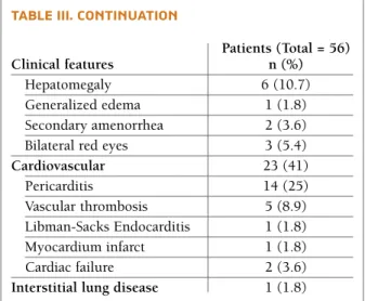
Respiratory system and gastrointestinal involvement were rare in our cohort of patients (Table III).
Immunological features and other laboratory fin-dings are discriminated in Tables IV and V.
Most patients received oral corticosteroid (87.5%) and chloroquine/hydroxychloroquine (85.7%) for treatment. Twenty-four (42.9%) were treated with in-travenous (IV) pulse methylprednisolone. 69.6% pa-tients were given immunosuppressive therapy with azathioprine in 55.4%, cyclophosphamide in 28.6%, mycophenolate mofetil in 14.3%, methotrexate in 14.3% and cyclosporine in 3.6%. Six patients were treated, with rituximab (four patients with neurolu-pus, one of them complicated by severe epilepsy and cerebrovascular infarct; five patients with severe he-matological involvement and four of them with
si-tAble i. MAin ePideMiologic feAtures of PAtients diAgnosed with jsle, 1987-2011
Characteristic
Total no. of patients 56 Female/male (no.) 46/10 Female (%) 82.1
Age, yr* at symptom onset 12.6 ± 4.04; 13,5 (1 – 17) Delay to diagnosis, months† 12.4 ± 19.0; 4 (0.5 – 84) Follow-up from disease 5.5 ± 5.4; 4 (0-24) diagnosis, yr*
Abbreviations: IQR = interquartile range *MeanT±SD; median (IQR)
†Delay to diagnosis from the onset of symptoms to the time of diagnosis of SLE. Mean T±SD; Median (IQR)
tAble ii. Presenting syMPtoMs
Patients (Total = 56) Category n (%) Arthritis/Arthralgia 23 (41.1) Myalgia 1 (1.8) Malar rash 6 (10.7) Photosensitivity 1 (1.8) Maculopapular rash 1 (1.8) Hematological disorder 8 (14.3) Thrombocytopenia 7 (12.5) Anemia 1 (1.8) Raynaud’s phenomenon 2 (3.6) Skin vasculitis 2 (3.6) Discoid rash 1 (1.8) Subcutaneous nodules 1 (1.8) Weight loss 2 (3.6) Fever 5 (8.9) Neurological disorder (intense headache) 1 (1.8) Serositis (Pericarditis) 1 (1.8) Lymphadenopathy 1 (1.8) Nephrotic syndrome 1 (1.8) Vascular thrombosis 1 (1.8)
multaneous renal involvement, all of them refractory to previous immunosuppressive therapy). Treatment also included IV immunoglobulin in 17.9%, intra-ar-ticular steroids in one, oral anticoagulants in 7.1% and as general maintenance therapy: NSAIDS, anti-hyper-tensive agents, calcium carbonate and Vitamin D. Mean (±SD) duration of steroid therapy was 47.6±59 months (range, <1 – 250 months/20.8 years) and for immunosuppressive agents it was 42.9±52.9 months (range, <1 – 190 months/15.8 years).
Disease activity was evaluated by SLEDAI and its average at the time of diagnosis was 11.0±9.1 (range, 0–44). Analyzing the maximum SLEDAI during fol-low up for each patient, its average was 14.5±10.6 (range, 0–47) and in the last evaluation it was 2.6±3.6 (range, 0–16).
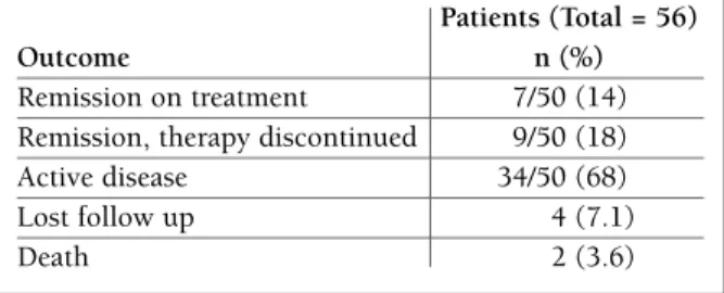
Improvement of clinical features, reflected by a de-creasing from the maximum to the final SLEDAI, was noted in 48/50 (96%) patients. Clinical, biological and immunological complete remission for more than 6 months was obtained in 16/50 (32%) patients (wi thout treatment: 9 (18%) patients). Disease was active in 34/50 (68%) and two (3.6%) patients died (Table VI). The causes of death were: 1) Sepsis and active SLE due to treatment discontinuation in one patient; 2) sudden *Miositis confirmed by muscular enzymes elevation and/or magnetic resonance
**Anemia (Hb<12g/dl); Thrombocytopenia
(Platelets<150.000/mm3); Leucopenia (Leucocytes<4.000/mm3); Lymphopenia (Lymphocytes<1500/ mm3)
***Persistent proteinuria >0.5g/24hours or cellular casts (granular, WBC and/or hematuria)
tAble iii. cuMulAtive frequencies of systeMic involveMent in our cohort
Clinical features Patients (Total = 56) n (%)
Musculoskeletal 49 (87.5)
Articular (arthritis and/or 48 (85.7) arthralgia) Myalgia / Miositis* 6 (10.7)/3 (5.4) Serositis 16 (28.6) Pericarditis 14 (25) Pleuritis 7 (12.5) Peritonitis 2 (3.6) Mucocutaneous 45 (80.3) Malar rash 30 (53.6) Discoid rash 1 (1.8) Skin vasculitis 10 (17.9) Urticariform vasculitis 1 (1.8) Mucocutaneous hemorrhage 11 (19.6) Photosensitivity 14 (25) Livedo reticularis 13 (23.2) Raynaud’s phenomenon 15 (26.8) Mechanic hands 1 (1.8) Oral and/or nasal ulcers 14 (25) Enantema 15 (26.8) Hematological disorder** 42 (75) Anemia 27 (48.2) Thrombocytopenia 21 (37.5) Leucopenia 29 (51.8) Lymphopenia 42 (80.8) Renal disease*** 26 (46.4) Neuropsychiatric disorder 12 (21.4) Lupus headache 7 (12.5) Seizure 3 (5.4) Psychosis 3 (5.4) Organic brain syndrome 4 (7.1) Cerebrovascular accident(s) 4 (7.1) Ataxia 1 (1.8) Other psychiatric symptoms 7 (12.5)
Miscellaneous Subcutaneous nodules 6 (10.7) Weight loss 17 (30.4) Astenia 28 (50) Fever 31 (55.4) Alopecia 3 (5.4) Hair loss 18 (32.1) Lymphadenopathy 8 (14.3)
continues on the next column
tAble iii. continuAtion
Patients (Total = 56)
Clinical features n (%)
Hepatomegaly 6 (10.7) Generalized edema 1 (1.8) Secondary amenorrhea 2 (3.6) Bilateral red eyes 3 (5.4)
Cardiovascular 23 (41) Pericarditis 14 (25) Vascular thrombosis 5 (8.9) Libman-Sacks Endocarditis 1 (1.8) Myocardium infarct 1 (1.8) Cardiac failure 2 (3.6)
sis may be considered as a complication associated with corticosteroids and/or a rare manifestation of jSLE, and it was observed in one patient. Besides renal involvement and NPSLE, complications were more fre-quent in patients with African-ancestry, anemia, leu-copenia and low C3 (Table VIII). Treatment adverse effects occurred in 50%, being visceral obesity the most prevalent (Table VII).
Contrary to expected, age below 10 year-old was not statistically associated with renal involvement, NPSLE, higher incidence of complications or higher maximum SLEDAI (Table VIII).
Considering correlations with patients’ ethnicity, re-nal or neuropsychiatric disorders were not more fre-quent in African-ancestry patients, but they had more complications (75% vs 28%, p=0.05) and a higher in-cidence of positive LAC (60% vs 14%, p=0.01) (Table VIII).
Comparing clinical and laboratory features in our cohort of patients, with and without renal involve-ment, showed that lupus nephritis was notably asso-ciated with malar rash, lower WBC count, higher ESR, lower Hb and C3 (Table VIII). From all immunologi-cal tests, only anti-dsDNA antibodies were significan-tly associated with renal disease. Also the incidence of complications was higher in patients with lupus ne -phritis (Table VIII). As we would expect, it was obser-ved a tendency for arterial hypertension in patients with nephritis (16% vs 3.6%), despite a not statisti-cally significant association being demonstrable, pro-bably because of the cohort’s dimension.
Neuropsychiatric manifestations were associated with lower lymphocytes count but not with lower WBC count, Hb or hypocomplementemia, contrarily to the observed with lupus nephritis. On the other hand, the incidence of complications, Sm, anti--SSA and anti-RNP antibodies were significantly hi gher in patients with NPSLE. However
antiphos-tAble iv. frequency of PAtients with Positive iMMunologicAl tests Patients (Total = 56) Immunological test (%) ANA 98.2 Anti-DNAds 71.4 Anti-Sm 12.5 Anti-RNP 21.4 Anti-SSA (RO) 23.2 Anti-SSB (La) 3.6 Anticardiolipin IgM/IgG 16 Anti-β2glycoprotein I 16 Low serum C3 71.4 Low serum C4 78.6
Abbreviations: ANA, antinuclear antibodies; Anti-Sm, anti-Smith, ESR, erythrocyte sedimentation rate
tAble vi. diseAse outcoMe in jsle PAtients
Patients (Total = 56)
Outcome n (%)
Remission on treatment 7/50 (14) Remission, therapy discontinued 9/50 (18) Active disease 34/50 (68) Lost follow up 4 (7.1) Death 2 (3.6)
Abbreviations: ESR, erythrocyte sedimentation rate; RV, reference value; *minimum absolute value; **maximum absolute value
tAble v. generAl lAborAtory findings
Laboratory findings Mean T ± SD IQR
Hemoglobin (g/dl)* 10.2±2.4 2.8-14.7 Platelets (x103/mm3)* 159±108 2-459
White blood cell count
(/mm3)* 3,794±1,604 1,400-10,600 Lymphocytes (/mm3)* 1,037±703 100-4,028 C3 (mg/dl)* (RV: 90-180) 67.9±36.5 8-230 C4 (mg/dl)* (RV: 20-50) 11.9±13.3 0.05-86 Elevated ESR (>20mm/ 74±40.9 5-155 1st hour)** C-reactive protein (mg/dl)** 4.5±7.2 0.01-36.2 Creatinine (mg/dl)** 0.91±0.3 0.48-2.1 Proteinuria (g/24hours)** 0.861±1.215 0.07-6
cardiac arrest secondary to ventricular fibrillation in a patient with sickle cell disease and severe cardiac com-plications inherent to SLE (Libman-sacks endocardi-tis; bacterial endocarditis, myocardium infarct) during recovery from pneumonia. Both patients who died had frequently an uncontrollable or progressive multisys-temic disease.
Complications and/or severe manifestations occur-red in 23.2%. Infection was the leading complication during treatment of jSLE (Table VII). Avascular
necro-pholipid antibodies were not statistically correlated, contrarily to the expected (Table VIII).
Anti-SSA antibodies were more frequent in patients with livedo reticularis, but the second was not statis-tically associated with antiphospholipid antibodies. On the other hand, anti-SSB antibodies were more fre-quent in patients with skin vasculitis (20% vs 0%, p=0.002) (Table VIII).
The mean SLEDAI at SLE diagnosis and the mean maximum SLEDAI were significantly higher in patients with lupus nephritis and neurolupus (Table VIII).
discussion
There have been several studies dealing with jSLE and their results suggested that age at onset modifies di-sease’s expression in terms of clinical presentation, pattern of organ involvement, and serological fin-dings6,7,16-23. However, the true prevalence of jSLE
among the SLE population is unknown7. One of the
reasons is that there is not a strict definition of jSLE. The most often used cut off ages are 14 or 16 years at onset of disease18–20or at diagnosis21. However, several
studies use a higher or lower cut off age. In our study we considered the onset age lower than 18 years for our cohort of jSLE, considering that in Portugal pe-diatric health care centers receive patients from 0 to 18 year-old.
A potential limitation of this study was the inclusion of a heterogeneous group of patients from different ethnic backgrounds. Other important limitations were the retrospective review and the small sample of pa-tients, which might have interfered with the results from statistical correlations. However, this is a true re-presentation of the general pediatric population living in Lisbon (centre and suburbs).
The female to male ratio in adult-onset SLE (aSLE) is generally found to be slightly more than 10:1.724,25.
A higher proportion of male patients is often reported in jSLE in some series17,21,22but not in others7. In our
cohort, male represented 17.9% of the cases with a fe-male to fe-male ratio of 4.6:1, which is the common in-cidence reported for cSLE26.
Generally, about 10% of SLE patients have familial SLE25, and it was corroborated in our study.
This group of patients had clinical presentation, de-mographic and laboratory data comparable to previous studies about jSLE, including the Portuguese cohorts of juvenile patients described by Costa MM et al and the adult patients described by Santos MJ et al3,6,7,16,17,19-23,27.
Similar to those studies, hematological and renal in-volvement were between the most common manifes-tations6,16,17,19,22,23. However, contrarily to the cohort of
childhood-onset (<16 years) described by Pusongchai
et al, and comparable to other studies including adult
patients, mucocutaneous and articular features were the commonest manifestations in our patients24,25,28.
Perhaps that’s because our cohort included not only children but also adolescents (<18 year-old), whose clinical presentation might be probably nearest to the aSLE.
The onset of SLE is rare before the age of 5
year-tAble vii. coMPlicAtions And/or severe MAnifestAtions develoPed during follow uP of the jsle PAtients
Patients (Total = 56) Outcome n (%) Infection 6 (10.7) Pneumonia 2 (3.6) Sepsis 1 (1.8) Bacterial endocarditis 1 (1.8) Cellulite 3 (5.3) Cognitive impairment 4 (7.1) ESRD 2 (3.6) On conservative therapy 1 (1.8) On dialysis 1 (1.8) Vascular thrombosis 5 (8.9) Cerebrovascular 4 (7.1) Cerebral hemorrhage 2 (3.6) Epilepsy 3 (5.3) Cardiac failure 2 (3.6) Myocardium infarct 1 (1.8) Arterial hypertension 5 (8.9) Antiphospholipid syndrome 2 (3.6) Macrophage activation syndrome 2 (3.6) Avascular necrosis 1 (1.8) Gastrointestinal tract bleeding 1 (1.8)
Treatment adverse effects 28 (50) Osteoporosis* 5 (8.9) Visceral obesity 25 (44.6) Growth delay 3 (5.3) Ophthalmological complications 6 (10.7) Cataract 4 (7.1) Hydroxychloroquine 2 (3.6) maculopathy
Abbreviations: ESRD, end stage renal disease *Confirmation with DEXA
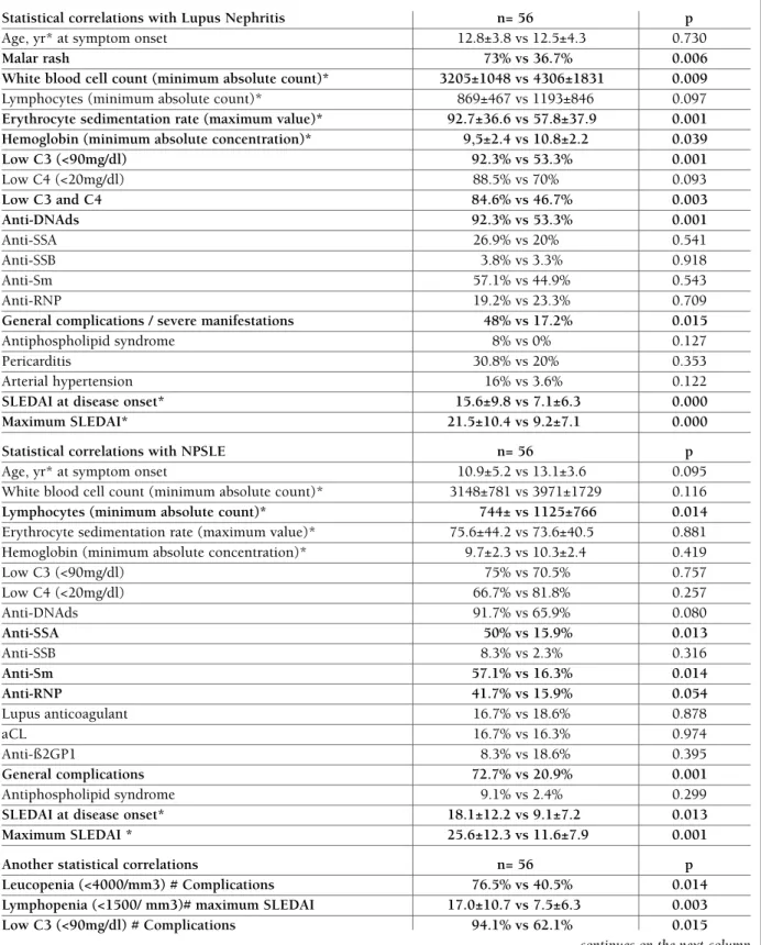
tAble viii. stAtisticAl correlAtions found with luPus nePhritis, nPsle And other significAnt stAtisticAl correlAtions
Statistical correlations with Lupus Nephritis n= 56 p
Age, yr* at symptom onset 12.8±3.8 vs 12.5±4.3 0.730
Malar rash 73% vs 36.7% 0.006
White blood cell count (minimum absolute count)* 3205±1048 vs 4306±1831 0.009
Lymphocytes (minimum absolute count)* 869±467 vs 1193±846 0.097
Erythrocyte sedimentation rate (maximum value)* 92.7±36.6 vs 57.8±37.9 0.001
Hemoglobin (minimum absolute concentration)* 9,5±2.4 vs 10.8±2.2 0.039
Low C3 (<90mg/dl) 92.3% vs 53.3% 0.001 Low C4 (<20mg/dl) 88.5% vs 70% 0.093 Low C3 and C4 84.6% vs 46.7% 0.003 Anti-DNAds 92.3% vs 53.3% 0.001 Anti-SSA 26.9% vs 20% 0.541 Anti-SSB 3.8% vs 3.3% 0.918 Anti-Sm 57.1% vs 44.9% 0.543 Anti-RNP 19.2% vs 23.3% 0.709
General complications / severe manifestations 48% vs 17.2% 0.015
Antiphospholipid syndrome 8% vs 0% 0.127 Pericarditis 30.8% vs 20% 0.353 Arterial hypertension 16% vs 3.6% 0.122
SLEDAI at disease onset* 15.6±9.8 vs 7.1±6.3 0.000
Maximum SLEDAI* 21.5±10.4 vs 9.2±7.1 0.000
Statistical correlations with NPSLE n= 56 p
Age, yr* at symptom onset 10.9±5.2 vs 13.1±3.6 0.095 White blood cell count (minimum absolute count)* 3148±781 vs 3971±1729 0.116
Lymphocytes (minimum absolute count)* 744± vs 1125±766 0.014
Erythrocyte sedimentation rate (maximum value)* 75.6±44.2 vs 73.6±40.5 0.881 Hemoglobin (minimum absolute concentration)* 9.7±2.3 vs 10.3±2.4 0.419 Low C3 (<90mg/dl) 75% vs 70.5% 0.757 Low C4 (<20mg/dl) 66.7% vs 81.8% 0.257 Anti-DNAds 91.7% vs 65.9% 0.080 Anti-SSA 50% vs 15.9% 0.013 Anti-SSB 8.3% vs 2.3% 0.316 Anti-Sm 57.1% vs 16.3% 0.014 Anti-RNP 41.7% vs 15.9% 0.054 Lupus anticoagulant 16.7% vs 18.6% 0.878 aCL 16.7% vs 16.3% 0.974 Anti-ß2GP1 8.3% vs 18.6% 0.395 General complications 72.7% vs 20.9% 0.001 Antiphospholipid syndrome 9.1% vs 2.4% 0.299
SLEDAI at disease onset* 18.1±12.2 vs 9.1±7.2 0.013
Maximum SLEDAI * 25.6±12.3 vs 11.6±7.9 0.001
Another statistical correlations n= 56 p
Leucopenia (<4000/mm3) # Complications 76.5% vs 40.5% 0.014
Lymphopenia (<1500/ mm3)# maximum SLEDAI 17.0±10.7 vs 7.5±6.3 0.003
Low C3 (<90mg/dl) # Complications 94.1% vs 62.1% 0.015
-old7. In this cohort, one patient presented the first
cli-nical manifestations of SLE at the age of 12 months. Ac-tually, it is known that the age at disease onset has an important impact on the clinical course and outcome of SLE. Previous reports comparing childhood with adult SLE reported that certain features of SLE usual-ly associated with severity, such as nephritis or central nervous system (CNS) dysfunction, are commonest in patients with cSLE. Therefore, it is assumed that cSLE is associated to a more severe disease course and wor-se prognosis3,7. Although renal and CNS involvement
were not statistically associated with age onset below 10 year-old, actually the younger patient in our study had a severe disease course, with renal involvement and severe neurolupus complicated by cerebrovascu-lar infarct26. However, we didn’t find a significant
sta-tistical association between age onset below 10 year--old and maximum SLEDAI or the occurrence of more complications/severe manifestations. In this way, the precise differences between child and adult-onset SLE in the prevalence of its manifestations may be still de-bated3. A meta-analysis conducted by Livingston B et al
found that malar rash, ulcers/mucocutaneous invol-vement, renal disease, neuropsychiatric manifesta-tions, thrombocytopenia, hemolytic anemia, fever and lymphadenopathy were more common in cSLE. On the other hand, Raynaud’s phenomenon, pleuritis, dis-coid rash and sicca symptoms were more common in aSLE3. Hematological abnormalities, specifically
he-molytic anemia and thrombocytopenia, fever and lymphadenopathy were more frequent in our cohort when compared to other series with a predominance of adult patients, as it was also reported by Font J et
al.7,24,25. Conversely, discoid lesions were really more
frequent in series of aSLE (our cohort had only one isolated case), but no significant differences were seen
considering frequencies of Raynaud’s and pleuritis7,24,25.
Renal involvement is one of the most important pre-dictors of a poor outcome. Its manifestations are va-riable, ranging from mild asymptomatic proteinuria to rapidly progressive glomerulonephritis leading to end--stage renal disease (ESRD)5, which was observed in
two patients in our study, despite their adequate treat-ment. Class IV nephritis was the most frequent class documented in our cohort, which is in agreement with most of the previous reports28. This is comparable to
other studies from United States29and Canada30, but
the frequency was lower than it was seen in cohorts from Iran28or Korea31. Aggressive treatment of lupus
nephritis, particularly class IV, have been recommen-ded to prevent disease progression; the low incidence of ESRD in the present study may reflect the adequa-te and prompt management of active renal disease in the three centers.
In our series, comparison of clinical and laboratory features of SLE patients with and without renal invol-vement showed that lupus nephritis was significantly associated with activity markers of autoimmunity, as moderate to severe anemia and leucopenia and eleva-ted ESRD. Anti-dsDNA antibodies and lower C3 were also associated with renal disease, as expected (Table VIII). A comparative European study of two groups of SLE patients with and without nephritis showed that patients with renal involvement suffered more com-monly from malar rash, pericarditis, arterial hyper-tension and antiphospholipid syndrome32. Contrarily
to this and other studies, lupus nephritis was not as-sociated with pericarditis, arterial hypertension or an-tiphospholipid syndrome in our study, but effectively, the statistical association with malar rash was verified (Table VIII).
The prevalence of neuropsychiatric manifestations
tAble viii. continuAtion
Statistical correlations with Lupus Nephritis n= 56 p
Anti-DNAds # Malar rash 83.3% vs 57.7% 0.034
Anti-SSA # Livedo reticularis 46.1% vs 16.3% 0.025
Anti-SSB # Skin vasculitis 20% vs 0% 0.002
African-descendants # lupus nephritis 40% vs 47% 0.763
# NPSLE 40% vs 19.6% 0.289
# Complications 75% vs 28% 0.051
# Lupus anticoagulant 60% vs 14% 0.011
in SLE patients varies widely among different series depending on inclusion criteria and ethnic origin. NPSLE have been reported to occur in 22–43% pe-diatric SLE patients32. In our study the frequency of
neuropsychiatric manifestations was at the lower end of the margin (21.4%). Probably, methodology diffe-rences are responsible for this discrepancy between studies. Cognitive disorder is diagnosed by cognitive complaints or objective cognitive dysfunction evalua-ted by standardized neuropsychological tests33, which
were not routinely performed in our patients. Other neuropsychiatric manifestations such as peripheral neuropathy or psychiatric disorders may also have not been documented without a prospective evaluation, which could explain its lower incidence in our cohort. Several previous studies showed that neuropsychiatric manifestations are associated with antiphospholipid antibodies, especially IgG aCL and LAC33-35. Our
pa-tients with neurolupus didn’t show such association, but revealed higher frequencies of severe lymphopenia, anti-Sm, anti-RNP and anti-SSA antibodies than did the patients without neuropsychiatric manifestations (Table VIII).
The mean SLEDAI at SLE diagnosis, the mean ma-ximum SLEDAI and the incidence of complications were markedly higher in patients with lupus nephritis and neurolupus, as observed in other studies (Table VIII)27.
Comparison of the autoantibodies profile showed that ANA and anti-dsDNA incidences were similar to the majority of the studies1,4,7,23-25,27. However, anti-RNP,
anti-Sm, anti-SSA and anti-SSB antibodies were found to have a lower incidence in our cohort, especially tho-se concerning aSLE1,7,23-25,27. We are unable to explain
these immunologic findings, but it may be due to in-ter-ethnic variation owing to genetic differences. Age group could also assign this difference, as the majori-ty of the other studies have predominantly adult pa-tients. However, Font J et al reported the presentation and the clinical course of SLE in a series of 430 pa-tients depending on their age at disease onset and no significant differences between cSLE and aSLE, except for aCL antibodies, were found among immunological tests7.
ANA are present in virtually all patients with SLE, so much so that the diagnosis is seriously in doubt in their absence; although clinically exceptional, most se-ries present a patient with negative ANA, as our co-hort. It was a 16-year-old female patient who presen-ted with fever, asthenia, polyarthritis, pericarditis,
lymphadenopathy, hepatomegaly and lupus nephritis confirmed by renal biopsy.
The prevalence of antiphospholipid antibodies in SLE is about 20–60%,13but in our cohort its
inciden-ce was lower for both aCL and anti-ß2GP1 antibodies (16.4%). In addition, and contrarily to several other se-ries, the frequency of neuropsychiatric disorders or vascular thrombosis was not higher in patients with positive aCL or anti-ß2GP1 antibodies and there was no association between anti-ß2GP1 antibodies and kidney disease (p=0.506) (Table VIII).
Pediatric SLE management is based on results from small pediatric series, clinical experience and large ran-domized controlled trials in adults. As a result of the shortage of clinical controlled trials in children, treat-ment protocols vary between different centers23. How
-ever, prognosis for jSLE has improved dramatically over the last 20 years attributable to early diagnosis and improved anti-inflammatory therapy5. It is of note
that therapy given to children in our study was simi-lar to that carried out in patients from aSLE series, ex-cept for a more common use of azathioprine in ours7,23,25,27. Rituximab has been used as an adjunctive
therapy with good results in aSLE patients with seve-re disease and seve-refractory to traditional immunosup-pressive drugs. However, studies demonstrating its sa-fety profile and optimal regimen in children are requi-red23,36,37. In this study, the authors report clinical
ex-perience on rituximab as an adjunctive therapy in six jSLE patients. It is likely that it has contributed to the good outcome of almost all these patients, reflected by a significant decrease in SLEDAI score in the six pa-tients. Nevertheless, renal function of one patient who developed ESRD did not improve despite treatment with this adjunctive agent.
Several agents used in jSLE therapy may produce severe complications, especially corticosteroids rela-ted adverse effects or gonadal toxicity and infections from cyclophosphamide. Infection remains the most common cause of morbidity and mortality in children and adults with SLE38, as it was observed in our
co-hort. Other common causes of morbidity in our pa-tients were the ophthalmologic complications, inclu-ding cataract as a common side effect of corticosteroids and chloroquine/hydroxychloroquine maculopathy, the last one observed in two patients from our cohort despite bi-annual retinal examination. No significant differences were detected between other series con-cerning side effects or drug toxicity.
of death in our cohort. As infections in SLE are often due to or influenced by the therapy employed, a ba-lance between benefits and side effects should be sidered very carefully when selecting treatment to con-trol jSLE.
The most severe manifestations, lupus nephritis and NPSLE, are associated with increased morbidity and mortality and poor long-term outcome39, which was
corroborated by our results, revealing a significant higher maximum SLEDAI score in these groups of pa-tients (Table VIII).
It is known that the clinical course of SLE may be different in different ethnic groups. For example, the-re is mothe-re the-renal disease in Asian than Caucasian pa-tients, whereas African-American acquire more renal damage than Asians3. In fact, recent studies
demons-trated that African-American patients are at increased risk for developing severe renal, hematologic and CNS involvement, with a poor long-term outcome, compa-red to Caucasian children40. In our cohort there were
five African-ancestry children and their ethnicity was, indeed, associated with an higher incidence of com-plications/severe manifestations and, actually, one of them died, although it was not associated with higher SLEDAI scores.
Finally, although the disease’s complete remission that was observed in 32% of our patients and the ex-traordinary improvement of clinical features, with a decreasing from the maximum to the final SLEDAI in 96% of the patients, we cannot assume that the present study demonstrated a good clinical outcome of our jSLE patients, considering the high rate of complica-tions and/or severe manifestacomplica-tions (23.2% of patients) and the occurrence of two deaths in a mean period of follow-up of 5.5±5.4 years.
corresPondence to Marta Cabral
Hospital Prof. Doutor Fernando Fonseca E.P.E . IC 19,
2720-276 Amadora, Portugal E-mail: [email protected] references
1. Cervera R, Khamashta MA, Font J et al. Systemic lupus eryt-hematosus: clinical and immunological patterns of disease ex-pression in a cohort of 1000 patients. The European working party on Systemic lupus erythematosus. Medicine (Baltimore) 1993; 72: 113-124
2. Bertsias G, Ioannidis JP, Boletis J et al; Task Force of the EULAR Standing Committee for International Clinical Studies Inclu-ding Therapeutics. EULAR recommendations for the manage-ment of systemic lupus erythematosus. Report of a Task Force
of the EULAR Standing Committee for International Clinical Studies Including Therapeutics. Ann Rheum Dis. 2008;67:195--205.
3. Livingston B, Bonner A and Pope J. Differences in clinical ma-nifestations between childhood-onset tal and adult-onset tal: a meta-analysis. Lupus 2011; 20: 1-11
4. Alonso MD, Llorca J, Martinez-Vazquez F et al. Systemic Lupus Erythematosus in Northwestern Spain. A 20-Year Epidemiolo-gic Study. Medicine 2011; 90: 350-358.
5. Bader-Meunier B. Lupus Éruthémateux Systémique. In Prieur AM, Quartier P, Bader-Meunier B, Glorion C. Maladies systé-miques et articulaires en rheumatologie pédiatrique. 2e Édi-tion. Paris: Médecine-Sciences, Flammarion, 2009: 119-129 6. Costa MM, Santos MJ, Teixeira da Costa JC, Romeu JC, Viana
Queiroz M. Lúpus eritematoso sistémico de início juvenil: re-visão de 45 casos clínicos e comparação com a doença inicia-da na iinicia-dade adulta. Rev Port Reumatol 1994;5: 999-1011 7. Font J, Cervera R, Espinosa G et al. Systemic lupus
erythema-tosus (SLE) in childhood: analysis of clinical and immunologi-cal findings in 34 patients and comparison with SLE characte-ristics in adults. Ann Rheum Dis 1998; 57: 456–459 8. Hopkinson N. Epidemiology of systemic lupus erythematosus.
Ann Rheum Dis 1992; 51: 1292-1294
9. Manzi S. Epidemiology of systemic lupus erythematosus. Am J Manag Care 2001; 7 (Suppl 16): S474-S479
10. Danchenko N, Satia JÁ, Anthony MS. Epidemiology of syste-mic lupus erythematosus: a comparison of worldwide disease burden. Lupus 2006; 15: 308-318
11. Tan EM, Cohen AS, Fries JF et al. The 1982 revised criteria for the classification of systemic lupus erythematosus. Arthritis Rheum 1982; 25: 1271-1277
12. Hochberg MC. Updating the American College of Rheumato-logy revised criteria for the classification of Systemic Lupus Erythematosus. Arthritis Rheum 1997; 40: 1725.
13. Miyakis S, Lockshin M, Atsumi T et al. International consen-sus statement on an update of the classification criteria for de-finite antiphospholipid syndrome (APS). J Thromb Haemost 2006; 4: 295-306
14. Bombardier C, Gladman DD, Urowitz MB, Caron D, Chang CH. Derivation of the SLEDAI. A disease activity index for lu-pus patients. The Committee on Prognosis Studies in SLE. Arth-ritis Rheum 1992; 35: 630–640.
15. Fitzgerald JD, Grossman JM. Validity and reliability of retros-pective assessment of disease activity and flare in observational cohorts of lupus patients. Lupus 1999; 8: 638–644. 16. Ting CK, Hsieh KH. A long term immunological study of
child-hood onset systemic lupus erythematosus. Ann Rheum Dis 1992;51: 45–51
17. Rosenberg AM. Systemic lupus erythematosus in children. Springer Semin Immunopathol 1994;16:261–79
18. Costallat LT, Coimbra AM. Systemic lupus erythematosus: cli-nical and laboratory aspects to age at disease onset. Clin Exp Rheumatol 1994;12: 603–7
19. Huong DLT, Weschler B, Piette JC et al. Clinical manifestations and outcome of childhood systemic lupus erythematosus: a re-trospective study of 50 cases. Eur J Int Med 1993;4: 15–22 20. Hashimoto H, Tsuda H, Hirano T, Takasaki Y, Matsumoto T,
Hi-rose S. Differences in clinical and immunological findings of systemic lupus erythematosus related to age. J Rheumatol 1987;14: 497–501
childhood-onset systemic lupus erythematosus: A comparison of onset, clinical features, serology, and outcome. Br J Rheu-matol 1995;34: 866–72
22. Meislin AG, Rothfield N. Systemic lupus erythematosus in childhood. Pediatrics 1989;83: 235–9
23. Pusongchai T, Jungthirapanich J, Khositseth S. Pediatric Syste-mic Lupus Erythematosus in Thammasat University Hospital. J Med Assoc Thai 2010; 93 (Suppl.7): S283-S293
24. AlSaleh J, Jassim V, ElSayed M, Saleh N, Harb D. Clinical and immunological manifestations in 151 SLE patients living in Du-bai. Lupus 2008; 17: 62-66
25. Al Arfaj AS, Khalil N. Clinical and immunological manifestations in 624 SLE patients in Saudi Arabia. Lupus 2009; 18: 465-473 26. Melo Gomes JA, Medeira A, Rodrigues ML, Santos I, Queiroz
MV. Lupus eritematoso sistémico iniciado aos 12 meses de ida-de. Acta Reumatol Port 1986; XI: 177-183
27. Santos MJ, Capela S, Figueira R et al. Characterization of a Por-tuguese population with systemic lupus erytematosus. Acta Reumatol Port. 2007;32: 153-161
28. Taheri S, Beiraghdar F. Lupus nephritis in Iranian children: a re-view of 60 patients. Renal Failure 2011; 33: 499-505 29. Barbano G, Gusmano R, Damasio B et al. Childhood-onset
lu-pus nephritis: A single-center experience of pulse intravenous cyclophosphamide therapy. J Nephrol 2002;15: 123–129 30. Hagelberg S, Lee Y, Bargman J et al. Long-term follow up of
childhood lupus nephritis. J Rheumatol 2002;29: 2635–2642 31. Lee BS, Cho HY, Kim EJ et al. Clinical outcomes of childhood lupus nephritis: A single center’s experience. Pediatr Nephrol 2007;22(2): 222–231
32. Huong DL, Papo T, Beaufils H et al. Renal involvement in sys-temic lupus erythematosus: a study of 180 patients from a sin-gle center. Medicine 1999; 78: 148-166
33. Yu HH, Lee JH, Wang LC, Yang YH, Chiang BL. Neuropsy-chiatric manifestations in pediatric systemic lupus erythema-tosus: a 20-year study. Lupus 2006; 15: 651–657
34. Afeltra A, Garzia P, Mitterhofer AP et al. Neuropsychiatric lu-pus syndromes: Relationship with antiphospholipid antibo-dies. Neurology 2003; 61: 108–110
35. Sanna G, Bertolaccini ML, Cuadrado MJ et al. Neuropsychia-tric manifestations in systemic lupus erythematosus: prevalen-ce and association with antiphospholipid antibodies. J Rheu-matol 2003;30: 985–992
36. Marks SD, Pattey S, Brogan PA. B lymphocyte depletion thera-py in children with refractory systemic lupus erythematosus. Arthritis Rheum 2005;52: 3168-3174
37. Podolskaya A, Stadermann M, Pilkington C, Marks SD, Tullus K. B cell depletion therapy for 19 patients with refractory sys-temic lupus erythematosus. Arch Dis Child 2008;93: 401-406 38. Wang LC, Yang YH, Lu MY, Chiang BL. Retrospective analysis of mortality and morbidity of pediatric systemic lupus erythe-matosus in the past two decades. J Microbiol Immunol Infect 2003; 36: 203-208
39. Tsutsumi A, Matsuura E, Ichikawa K et al. Antibodies to ß2-glycoprotein I and clinical manifestations in patients with sys-temic lupus erythematosus. Arthritis Rheum 1996; 39: 1466–1474
40. Vyas S, Hidalgo G, Baqi N, Gizyki HV, Singh A. Outcome in African-American children of neuropsychiatric lupus and lupus nephritis. Pediatr Nephrol 2002; 17:45–49.