422 Food Sci. Technol, Campinas, 34(2): 422-430, Apr.-June 2014
ISSN 0101-2061 Food Science and Technology
Received 4 Apr., 2014 Accepted 25 May, 2014 (006335)
1 Department of Molecular Biology and Genetic, Faculty of Science, Istanbul University, Vezneciler, Istanbul, Turkey, e-mail: [email protected] 2 Department of Molecular Biology and Genetic, Faculty of Science and Letters, Istanbul Kultur University, Ataköy, Istanbul, Turkey
*Corresponding author
Evaluation of biological activities of
Physalis peruviana
ethanol extracts
and expression of
Bcl-2
genes in HeLa cells
Özgür ÇAKIR1*, Murat PEKMEZ1, Elif ÇEPNİ1, Bilgin CANDAR2, Kerem FİDAN1
1 Introduction
Medicinal plants are important therapeutic agents for medicine due to their alternativity to chemical products and most of them have valuable bioactive phytochemicals compounds (Brusotti et al., 2014; Agbor et al., 2005). These phytochemicals have physiological effects on humans due to antioxidant, antibacterial and antifungal activities of flavonoids, terpenoids, vitamins and alkaloids. Many compounds of the medicinal plants have been isolated and identified as valuable drugs in modern medicine system (Brusotti et al., 2014) and researchers still make an effort on screening of plant extracts to discover new compounds.
Apoptosis or programmed cell death is a highly organized process defined by a number of morphological and biochemical changes including blebbing, cell shrinkage, chromatin condensation, and nuclear fragmentation (Elmore, 2007). Two different pathways, extrinsic and intrinsic, mainly play a role in mechanisms of apoptosis. Also in an other pathway, the proteins Bax and Bcl-2, which both belong to the Bcl-2 family, play adverse roles in apoptosis with their ratio being an important determinant of the susceptibility to apoptosis by regulating of proapoptotic and antiapoptotic proteins. Bcl-2 family proteins govern mitochondrial outer membrane permeabilization (Chang & Yang, 2000; Evan & Vousden, 2001; Bhalla, 2003). Apoptosis-induced proteins are used in cancer therapy in recent years and plants are important source of natural compounds that can trigger apoptosis in human cancers. For this reason it is necessary to search for apoptosis- inducing novel compounds as postulant anticancer agents. Generation
of reactive oxygen species (ROS) such as superoxide anion (.O
2), hydrogen peroxide (H2O2), hydroxyl radical (
.OH) and
singlet oxygen are accepted as mediators of the apoptosis and these radicals can damage to lipids, proteins and DNA and decrease cell viability (Hamad et al., 2010). Epidemiological studies approved that exogenous antioxidants is effective in preventing many diseases, for this reason, there is an interest in natural phenolic antioxidants existing in plants that might help decreasing oxidative damages without causing side effects. (Silva et al., 2005).
Physalis peruviana (golden berry) is a member of
Solanaceae family (Martinez, 1998). The fruits of plant have high nutritional value because of possessing high vitamins content, minerals and antioxidants. These plants have also potential medicinal properties like anti-bacterial, antiinflammatory, and antioxidant properties (Caceres et al., 1991; Chiang et al., 1992; Dimayuga et al., 1998; Kennelly et al., 1997; Yen et al., 2010). Besides this plant is known to induce apoptosis in different phases and has anti-cancer activity (Yen et al., 2010). Therefore, the interest in this genus has grown in many regions of the world in recent decade.
In this study we aimed to examine the antioxidant and antibacterial activities of potential anticancer plant, Physalis peruviana on leaves and shoots. Then we investigated the effects of leaf and shoot extracts on DNA damage prevention. Cytotoxicity and apoptotic genes expression levels on HeLa cell line were also determined.
Abstract
Physalis species are used in folk medicine for phytotherapeutic properties. The extracts of medicinal plants are known to possess cytotoxic and chemopreventative compounds. In this study we investigated antibacterial, antioxidant, DNA damage preventative properties of Physalis peruviana (golden berry) on leaf and shoot ethanol extracts and their effects on cytotoxicity of HeLa cells and expression of apoptotic pathway genes. Among the tested bacteria for antibacterial activity, maximum inhibition zone was determined in Lactococcus lactis. The phenolic content was found higher in leaf extracts than shoot extracts. The antioxidant activity showed the highest TEAC values of the leaf (2 mg/mL) and the shoot (0.5 mg/mL) extracts as 0.291±0.04 and 0.192±0.015, respectively. In DNA damage prevention assay both leaf and shoot extracts, especially 30 and 20 µg/mL concentrations, exhibited significant protection against DNA damage-induced by hydroxyl radical generated by Fenton reaction. Our results suggest that leaf and shoot extracts possess cytotoxic effect on HeLa cells when applied as 100 µg/mL concentration. Also mRNA expression analysis showed the alteration of antiapoptotic genes, so the results suggest that P. peruviana ethanol extracts induce apoptotic cell death and should be investigated for identification of active compounds and their mechanisms of action.
Keywords:Bcl-2; antibacterial; antioxidant; DNA damage; HeLa; Physalis. OI:
divided by the εT, and TEAC value was calculated as mmol TE/g dry weight of extract.
2.4 DNA damage prevention assay
pIVEX plasmid DNA (0.2 µg, Roche Applied Sciences, Cat. No. 3 253 538) was diluted to a final volume of 10 µl in sterile H2O. The DNA was mixed with 1 mM of H2O2 and 0.05 mM of FeCl2. The extracts (10, 20, 30 µg/mL) were added in the reaction mixture and further incubated at 37°C for 1 h. Electrophoresis was immediately performed in 0.8% agarose gel (Park et al., 2009). The bands in the gels were observed on transilluminator, and agarose gels were analysed on densitometer (GS-800 Calibrated Densitometer, BIO-RAD) with ‘Quantity OneR SW’ software. The ratio between supercoiled and relaxed form of plasmids were calculated.
2.5 Determination of total phenolic contents
Folin-Ciocalteu reagent (Sigma, F9252) was used to determine the amount of total phenolics in extracts. Gallic acid (GAE, Sigma G7384) was used as a standard and the total phenolics were expressed as mg/mL gallic acid equivalents (Lim et al., 2009). Gallic acid was prepared at concentrations of 0.01, 0.02, 0.03, 0.04 and 0.05 mg/mL in ethanol. Plant extracts of 0.1 to 4 mg/mL concentrations were also prepared in ethanol and 0.5 mL of each sample was added to test tubes and mixed with 10 fold diluted 2.5 mL Folin-Ciocalteu reagent and 2 mL of 7.5% sodium carbonate. Samples were covered with parafilm and allowed to stand in dark for 30 minutes at room temperature. Absorbance was read at 760 nm.
2.6 Cell culture and ın vitro cytotoxicity assay
HeLa cells were grown and maintained in Eagle’s minimum essential medium (EMEM) with Earle’s saline, supplemented with an antibiotic-antimycotic mixture [penicillin (100 U mL–1), streptomycin (100 µg mL–1), amphotericin B (0.25 µg mL–1)], and 10 % fetal calf serum. Cells were maintained in a humidified atmosphere, containing 5% CO2 at 37°C in 96 well-plates (200 µL/well at a density of 105 cells/mL). After reaching confluent, the cells were treated with the P. peruviana extracts. The experiment was allowed to proceed for 48 h at 37°C in a humidified 5% CO2 atmosphere. The cytotoxicity assays were performed according to the microculture MTT method (Mosmann, 1983). The cells were harvested (105 cells/mL) and inoculated in 96 well microtiter plates. The cells were washed with phosphate buffered saline (PBS) and the cultured cells were then inoculated with and without the extract. After 48 h incubation, at the end of this period, the culture medium was removed and the cells were washed with PBS, and then 30 µL of MTT solution (5 mg/mL, Sigma M5655) was added to each well and incubated at 37°C for 4 h. After incubation, 200 microliters of DMSO (Sigma, D2650) was added in order to dissolve the water-insoluble purple formazan crystals which formed in the solution and followed by gentle shaking to solubilize the formazan dye for 30 min. Absorbance was recorded at 540 nm and surviving cell fraction was calculated. Cytotoxicity was calculated according to this Equation 1:
2 Materials and methods
2.1 Plant material, culturing conditions, and preparation of plant extracts
Surface sterilized P. peruviana seeds were incubated in growth regulator-free MS (Murashige & Skoog, 1962) medium supplemented with 3% (w/v) sucrose and solidified with 0.8% agar (w/v). Petri dishes were incubated in a growth chamber illuminated with fluorescent light (ca. 1400 μmol-2ms–1) over a 16 h light, 8 h dark photoperiod at 25 ± 2 °C. Plantlets which grown in the conditions described, were used for preparation of extracts. One hundred grams of P. peruviana sawdust was soaked with 1 L of 95% ethanol at room temperature for 6 d. The extract was centifuged while the residue was further extracted under the same conditions twice. The supernatant collected from three separate extractions were combined and evaporated to dryness under vacuum. Extracts were dissolved in Eagle’s minimum essential medium (EMEM). This was used as stock solution and diluted to final concentrations described at analysis.
2.2 Antibacterial activity test
Six bacterial strains were used in this study. Three of them are Gram-positive (Staphylococcus aureus A950277, Staphylococcus epidermidis 14990 and Lactococcus lactis ATCC 11454) and other strains are Gram-negative (Chromobacterium violaceum 12472, Escherichia coli DH5-α and Erwinia herbicola pv. gypsopholia-824). The antibacterial activities of ethanolic extracts of Physalis peruviana were evaluated by a modified disk diffusion assay (National Committee for Clinical Laboratory Standarts, 2003). Five mililiters of molten tyriptic soy agar (1% w/v) inoculated with the bacterial culture (108 cfu/mL) and was poured over the surface of TSA plates. Sterile discs (Whatman filter paper, 6 mm) were impregnated with different amount of the extracts (100-1000 µg) and were placed onto the plates. These plates were incubated at 30 °C for 24 h except for E. coli (at 37°C). Ampicillin (10 µg, Oxoid) and geneticin (10 µg, Oxoid) were used as standard antibiotics. Minimum inhibitory concentrations (MIC’s) were determined by the disk diffusion assay using same conditions. The MIC was defined as the lowest concentration that inhibited the growth of microorganisms. Antibacterial activity was evaluated by measuring the zone of inhibition against the test organisms. These values were calculated with GraphPad Prism software and determined as mean ± SD.
2.3 Determination of antioxidant capacity of extracts
Food Sci. Technol, Campinas, 34(2): 422-430, Apr.-June 2014 424
Biological activities of Physalis peruviana ethanol extracts
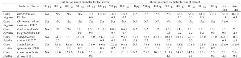
pv. gypsopholia-824. However, Chromobacterium violaceum 12472 was found to be insensitive to the extracts. The shoot and leaf extracts of Physalis peruviana showed high activity against the Gram-positive bacterium Lactococcus lactis ATCC 11454 compared with the geneticin (Table 1). Although the lowest MIC value was 100 µg/disc for Staphylococcus aureus A950277, the highest MIC value was 700 µg/disc for Escherichia coli DH5-α (Table 2). Inhibition zone on Escherichia coli DH5-α and Erwinia herbicola pv. gypsopholia-824, were not concentration dependent while Staphylococcus sp. and Lactococcus lactis ATCC 11454 inhibition zones were effected positively by increasing concentration of extracts (Table 1). In previous studies, extractions of the P. peruviana tissues with several different polar solvents showed that ethanol extracts provided the highest activity levels in response to their diffusion in agar (Acar & Goldstein, 1986; Barry, 1986). According to our results, extracts of P. peruviana did not demonstrate the highest antibacterial activity compared with positive controls (geneticin and ampicillin) except L. lactis strain. To our knowledge, this is the first demonstration of antibacterial activity from this species of Physalis grown in tissue culture. Some compounds such as withanolides from Physalis species were shown to be effective antibacterials in vitro (Kathleen et al., 2012). These results suggest that further studies should be done for identifying these compounds from tissue cultures of Physalis peruviana.
Apak et al. (2004) developed CUPRAC method to measure antioxidant capacity index for polyphenols and plant materials. CUPRAC was expressed as mmol Trolox equivalents (TE)/g extract. In our study, leaf extracts showed a higher total antioxidant capacity than shoot extracts. This may be related to the higher total phenolic contents of leaves (Kathleen et al., 2012). In our study the phenolic content was found higher in leaf extracts than shoot for both tested three concentrations. As seen from the Table 2, the highest TEAC values of the leaf (2 mg/mL) and the shoot (0.5 mg/mL) extracts are 0.291±0.04 and 0.192±0.015 respectively. Total amount of phenolics were determined with Folin-Ciocalteu reagent. Phenolic contents and antioxidant capacity of extracts were given at Table 3. The antioxidant activity of the P. physalis plant extracts is attributed to their bioactive compounds, mostly phenolics, because of their ability to scavenge free radicals (Casanova et al., 2008, Saito et al., 2008). Similarly, Rodrigues et al. (2011) confirmed that the blueberry (Vaccinium sp.) produced in Brazil is a good source of phenolic compounds, has high antioxidant activity. Although the antioxidant capacity of extracts were not very remarkable, determination of the antioxidant status of P. physalis will promote research on the identification and quantitation of active constituents of this plant.
The effect of P. peruviana extract on DNA damage protection was assessed by agarose gel electrophoresis (Figure 1). Extracts at a concentration of 30, 20 µg/mL protected pIVEX plasmid DNA from damage induced by Fe (II) plus H2O2. It can be observed from the figure that the control plasmid DNA (without Fe (II) and H2O2) contains supercoiled form small and relaxed form (lane A). At the fixed Fe (II) concentration (50 µM) and H2O2 concentration (1 mM), it could be observed that bacterial plasmid DNA breaks down by oxidative stress. H2O2 and FeCl2 induce oxidative stress by generating hydroxyl radicals which Cytotoxicity (%) = [(Absorbance of untreated group-
absorbance of treated group)/Absorbance of Untreated group] x 100 (1)
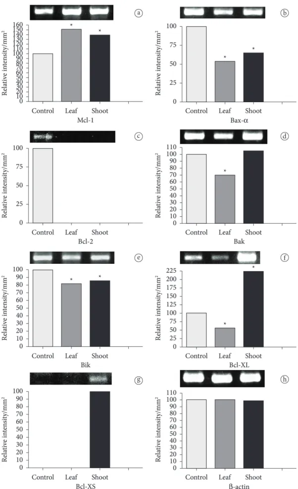
2.7 Expression analysis of Bcl-2 family genes
Cells which were grown and incubated with and without extracts in the concentration of 100 μg/mL as described before, were used for RNA isolation according to the manufacturer’s instructions (GenElute Mammalian Total RNA Purification Sigma, RTN70). Total RNA was extracted from HeLa cells, and was reverse-transcribed to the first cDNA strand (Fermentas, K1621). The amplification for Bcl-2 family genes (Mcl-1, Bfl-1, Bax-α, Bcl-2, Bak, Bik, Bcl-XL, Bcl-XS) and actin gene were performed using primers from Takara Apoprimer set (Catalog No: 6623). The amplification reaction was carried out in a 25 µl volume containing 10-20 ng of cDNA. The PCR mixture consisted of 1×PCR buffer (50 mM KCl, 20 mM Tris-HCl, pH 8.4), 1.5 mM MgCl2, 10 mM dNTPS, 20 µM of each primer, and 1 units of Taq DNA polymerase. Sterile distilled water was used to complete the volume to 25 µl and the reaction was performed using the thermal cycler (Biorad, T-100). The PCR mixture was incubated at 94 °C for 2 min prior to amplification for 30 cycles. Each cycle consisted of denaturation at 94 °C for 30 sec, annealing at 60 °C for 30 sec, and elongation at 72 °C for 30 sec. The PCR product was subjected to 1.2% agarose gel electrophoresis and observed under ultraviolet light. Agarose gels were imaged on densitometer (GS-800 Calibrated Densitometer, BIO-RAD) with ‘Quantity One® SW’ software.
2.8 Statistical analysis
All experiments were done as three replicates. Analysis of variance was used to distinguish the difference among groups. Significance was assumed at 5% level. For detecting the point of difference the post Tukey test was used. Data is presented as means ± S.D. Graphpad prism trial version is used for statistical analysis.
3 Results and discussion
Plants are important source of potentially new chemotherapeutic agents. A lot of reports are available on biological properties of plants (Chew et al., 2012; Kováts et al., 2010; Najjaa et al., 2011; Xia et al., 2011). These studies resulted in isolating and identifying the new active compounds responsible for biological activities and in developing new drugs for the therapeutic use. It is well known that Physalis peruviana has many pharmaceutical beneficial effects. A variety of molecules from P. peruviana, were reported to be medically beneficial (Chiang et al., 1992; Dimayuga et al., 1998; Kennelly et al., 1997).
o d S ci . T ec hn ol, C am p in
as, 34(2): 422-430, A
p r.-J un e 2014 425 Ç ak
ır et a
l.
Table 1. Antibacterial activity of etanolic extracts of Physalis peruviana.
Inhibition zones diameter for leaf extract Inhibition zones diameter for shoot extract
Bacteriall Strains 100 µg 200 µg 400 µg 600 µg 700 µg 800 µg 900 µg 1000 µg 100 µg 200 µg 400 µg 600 µg 700 µg 800 µg 900 µg 1000 µg Gen 10µg Amp 10µg Gram Negative Escherichia coli DH5-α
NA NA NA NA 8 ± 0.8
8 ± 0.8 7.6 ± 0.5
7.6 ± 0.5
NA NA NA NA 7.5 ± 1.3
8.5 ± 1.3
6.6 ± 0.5
7 ± 1 18.3 ± 1,5 21.3 ± 0.5 Gram Negative Chromobacterium violaceum 12472
NA NA NA NA NA NA NA NA NA NA NA NA NA NA NA NA 17 ± 0
-Gram Negative
Erwinia herbicola
pv. gypsopholia-824
NA NA NA 8.6 ± 0.5
7.5 ± 0.8
8 ± 0.8 8.8 ± 0.7
8.6 ± 0.7
NA NA NA 9.6 ± 0.5 9.1 ± 0.2 7.8 ± 0.2 9.6 ± 0.2 9.3 ± 0.5 19.3 ± 0.5 26 ± 2.3 Gram Positive Staphylococcus aureus A950277
NA 7.1 ± 0.2
8 ± 1 8.5 ± 0 10 ± 0 9.6 ± 0.5 10.1 ± 0.2 9.3 ± 0.5 7.5 ± 0.8 7.8 ± 0.2
8.8 ± 1 10.3 ± 0.5
10 ± 0 9.3 ± 0.5
10 ± 0 10 ± 0 22.6 ± 0.5 16.3 ± 1 Gram Positive Staphylococcus epidermidis 14990
NA 7.3 ± 0.5
8.1 ± 0.2
8.8 ± 0.2
10 ± 0 9.6 ± 0.5
10.1 ± 0.2
9.8 ± 0.7
NA 7.3 ± 0.5 8.1 ± 0.2 9.6 ± 0.5 9.8 ± 0.2
10 ± 0 10.3 ± 0.2
10.1 ± 0.2
22 ± 0 16 ± 0
Gram Positive
Lactococcus lactis
ATCC 11454
NA 8 ± 0 12 ± 0 12 ± 0 15.6 ± 0.5
17 ± 1 17 ± 1 16 ± 1 NA 7 ± 0 10 ± 0 11.3 ± 1.1
14 ± 0 14.3 ± 0.5 15.3 ± 1.1 15.6 ± 0.5 16.3 ± 2.3 28.6 ± 0.5
Food Sci. Technol, Campinas, 34(2): 422-430, Apr.-June 2014 426
Biological activities of Physalis peruviana ethanol extracts
hepatoma cells lines, the IC50 values were found 9.43±0.30, 41.25±1.40, >100µg/mL, respectively.
Bcl-2 family proteins regulate apoptosis by governing mitochondrial outer membrane permeabilization. Bcl-2, Bcl-Xl and Mcl-1 Of Bcl-2 family members are antiapoptotic and arrest apoptosis. Bax, Bak, Bid, Bik, Bim, and Bad are proapoptotic and trigger apoptosis (Chang & Yang, 2000; Evan & Vousden, 2001; Bhalla, 2003). These proteins generate heterodimers and they share high sequences homology (BH1-4 regions). While antiapoptotic proteins share all these four homolog regions, break down supercolied form of plasmid. All extracts exhibited
significant protection against DNA damage-induced by hydroxyl radical generated by Fenton reaction. Table 4 exhibited ratio between supercoiled and relaxed form DNA. The protective effects of extracts on DNA damage were significantly increased concentration-dependent manner, especially in leaf extracts. Furthermore, the extracts exhibited antioxidant activity and a high amount of phenolic content. These findings indicate that the amount of phenolic contents of extracts could be contributed to potential antioxidant activities.
In this study, we aimed to investigate the changes on cytotoxicity and apoptotic genes expression levels by applying P. peruviana extracts to HeLa cell lines. We evaluated the effects of P.peruviana extracts on Hela cells by MTT assay to investigate the cytotoxic activity. As shown in Figure 2, the ethanolic extracts of leaf and shoot exhibits low cytotoxic activity against HeLa cells for low concentrations, however it was shown that both extracts have an IC50 value of 100 μg/mL and this concentration was used for further experiments. The most effective value of extracts was found to be the 200 µg/mL in both shoot and leaf ethanol extracts (Figure 2). But 100 µg/mL value was chosen for further studies, because it is the value that inhibition of viability is almost fifty percent. Wu et al., (2004) also performed cytotoxic assays with ethanolic extracts of P. peruviana on the Hep G2, Hep 3B, and PLC/PRF/5 human
Table 2. Determination of minimum inhibitory concentration (MIC) of extracts as µg/disc.
Minimum inhibitory concentration (MIC) as µg/disc
Extracts Escherichia coli DH5-α
Chromobacterium violaceum 12472
Erwinia herbicola pv. gypsopholia-824
Staphylococcus aureus A950277
Staphylococcus epidermidis 14990
Lactococcus lactis ATCC 11454
Physalis peruviana leaf extract
700 ND 450 200 200 200
Physalis peruviana shoot extract
700 ND 450 100 200 200
ND: Not detected.
Table 3. Total antioxidant capacity and total phenolic content of P. peruviana extracts.
Leaf Extract Shoot Extract
0.5 mg/mL 1 mg/mL 2mg/mL 0.5 mg/mL 1 mg/mL 2mg/mL
TEAC* 0.245±0.015 0.255±0.01 0.291±0.04 0.192±0.015 0.175±0.009 0.154±0.003
TPC** (mg/mL) 9.26±0.003 18.54±0.38 40.69±0.21 7.59±0.28 15.21±0.46 30.21±0.71
*trolox equivalent antioxidant capacity. **TPC (Total Phenolic Content). Antioxidant capacity were expressed as mean ±SD.
Figure 1. Electrophoresis picture of pIVEX plasmid DNA. The sample was treated by 5 µM Fe(II) and 1 mM H2O2 for 60 min with different concentrations of P. peruviana (A) control without Fe(II) and H2O2; (B) 30 µg/mL Shoot (C) 20 µg/mL Shoot; (D) 10 µg/mL Shoot; (E) 30 µg/ mL Leaf; (F) 20 µg/mL Leaf (G) 10 µg/mL Leaf and (H) control with Fe(II) and H2O2.
Table 4. Ratio between supercoiled and relaxed form DNA after treatment with hydroxyl radical and P.peruviana extract.
Concentration (µg/ml) Ratio between supercoiled and relaxed
form DNA
Shoot Extracts 10 1.33*
20 1.40*
30 1.33*
Leaf Extracts 10 1.29*
20 1.35*
30 1.36*
Control without Fe(II) and H2O2 1.46*
Control with Fe(II) and H2O2 1.13
was presented with significant decrease when compared to the control. We found that proapoptotic Bax gene was expressed decreasingly in exract treated conditions. In previous studies it was considered that ratio of Bax to Bcl-2, was the mark of the susceptibility of cells to death signals (Marzo & Naval, 2008). For this reason, Bcl-2 proteins are an attractive target for the development of novel anticancer drugs (Chang et al., 2005). In numerous studies it was determined that alterations in Bcl-2/Bax ratio were caused by downregulation of Bcl-2, Bax (Cha et al., 2004; Paris et al., 2007; Mohammad et al., 2008) and downregulation of Bcl-2 with no change in the level of Bax (Han et al., 2008). In our study we determined no Bcl-2 expression of extract treated cells, Bax-α expression level was also decreased but still ongoing. These results suggest that P. peruviana leaf and shoot extracts might have induced apoptosis by the regulation of this ratio and the extracts damage mitochondrial membrane potential instead of protecting it.
4 Conclusion
It is still important to investigate and explore natural sources for the products that are candidates of new anticancer drugs. The last few decades, screening of medicinal plants for potential anticancer properties has increased greatly. Results that obtained from this study is thought to be encouraging for the forthcoming studies about medicinal plant extracts. The active components and mechanism(s) of action of P. peruviana leaf and shoot extracts should also be investigated for further studies in both in vitro and in vivo models. It may also be useful for investigations that focus on treating cancer with plant based natural products.
Acknowledgements
Research supported by the Research Fund of the Istanbul University (#36493).
proapoptotic ones share only three of them or just BH-3 region. Due to these homolog regions, these proteins act pro- or antiapoptotic (Shamas-Din et al., 2011). It is well known that pro- and anti-survival Bcl-2 family proteins decide whether the cell will die- if more Bcl-xL is existing, pores of the cell are non-permeable and the cell remains alive. We examined effects of P. peruviana shoot and leaf ethanol extracts on expression rates of Bcl-2 gene family to investigate molecular mechanism of extract stimulated apoptosis. For 48 h application, expression rates of proapoptotic (Bax), antiapoptotic (Bcl-2) genes of Bcl-2 family and β-actin as internal standard are presented on Figure 3. Bcl-2 is a regulator component of apoptosis which acts as an antiapoptotic protein. Azizi et al. (2009) determined that in the presence of Astrodaucus persicus plant extracts p53 gene expression increased significantly but Bcl-2 expression decreased in human breast cancer T47D cells. Also, treatment of T47D cells with plant extracts reduced the nuclear staining of p53 and cytoplasmic staining of Bcl-2 proteins. Azizi et al. (2009) stated that, the methanolic extracts of Astrodaucus persicus might contain bioactive compounds. In our study, Bcl-2 gene was not expressed in both P. peruviana leaf and shoot extracts treated HeLa cells when compared to the control cells. Shafi et al.(2009) demonstrated that Bcl-2 and bcl-XL expression was significantly inhibited while p53 and caspase -3,-8 &-9 expression was increased when HeLa cells treated with Nigella sativa chloform fraction of seed extracts. They showed that the Nigella sativa induced apoptosis by regulating apoptotic genes. In our study, antiapoptotic Bcl-XL expression decreased in leaf but increased in shoot extract treated HeLa cells. Interestingly, in shoot extract treated cells proapoptotic Bcl-XS was expressed slightly. Furthermore, Bfl-1 gene which is an antiapoptotic member of Bcl-2 family was not expressed at all. On each experimental group, we determined that proapoptotic bak gene was expressed, antiapoptotic Mcl-1 was also expressed in HeLa cells when extracts applied. It was found that Mcl-1 expression was increased slightly. Expression rates of proapoptotic bik gene
Food Sci. Technol, Campinas, 34(2): 422-430, Apr.-June 2014 428
Biological activities of Physalis peruviana ethanol extracts
Chiang, H. C., Jaw, S. M., Chen, C. F., & Kan, W. S. (1992). Antitumor agent, physalin F from Physalis angulata L. Anticancer Research, 12(3), 837-843. PMid:1622143.
Dimayuga, R. E., Virgen, M., & Ochoa, N. (1998). Antimicrobial activity of medicinal plants from Baja California Sur (México). Pharmaceutical Biology, 36(1), 33-43. http://dx.doi.org/10.1076/ phbi.36.1.33.4625
Elmore, S. (2007). Apoptosis: a review of programmed cell death. Toxicologic Pathology, 35(4), 495-516. PMid:17562483 PMCid:PMC2117903. http://dx.doi.org/10.1080/01926230701320337
Evan, G. I., & Vousden, K. H. (2001). Proliferation, cell cycle and apoptosis in cancer. Nature, 411, 342-347. PMid:11357141. http:// dx.doi.org/10.1038/35077213
Hamad, I., Erol, O., Pekmez, M., Ucar, E., & Arda, E. S. N. (2010). Antioxidant and cytotoxic activities of aphanes arvensis extracts. Plant Foods for Human Nutrition, 65(1), 44-49. PMid:20108047. http://dx.doi.org/10.1007/s11130-009-0151-y
Han, M. H., Yoo, Y. H., & Choi, Y. H. (2008). Sanguinarine-induced apoptosis inhuman leukemia U937 cells via Bcl-2 downregulation and caspase-3 activation. Chemotherapy, 54(3), 157-165. PMid:18560221. http://dx.doi.org/10.1159/000140359
Kathleen, A., Gibson, R., Reese, N., Halaweish, F. T., & Ren, Y. (2012). Isolation and characterization of a bactericidal withanolide from Physalis virginiana. Pharmacognosy Magazine, 8(29), 22-28. PMid:22438659 PMCid:PMC3307198. http://dx.doi. org/10.4103/0973-1296.93307
Kennelly, E. J., Gerhaeuser, C., Song, L. L., Graham, J. G., Beecher, C. W., Pezzuto, J. M., & Kinghornet, A. D. (1997). Induction of quinone reductase by withanolides isolated from Physalis philadelphica (tomatillos). Journal of Agricultural Food Chemistry, 45(10), 3771-3777. http://dx.doi.org/10.1021/jf970246w
Kováts, N., Gölöncsér, F., Ács, A., & Refaey, M. (2010). Quantification of the antibacterial properties of Artemisia absinthium, A. vulgaris, Chrysanthemum leucanthemum and Achillea millefolium using the Vibrio fischeri bacterial bioassay. Acta Botanica Hungarica, 52(1-2), 137-144. http://dx.doi.org/10.1556/ABot.52.2010.1-2.e2 Lim, Y. Y., Lim, T. T., & Jing, J. (2009). Antioxidant properties of
guava fruit: comparison with some local fruits. Sunway Academic Journal, 3, 9-20.
Martinez, M. Revision of Physalis section Epeteiorhiza (Solanaceae). (1998). Anales del Instituto de Biología. Serie Zoología, 69, 71-117. Marzo, I., & Naval, J. (2008). Bcl-2 family members as molecular
targets incancer therapy. Biochemical Pharmacology, 76(8), 939-946. PMid:18638457. http://dx.doi.org/10.1016/j.bcp.2008.06.009 Mohammad, R., Giri, A., & Goustin, A. S. (2008). Small-molecule
inhibitors of Bcl-2 family proteins as therapeutic agents in câncer. Recent Patents on Anti-Cancer Drug Discovery, 3(1), 20-30. PMid:18289121. http://dx.doi.org/10.2174/157489208783478676 Mosmann, T. (1983). Rapid colorimetric assay for cellular growth
and survival: application to proliferation and cytotoxicity assays. Journal of Immunological Methods, 65(1-2), 55-63. http://dx.doi. org/10.1016/0022-1759(83)90303-4
Murashige, T., & Skoog, F. (1962). A revised medium for rapid growth and bioassays with tobacco tissue cultures. Plant Physiology, 15(3), 473-497. http://dx.doi.org/10.1111/j.1399-3054.1962.tb08052.x Najjaa, H., Zerria, K., Fattouch, S., Ammar, E., & Neffati, M. (2011).
Antioxidant and antimicrobial activities of Allium roseum L. “Lazoul,” a wild edible endemic species in North Africa. International Journal of Food Properties, 14(2), 371-380. http:// dx.doi.org/10.1080/10942910903203164
References
Acar, J. F., & Goldstein, F. W. (1986). Disc susceptibility test. In L. Lorian (Ed.), Antibiotics in laboratory medicine (2nd ed., pp. 27-63). Baltimore: Williams and Wilkins.
Agbor, G. A., Oben, J. E., Ngogang, J. Y., Xinxing, C., & Vinson, J. A. (2005). Antioxidant capacity ofsome herbs/spices from Cameroon: a comparative study of two methods. Journal of Agricultural Food Chemistry, 53(17), 6819-6824. PMid:16104805. http://dx.doi. org/10.1021/jf050445c
Apak, R., Guçlu, K., Ozyurek, M., & Karademir, S. E. (2004). Novel total antioxidant capacity index for dietary polyphenols and vitamins C and E, using their cupric ion reducing capability in the presence of neocuproine: CUPRAC method. Journal of Agricultural Food Chemistry, 52(26), 7970-7981. PMid:15612784. http://dx.doi. org/10.1021/jf048741x
Azizi, E., Abdolmohammadi, M. H., Fouladdel, S., Shafiee, A., Amin, G., & Ghaffari, S. M. (2009). Evaluation of p53 and Bcl-2 genes and proteins expression in human breast cancer T47D cells treated with extracts of Astrodaucus persicus (Boiss.) Drude in comparison to Tamoxifen. DARU Journal of Pharmaceutical Sciences, 17(3), 181-186.
Barry, A. L. (1986). Procedure for testing antimicrobial agents in agar media: theoretical considerations. In L. Lorian (Ed.), Antibiotics in laboratory medicine. (2nd ed., pp. 1-26). Baltimore: Williams and Wilkins. PMid:3088028 PMCid:PMC268820.
Bhalla, K. N. (2003). Microtubule-targeted anticancer agents and apoptosis. Oncogene, 22, 9075-9086. PMid:14663486. http://dx.doi. org/10.1038/sj.onc.1207233
Brusotti, G., Cesari, I., Dentamaro, A., Caccialanza, G., & Massolini, G. (2014). Isolation and characterization of bioactive compounds from plant resources: The role of analysis in the ethnopharmacological approach. Journal of Pharmaceutical and Biomedical Analysis, 87, 218-228. PMid:23591140. http://dx.doi. org/10.1016/j.jpba.2013.03.007
Caceres, A., Alvarez, A. V., Ovando, A. E., & Samayoa, B. E. (1991). Plants used in Guatemala for the treatment of respiratory diseases. Screening of 68 plants against gram-positive bacteria. Journal of Ethnopharmacology, 31(2), 193-208. http://dx.doi. org/10.1016/0378-8741(91)90005-X
Casanova E., Garcia-Mina, J. M., & Calvo, M. I. (2008). Antioxidant and antifungal activity of Verbena officinalis L. leaves. Plant Foods For Human Nutrition, 63(3), 93-145. PMid:18498054. http://dx.doi. org/10.1007/s11130-008-0073-0
Cha, Y. Y., Lee, E. O., Lee, H. J., Park, Y. D., Ko, S. G., Kim, D. H., Kim, H. M., Kang, I. C., & Kim, S. H. (2004). Methylenechloride fraction of Scutellaria barbata induces apoptosis inhuman U937 leukemia cells via the mitochondrial signaling pathway. Clinica Chimica Acta, 348(1-2), 41-48. PMid:15369734. http://dx.doi.org/10.1016/j. cccn.2004.04.013
Chang, H. Y., & Yang, X. (2000). Proteases for cell suicide: functions and regulation of caspases. Microbiology and Molecular Biology Reviews, 64(4), 821-826. PMid:11104820 PMCid:PMC99015. http:// dx.doi.org/10.1128/MMBR.64.4.821-846.2000
Chang, J., Hsu, Y., Kuo, P., Kuo, Y., Chiang, L., & Lin, C. (2005). Increase of Bax/Bcl-XL ratio and arrest of cell cycle by luteolin in immortalized human hepatoma cell line. Life Science, 76(16), 1883-1893. PMid:15698865. http://dx.doi.org/10.1016/j.lfs.2004.11.003 Chew, A. L., Jessica, J. J. A., & Sasidharan, S. (2012). Antioxidant and
Food Sci. Technol, Campinas, 34(2): 422-430, Apr.-June 2014 430
Biological activities of Physalis peruviana ethanol extracts
Shamas-Din, A., Brahmbhatt, H., Leber, B., & Andrews, D. W. (2011) BH3-only proteins: orchestrators of apoptosis. Biochimica et Biophysica Acta, 1813(4), 508-520. PMid:21146563. http://dx.doi. org/10.1016/j.bbamcr.2010.11.024
Silva, B. A., Ferreres, F., Malva, J. O., & Dias, A. C. P. (2005). Phytochemical and antioxidant characterization of Hypericum perforatum alcoholic extracts. Food Chemistry, 90(1-2), 157-167. http://dx.doi.org/10.1016/j.foodchem.2004.03.049
Xia, D. Z., Yu, X. F., Zhu, Z. Y., & Zou, Z. D. (2011). Antioxidant and antibacterial activity of six edible wild plants (Sonchus spp.) in China. Natural Products Research, 25(20), 1893-1901. PMid:21793765. http://dx.doi.org/10.1080/14786419.2010.534093 Wu, S. J., Ng, L. T., Chen, C. H., Lin, D. L., Wang, S. S., & Lin, C. C.
(2004). Antihepatoma activity of Physalis angulata and P. Peruviana extracts and their effects on apoptosis in human Hep G2 cells. Life Science, 74(16), 2061-2073. PMid:14967200. http://dx.doi. org/10.1016/j.lfs.2003.09.058
Yen, C. Y., Chiu, C. C., Chang, F. R., Chen, J. Y., Hwang, C. C., Hseu, Y. C., Yang, H. L., Lee, A. Y., Tsai, M. T., Guo, Z. L., Cheng, Y. S., Liu, Y. C., Lan, Y. H., Chang, Y. C., Ko, Y. C., Chang, H. W., & Wu, Y. C. (2010). 4b-Hydroxywithanolide E from Physalis peruviana (golden berry) inhibits growth of human lung cancer cells through DNA damage, apoptosis and G2/M arrest. BMC Cancer, 10(46). PMid:20167063 PMCid:PMC2830937. http://dx.doi. org/10.1186/1471-2407-10-46
National Committee for Clinical Laboratory Standards - NCCLS. (2003). NCCLS M2-A7: methods for disc susceptibility tests for bacteria that grow aerobically. Wayne: NCCLS.
Paris, C., Bertoglio, J., & Breard, J. (2007). Lysosomal and mitochondrial pathwaysin milterfosine-induced apoptosis in U937 cells. Apoptosis, 12(7), 1257-1267. PMid:17347868. http://dx.doi. org/10.1007/s10495-007-0052-1
Park, B. J., Lim, Y. S., Lee, H. J., Eum, W. S., Park, J., Han, K. H., Choi, S. Y., & Lee, K. S. (2009). Anti oxidative effects of Phellinus linteus and red ginseng extracts on oxidative stress-induced DNA damage. BMB Reports, 42(8), 500-505. PMid:19712586. http://dx.doi.org/10.5483/ BMBRep.2009.42.8.500
Rodrigues, E., Poerner, N., Rockenbach, I. I., Gonzaga, L. V., Mendes, C. R., & Fett, R. (2011). Phenolic compounds and antioxidant activity of blueberry cultivars grown in Brazil. Ciência e Tecnologia de Alimentos, 31(4), 911-917.
Saito, K., Kohno, M., Yoshizaki, F., & Niwano, Y. (2008). Extensive screening for edible herbal extracts with potent scavenging activity against superoxide anions. Plant Foods For Human Nutrition, 63(2), 65-70. PMid:18236159. http://dx.doi.org/10.1007/ s11130-008-0071-2