Article
ISSN 0102-695X
http://dx.doi.org/10.1590/S0102-695X2011005000176 Received 18 Oct 2010 Accepted 12 Jan 2011 Available online 23 Sep 2011
genotoxicity and free radical scavenging
activities of
Stryphnodendron adstringens
(“barbatimão”) leaf extracts
Plínio R. dos Santos Filho,
1Lidiane A. Ferreira,
2Cibele M. C.
Paiva Gouvêa
*,21Departamento de Biologia Vegetal, Laboratório de Bioquímica de Plantas, Instituto
de Biologia, Universidade Estadual de Campinas, Brazil,
2Instituto de Ciências da Natureza, Universidade Federal de Alfenas, Brazil.
Abstract: The present study describes for the first time the antigenotoxic and antioxidant properties of Stryphnodendron adstringens (Mart.) Coville, Fabaceae (“barbatimão”) leaves. The aqueous (AEB), water fraction (WFB) and ethanolic (EEB) extracts of leaves were obtained and they presented high content of phenolic compounds, flavonoids and proanthocyanidins. WFB and EEB inhibited the genotoxicity induced by cyclophosphamide. WFB inhibited the DNA lesion formation and both WFB and EEB decreased significantly (p<0.05) micronucleus formation. All extracts also showed high DPPH radical scavenging activity and reducing power. In conclusion all extracts presented antioxidant activity and WFB and EEB protected cells from genotoxicity induced by cyclophosphamide in rat bone marrow cells. Thus, our results support the beneficial effects of the barbatimão extracts as an anticarcinogenic agent.
Keywords:
antioxidant apoptosis cytotoxicity DNA ladder micronucleus
Introduction
The use of herbal medicine (leaves, l owers,
roots, stems and berries) is a traditional form of healthcare (Diwanay et al., 2004). In this context, an important focus of the search is to understand the true
efi cacy of plant materials and determine its mechanism
of action in different experimental models (Romero-Jiménez et al., 2005). Although human body is equipped
with self defense mechanisms such as detoxii cation
process through various enzymes, serious exposure to dangerous chemicals can cause changes in the genetic material (mutations) leading to carcinogenic events (Edenharder et al., 1998; Belitsky & Yakubovskaya, 2008). Cyclophosphamide (CP), an antineoplastic agent, and its metabolites can bind DNA, causing damage that may result in chromosome breaks, micronucleus formation and cell death (Murata et al., 2004). Beyond that, CP alters the redox balance of tissues suggesting that physiological and biochemical disturbances may result from oxidative stress (Sugumar et al., 2007).
Oxidative stress is an important factor in the genesis of several pathologies, from cancer to cardiovascular and degenerative diseases. Human cells are constantly exposed to reactive oxygen species (ROS) generated by a number of biotic and abiotic factors that
can harm the cells. Antioxidants provide protection to living organisms from damage caused by uncontrolled production of ROS and the concomitant oxidation of nucleic acids, proteins, lipids and carbohydrates, which may subsequently determine mutagenesis and diseases related to DNA damage (Halliwell, 2007a; 2007b). Anticarcinogens/anti-mutagens, often used in traditional medicine, can prevent or revert some of the effects produced by carcinogens. However, knowledge about the mechanism of action of many anti-carcinogens, e.g., phytochemicals, is still poor (Paolini & Nestle, 2003).
Because of their wide spectrum of activities,
constituents such as phenolic compounds and l avonoids
have been the subject of many investigations. An important but often neglected group of polyphenolic compounds is that of the tannins, which are widely distributed in higher
plants and they have proven to possess health benei ts
(Okuda, 2005).
Stryphnodendron adstringens (Mart.) Coville, Fabaceae, known as ‘barbatimão’, occurs in the central savannah region of Brazil and is a rich source of tannins (Santos et al., 2002) and proanthocyanidins (Mello et al., 1996a; 1996b; 1999; Ishida et al., 2006; Lopes et al., 2008). Stem bark is the main part of the plant used
popularly as an anti-inl ammatory, astringent, in the
al., 1988; Lopes et al., 2005). Several reports have proven
the anti-ulcerogenic, anti-inlammatory, antinociceptive
and antimicrobial (Lima et al., 1998; Audi et al., 1999; Ishida et al., 2006; Ishida et al., 2009; Melo et al., 2007) activities of bark extract of S. adstringens. However there are few reports on the toxicity of barbatimão leaf extracts (Almeida et al., 2010). Thus, the aim of the present study was to determine the antigenotoxic activity of S. adstringens leaf extracts on genotoxicity induced by CP in rat bone marrow cells and the in vitro antioxidant potential of the extracts.
Materials and Methods
Plant material
Leaves of Stryphnodendron adstringens
(Mart.) Coville, Fabaceae, were collected in Alfenas (Minas Gerais, Brazil) in Julho/2007 and deposited at the Herbarium of the Universidade Federal de Alfenas, Brazil (voucher specimen N. 196).
Preparation of leaf extract
The leaves were dried at 30 °C and then pulverized. The aqueous extract (AEB) was obtained by decoction of 10 g of leaves in 100 mL of deionized water,
at 90 °C, for 10 min and iltered. The water fraction
(WFB) remained after powder leaf extraction (100 g) with 70% (v/v) acetone aqueous solution (1 L) and partitioning with ethyl acetate. The ethanol extract (EEB) was obtained by maceration of 100 g of powder in 1 L of 70% (v/v) ethanol aqueous solution and the solvent was evaporated in a rotavapor under reduced pressure. All extracts were lyophilized and stored at -20 °C, until use. For use the extracts were dissolved in deionized water.
Determination of phenolic, lavonoid and
proanthocyanidins content
The total phenolic compounds were determined using Folin-Ciocalteau’s method (Rai et al., 2006),
total lavonoids using aluminum chelating method
(Ebrahimzadeh et al., 2008) and proanthocyanidins content was obtained after acid depolymerization to the corresponding anthocyanidins (Rösch et al., 2003).
Animals and treatment
Male Wistar rats (120±20 g) were housed with access to food and water ad libitum and light-dark cycles of 12 h were used. The animals were divided into eight groups (each group consisted of six rats). Control group received water; CP group received cyclophosphamide (40 mg/kg, per gavage), 24 h before euthanasia; extract
groups received 500 mg/kg body wt, per gavage, for 7 days and extract+CP groups received the extract for 7 days and CP 24 h before euthanasia. After euthanasia, with overdose of anesthesia, the femur was quickly removed and the bone marrow cells collected.
DNA isolation and electrophoresis
The DNA of the bone marrow cells from the left femur was isolated using the SDS/Proteinase K/RNase A extraction method (DeSierve et al., 2002). Samples were electrophoresed in 0.8% (w/v) agarose gel and DNA was visualized by ethidium bromide staining. The presence of apoptosis was indicated by the appearance of a ladder of oligonucleosomal DNA fragments that are approximately 180-200 bp multiples and the necrosis by the appearance of a smear on the agarose gel.
Micronucleus assay
This assay was performed according to
Nakagawa et al. (2008), with modiications. Bone marrow cells of the right femur of each animal was lushed out as a ine suspension with 2.5 mL fetal bovine serum
(FBS) and centrifuged at 1,000 x g for 5 min. The pellet was resuspended in 0.5 mL saline. The suspension was smeared onto the precleaned air-dried slides following
ixing in 100% methanol for 5 min and stained with
Giemsa. One thousand total erythrocytes (polychromatic and normochromatic) were scored per animal. The frequency of micronucleated polychromatic erythrocytes (MNPCE) and the total PCE and NCE were determined.
Antioxidant assays
Total antioxidant capacity
The total antioxidant capacity was determined (Prieto et al., 1999), and the results were expressed as equivalents of ascorbic acid.
DPPH assay
The hydrogen atoms or electron donation ability of the extracts was measured from the bleaching of purple colored methanol solution of DPPH according to Yen & Wu (1999). L-ascorbic acid and BHT were used as the reference standards.
Reducing power
Statistical analysis
Data were analyzed using ANOVA with post hoc analysis by Tukey test, when p<0.05. Results are expressed as mean±standard error of the mean (SE).
Results and Discussion
In our study, the chemopreventive and antioxidant properties of barbatimão leaf extracts were evaluated. We used cyclophosphamide (CP), a genotoxic agent widely used both, in clinical treatment and experimentally (Murata et al., 2004; Nagar, 2010). CP belongs to the group of N-lost compounds. It requires CYP450-dependent activation, yielding the active compound 4-hydroxy-cyclophosphamide. This molecule undergoes spontaneous decomposition to the biologically active alkylating species acrolein and phosphoramide mustard, which preferentially alkylates the N7 position of guanine. Since CP is a bifunctional compound, DNA alkylation gives rise to interstrand cross-links that are thought to be responsible for the induction of cell death (Murata et al., 2004). From the DNA fragmentation analysis, it can be seen that CP induced apoptosis, confirmed by the DNA ladder pattern (Figure 1). The AEB and WFB were not genotoxic, as they did not induce DNA lesion. The WFB was antigenotoxic, as it protected cells from the apoptotic effect of CP and it could be postulated that proanthocyanidins contributed to its activity, once, amongst the studied extracts, WFB presented the highest content of these compounds.
Figure 1. Agarose gel electrophoresis of bone marrow cells genomic DNA of control and treated rats. 1, 123 bp ladder marker; 2, Control; 3, Cyclophosphamide (CP); 4, Aqueous extract (AEB); 5, AEB plus CP; 6, Water fraction (WFB); 7, WFB plus CP; 8, ethanolic extract (EEB); 9, EEB plus CP. DNA ladder formation indicates apoptosis as seem in lanes 3 and 5 and DNA smear as seen in lanes 8 and 9 indicates necrosis. The results are representative of ive independent experiments.
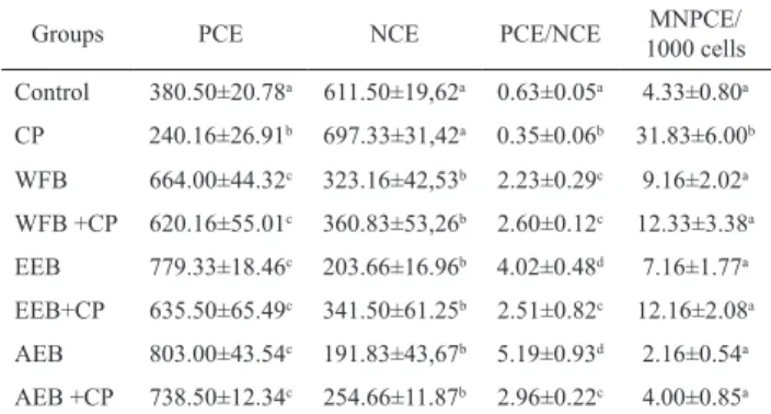
The quantitative assessment of micronucleus containing cells serves as a good indicator for the induction of structural and numerical chromosomal aberrations. Breakage of chromatids or chromosomes can result in micronuclei formation if an acentric fragment is produced; therefore assays detecting either chromosomal aberration or micronuclei are acceptable for detecting clastogenicity (Natarajan, 2002) and the micronucleus test is recommended by the Brazilian Health Surveillance Agency for phytotherapic preclinical toxicity studies (Anvisa, 2004). The results of micronucleus assay are shown in Table 1. The data of this test corroborates the
indings of the DNA fragmentation test. A signiicant
(p<0.05) increase in the percentage of micronuclei was observed after treatment with CP as compared to the control and treated groups. Administration of CP in combination
with extracts showed signiicant decrease (p<0.001) in the percentage of micronuclei as compared to CP treated rats. In the groups treated with extracts the PCE/NCE ratio increased, indicating, possibly, a cell proliferation stimulation activity of the extracts. These results further
conirm that the extracts were antigenotoxic. Our results
are in agreement with Costa et al. (2010) who observed no genotoxic effect of the proanthocyanidin polymer-rich fraction of stem bark of S. adstringens. This work
reported no signiicant differences in the frequency
of MNPCE induced by the fraction treatment when compared to the negative control. The fraction was also antimutagenic and this activity could be attributed to the tannin content and to its antioxidant activity, due to its ability to form complexes with other macromolecules. In the present work we observed an antigenotoxic activity of all extracts and that all of them exhibited antioxidant activity.
Table 1. Frequencies of micronucleated polychromatic erythrocytes (MNPCE), polychromatic erythrocytes (PCE) and normochromatic erythrocytes (NCE) in mouse bone marrow cells of control and treated groups.
Groups PCE NCE PCE/NCE MNPCE/
1000 cells
Control 380.50±20.78a 611.50±19,62a 0.63±0.05a 4.33±0.80a
CP 240.16±26.91b 697.33±31,42a 0.35±0.06b 31.83±6.00b
WFB 664.00±44.32c 323.16±42,53b 2.23±0.29c 9.16±2.02a
WFB +CP 620.16±55.01c 360.83±53,26b 2.60±0.12c 12.33±3.38a
EEB 779.33±18.46c 203.66±16.96b 4.02±0.48d 7.16±1.77a
EEB+CP 635.50±65.49c 341.50±61.25b 2.51±0.82c 12.16±2.08a
AEB 803.00±43.54c 191.83±43,67b 5.19±0.93d 2.16±0.54a
AEB +CP 738.50±12.34c 254.66±11.87b 2.96±0.22c 4.00±0.85a
AEB, aqueous extract; WFB, water fraction and EEB, ethanolic extract of barbatimão leaves; CP, cyclophosphamide. N=6. A total of 6,000 cells were counted per group (1,000 cells per animal). The results are mean±SE. Different letters in the same column indicate signiicant difference (p<0.05) by the Tukey test.
The determination of main components of the extracts (Table 2) showed that EEB presented the
higher phenolics and lavonoids content while the WFB
presented the higher proanthocyanidins content. Phenolic compounds are regarded to be the most important antioxidative constituent of plants; hence correlations between the concentrations of plant phenolics and the total antioxidant capacities have been well reported (Gulcin et al., 2002; Oktay et al., 2003). The content of phenolic compounds in the extracts was higher than that reported to other previously examined sources of antioxidants (Hu et al., 2000; Mathew & Abraham, 2004; Basile et al., 2005). It is important to point out that the WFB presented the highest amount of proanthocyanidins and it was expected since we used a recommend solvent system to better extract this class of compound (Foo &
Porter, 1980). We further conirm the proanthocyanidin
content of the WFB by heating it with 5% HCl in tert-butanol which yielded cyanidin as the principal pigment.
Table 2. Barbatimão leaf extracts total phenolic compounds, lavonoids and proanthocyanidins content.
Extracts Content
Phenolics (%) Flavonoids (%) Proanthocyanidins (µM)
AEB 32,00±2,05a 5,02±0,29ab 23.32±1.75a
WFB 23,93±1,29b 3,63±0,22a 35.40±2.10b
EEB 34,94±2,95a 6,44±0,19b 24.75±1.28a
AEB, aqueous extract; WFB, water fraction and EEB, ethanolic extract of barbatimão leaves. The results are mean±SE of three independent experiments (N=5). Different letters in the same column indicate signiicant difference (p<0.05) by the Tukey test.
It was observed that the amount of lavonoids
in the analyzed barbatimão leaf extracts showed a high correlation with the total amount of phenolics and antioxidant activity. Extracts with high content of phenols
and lavonoids are known to exhibit high antioxidant
activity (Boudet, 2007).
All of the extracts tested presented antioxidant activity, and it could be attributed to the extract constituents. The total antioxidant capacity of the extracts was: 147.85±12.68, 124.36±7.18 and 148.64±8.41 mg ascorbic acid/g extract, for AEB, WFB and EEB respectively. The extracts also showed strong DPPH scavenging activity (Figure 2A). The free radical DPPH scavenging activity of the extracts was concentration-dependent and the maximum activity was achieved with 62.00±0.91 µg/ mL. The extracts showed a similar scavenging activity to ascorbic acid and higher than BHT. DPPH assay evaluates the ability of antioxidants to scavenge free radicals. Hydrogen-donating ability is an index of the primary antioxidant activity. In our study WFB, that had the highest content of proanthocyanidins, showed a similar radical scavenging activity to the extracts (AEB and EEB)
with higher phenols and lavonoids content than WFB.
Proanthocyanidins certainly contributed to the free radical scavenging activity of the extracts, as demonstrated by others (Kimura et al., 2008; Spranger et al., 2008). The extracts presented Fe3+ reducing capacity similar to ascorbic acid and higher than BHT and it was also concentration-dependent. The maximum reducing power was achieved
with extracts at 250 μg/mL (Figure 2B).
0 10 20 30 40 50 60 70 0
20 40 60 80 100
AEB WFB EEB AA BHT
DPPH scavenging activity (%)
Concentration (µg/mL)
0 100 200 300 400 500
2.0
1.0 1.5
0.5
0.0
AEB WFB EEB AA BHT
ABS 700
Concentration (µg/mL)
Figure 2. Antioxidant activity of barbatimão leaf extracts. A, DPPH scavenging activity; B, reducing power. AEB, aqueous extract; WFB, water fraction; EEB, ethanolic extract; AA, acid ascorbic (standard); BHT, butylated hydroxytoluene (standard). The results are mean±SE of ive independent experiments performed in triplicate.
The interplay between free radicals and antioxidants is important in maintaining health and preventing the onset and progression of several pathologies (Halliwell, 2007a; Halliwell, 2007b).
The antioxidant activity of plant compounds has been attributed to various mechanisms, including prevention of chain initiation, binding of transition metal ion catalysts, decomposition of peroxides, prevention of continued hydrogen abstraction, reductive capacity and radical scavenging (Cos et al., 2003). The results of the present work indicate that the extracts act as antioxidants. The most important target for ROS in the carcinogenesis process is probably DNA. It is believed that antioxidant
A
Belitsky GA, Yakubovskaya MG 2008. Genetic polymorphism and variability of chemical carcinogenesis. Biochemistry-Moscow 73: 543-554.
Boudet AM 2007. Evolution and current status of research in phenolic compounds. Phytochemistry 68: 2722-2735. Bruni R, Rossi D, Muzzoli M, Romagnoli C, Paganetto G, Besco
E, Choquecillo F, Peralta K, Lora WS, Sacchetti G 2006. Antimutagenic, antioxidant and antimicrobial properties of Maytenus krukovii bark. Fitoterapia 77: 538-545. Cos P, De Bruyne T, Hermans N, Apers S, Berghe DV, Vlietinck
AJ 2003. Proanthocyanidins in health care: current and new trends. Curr Med Chem 11: 1345-1359.
Costa MA, Ishida K, Kaplum V, Koslyk EDA, Mello JCP, Ueda-Nakamura T, Dias Filho BP, Nakamura CV 2010. Safety evaluation of proanthocyanidin polymer-rich fraction obtained from stem bark of Stryphnodendron adstringens (Barbatimao) for use as a pharmacological agent. Regul Toxicol Pharmacol 58: 330-335.
DeSiervi A, Vazquez ES, Rezaval C, Rossetti MV, Del Batlle AM 2002. Delta-aminolevulinic acid cytotoxic effects on human hepatocarcinoma cell lines. BMC Cancer 2: 6. Diwanay S, Chitre D, Patwardhan B 2004. Immunoprotection
by botanical drugs in chemotherapy. J Ethnopharmacol 90: 49-55.
Ebrahimzadeh MA, Pourmorad F, Bekhradnia AR 2008. Iron chelating activity, phenol and lavonoid content of some medicinal plants from Iran. African J Biotech 7: 3188-3192.
Edenharder R, Frangart J, Hager M 1998. Protective effects of fruits and vegetables against in vivo clastogenicity of cyclophosphamide or Benzo[α] pyrene in mice. Food Chem Toxicol 36: 637-645.
Foo LY, Porter LJ 1980. The phytochemistry of proanthocyanidin polymers. Phytochemistry 19: 1747-1754.
Gulcin I, Oktay M, Kufrevioglu OI, Aslan A 2002. Determination of antioxidant activity of lichen Cetraria islandica (L.) Ach. J Ethnopharmacol 79: 325-329.
Halliwell B 2007a. Oxidative stress and cancer: have we moved forward? Biochem J 401: 1-11.
Halliwell B 2007b. Biochemistry of oxidative stress. Biochem Soc Trans 35: 1147-1150.
Hosseinimehr SJ, Azadbakht M, Abadi AJ 2008. Protective effect of hawthorn extract against genotoxicity induced by cyclophosphamide in mouse bone marrow cells. Environ Toxicol Pharmacol 25: 51-56.
Hu C, Zhang Y, Kitts DD 2000. Evaluation of antioxidant and prooxidant activity of bamboo Phyllostachys nigra var. henonis leaf extract in vitro. J Agr Food Chem 48: 3170-3176.
Ishida K, Mello JC, Cortez DA, Filho BP, Ueda-Nakamura T, Nakamura CV 2006. Inluence of tannins from Stryphnodendron adstringens on growth and virulence factors of Candida albicans. J Antimicrob Chemother 58: 942-949.
Ishida K, Rozental S, de Mello JC, Nakamura CV 2009. Activity
properties of these phytochemical materials protect cells from ROS-mediated DNA damage that can result in mutation and subsequent carcinogenesis and they may donate hydrogen to free radicals, leading to non-toxic species and therefore to inhibition of the propagation phase of lipid oxidation (Hosseinimehr et al., 2008).
The barbatimão leaf extracts might be reducing the micronuclei formation by scavenging the DNA damaging free radicals generated following CP exposure. Another possible extracts mechanism of action might involve selective removal of cells with DNA damage by apoptosis, which seems to be case of EEB. Proanthocyanidins and flavonoids are known to form a complex with proteins and monooxygenases metabolizing xenobiotics, due to the fact that the formers can act as multidentate ligands and thus are able to bind simultaneously to more than one point of the protein surface. The more the oligomeric nature, the more its association produces an increased antimutagenic activity towards the inhibition of the enzyme mediated genotoxicity of indirect acting mutagens (Bruni et al., 2006).
In conclusion, the present study demonstrates that WFB and EEB protected cells from the CP chemical-induced genotoxicity and all extracts presented powerful
antioxidant activity. Thus, our results support the beneicial
effects of the barbatimão extracts as anticarcinogenic agent.
Acknowledgments
We gratefully acknowledge inancial support
(Process 1305-06) and fellowship (PRSF and LAF) received from FAPEMIG.
References
Almeida AC, Sobrinho EM, de Pinho L, Souza PNS, Martins ER, Duarte ER, Santos HO, Brandi IV, Cangussu AS, Costa JPR 2010. Toxicidade aguda dos extratos hidroalcoólicos das folhas de alecrim-pimenta, aroeira e barbatimão e do farelo da casca de pequi administrados por via intraperitoneal. Cienc Rural 40: 200-203. Anvisa 2004. Resolução-RE No. 90 da Agência Nacional de
Vigilância Sanitária. Guia para a realização de estudos de toxicidade pré-clínica de itoterápicos.
Audi EA, Toledo DP, Peres PG, Kimura E, Pereira WK, Melo JC, Nakamura C, Alves-do-Prado W, Cumam RK, Bersani-Amado CA 1999. Gastric antiulcerogenic effects of Stryphnodendron adstringens in rats. Phytother Res 13: 264-266.
of tannins from Stryphnodendron adstringens on Cryptococcus neoformans: effects on growth, capsule size and pigmentation. Ann Clin Microbiol Antimicrob 8: 29.
KimuraY, Ito H, Kawaji YM, Ikami T, Hatano T 2008. Characterization and antioxidative properties of oligomeric proanthocyanidin from prunes, dried fruit of Prunus domestica L. Biosci Biotechnol Biochem 72: 1615-1618.
Lima JCS, Martins DTO, De Souza PT, 1998. Experimental evaluation of stem bark of Stryphnodendron adstringens (Mart.) Coville for anti-inlammatory activity. Phytother Res 12: 218-220.
Lopes GC, Sanches ACC, Nakamura CV, Dias Filho BP, Hernandes L, de Mello JCP 2005. Inluence of extracts of Stryphnodendron polyphyllum Mart. and Stryphnodendron obovatum Benth. on the cicatrisation of cutaneous wounds in rats. J Ethnopharmacol 99: 265-272.
Lopes GC, Machado FAV, Toledo CEM, CM, Mello JCP 2008. Chemotaxonomic signiicance of 5-deoxyproanthocyanidins in Stryphnodendron species. Bioch Syst Ecol 36: 925-931.
Mathew S, Abraham ET 2004. In vitro antioxidant activity and scavenging effects of Cinnamomum verum leaf extract assayed by different methodologies. Food Chem Toxicol 44: 198-206.
Melo JO, Endo TH, Bersani-Amado LE, Svidzinski AE, Baroni S, Mello JCP, Bersani-Amado CA 2007. Effect of Stryphnodendron adstringens (barbatimão) bark on animal models of nociception. Rev Bras Ciênc Farm 43: 465-469.
Mello JCP, Petereit F, Nahrstedt A 1996a. Flavan-3-ols and prodelphinidins from Stryphnodendron adstringens. Phytochemistry 41: 807-813.
Mello JCP, Petereit F, Nahrstedt A 1996b. Prorobinetinidins from Stryphnodendron adstringens. Phytochemistry 42: 857-862.
Mello JCP, Petereit F, Nahrstedt A 1999. A dimeric proanthocyanidin from Stryphnodendron adstringens. Phytochemistry 51: 1105-1107.
Murata M, Suzuki T, Midorikawa K, Oikawa S, Kawanishi S 2004. Oxidative DNA damage induced by a hydroperoxide derivative of cyclophosphamide. Free Radic Biol Med 37: 793-802.
Nagar S 2010. Pharmacokinetics of anti-cancer drugs used in breast cancer chemotherapy. Adv Exp Med Biol 678:124-132.
Nakagawa K, Hidaka T, Kitano M, Asakura M, Kamigaito T, Noguchi T, Hosoe K 2008. Genotoxicity studies on licorice lavonoid oil (LFO). Food Chem Toxicol 46: 2525-2532.
Natarajan AT 2002. Chromosome aberrations: past, present and future. Mut Res 504: 3-16.
Oktay M, Gulçin I, Kufrevioglu OI 2003. Determination of in vitro antioxidant activity of fennel (Foeniculum
vulgare) seed extracts. Lebenson Wiss Technol 36: 263-271.
Okuda T 2005. Systematics and health effects of chemically distinct tannins in medicinal plants. Phytochemistry 66: 2012-2031.
Oyaizu M 1986. Studies on product of browning reaction prepared from glucose amine. Jpn J Nutr 44: 307-315. Panizza S, Rocha AB, Gecchi, R, Silva RAPS 1988.
Stryphnodendron barbadetiman (Vellozo) Martius: teor em tanino na casca e sua propriedade cicatrizante. Rev Ciênc Farmac 10: 101-106.
Paolini M, Nestle M 2003. Pitfalls of enzymes-based molecular anticancer dietary manipulations: food for thought. Mut Res 543: 181-189.
Prieto P, Pineda M, Aguilar M 1999. Spectrophotometric quantitation of antioxidant capacity through the formation of a phosphomolybdenum complex: speciic application to the determination of vitamin E. Anal Biochem 269: 337-341.
Rai S, Wahile A, Mukherjee K, Saha B P, Mukherjee PK 2006. Antioxidant activity of Nelumbo nucifera (sacred lotus) seeds. J Ethnopharmacol 104: 322-327.
Romero-Jiménez M, Campos-Sánchez J, Analla M, Muñoz-Serrano A, Alonso-Moraga Á 2005. Genotoxicity and anti-genotoxicity of some traditional medicinal herbs. Mut Res 585: 147-155.
Rosch D, Bergmann M, Knorr D, Kroh LW 2003. Structure-antioxidant eficiency relationships of phenolic compounds and their contribution to the antioxidant activity of sea buckthorn juice. J Agric Food Chem 51: 4233-4239.
Santos SC, Costa WF, Ribeiro JP, Guimarães DO, Ferri PH, Ferreira HD, Seraphin JC 2002. Tannin composition of barbatimão species. Fitoterapia 73: 292-299.
Spranger I, Sun B, Mateus AM, Freitas V, Ricardo-da-Silva JM 2008. Chemical characterization and antioxidant activities of oligomeric and polymeric procyanidin fractions from grape seeds. Food Chem 108: 519-532. Sugumar E; Abraham P, Kanakasabapathy I 2007. Normal
plasma creatinine level despite histological evidence of damage and increased oxidative stress in the kidneys of cyclophosphamide treated rats. Clin Chim Acta 376: 244-245.
Yen G, Wu J 1999. Antioxidant and radical scavenging properties of extracts from Ganoderma tsugae. Food Chem 65: 375-379.
*Correspondence
Cibele M. C. Paiva Gouvêa
Instituto de Ciências da Natureza, Universidade Federal de Alfenas
Rua Gabriel Monteiro da Silva, 700, 37130-000 Alfenas-MG, Brazil