japonica
): Characterization of a Naturally
Occurring Infection in a Commercial Rearing
Farm
1Marcel Teixeira
Laboratório de Coccídios e Coccidioses Projeto Sanidade Animal/Embrapa Br 465, km 07
23.890-000 - Seropédica, RJ. Fone 55 21 2682 2940 (ramal 34) E-mail: teixeira@ufrrj.br
Mail Address
Keywords
Coccidiosis, Coturnix japonica, japanese quails, natural infection.
1 Supported by CNPq. Teixeira M2
Teixeira Filho WL3 Lopes CWG3
2 Curso de Pós-Graduação em Ciências Veterinárias, UFRRJ.
3 Departamento de Parasitologia Animal, Instituto de Veterinária, UFRRJ.
Author(s)
Arrived: november / 2003 Approved: june / 2004
ABSTRACT
A study about coccidiosis in Japanese quails was carried out in order to identify species of the genus Eimeria and characterize a naturally occurring infection in a commercial rearing farm. For this purpose, fecal exams, oocyst counting and morphological study were performed, besides necropsy and histopathology to confirm diagnosis. Three species of the genus Eimeria were found and identified as E. tsunodai, E. uzura
and E. bateri. The natural infection was characterized as subclinical because of the mild and nonspecific clinical signs. Nevertheless, coccidiosis was considered an important disease because endogenous stages of the parasites and a high number of oocysts in feces were associated with intestinal lesions. The results suggest that such infection might represent a limiting factor to this branch of the modern poultry industry.
INTRODUCTION
Quail production can be considered a branch of the modern poultry industry. Similar to the majority of animal production systems, it demands constant improvements on the application of new technologies and sanitary control. However, most studies have been published on Japanese quail nutrition (Garcia et al., 2002) and few have been done
concerning quail diseases (Bigland et al., 1964). Among the avian
diseases, coccidiosis affect bird development as well as production. Since quails are reared mostly in battery, there is no report in the literature on this disease, especially in this country.
Thus, this study presents an evaluation of coccidiosis in naturally infected Japanese quails from a commercial rearing farm.
MATERIAL AND METHODS
Birds
Japanese quails were reared in a farm located in the municipality of Seropédica, Rio de Janeiro, Brazil. There were approximately 27,000 birds in the farm, including young male and female quails (from 1 to 35 days of age) and mature females (above 35 days of age). Young birds were housed on litter bed and mature birds in cages for egg production.
Fecal samples
Preparation of oocysts
Samples were diluted into 2.5% aqueous potassium dichromate (K2Cr2O7) and kept in Petri dishes for sporulation at room temperature. After sporulation, oocysts were recovered by centrifugation with saturated sugar solution as described by Duszynski & Wilber (1997) and used in subsequent analysis.
Morphological diagnosis and oocyst counting
A Carl Zeiss binocular microscope with immersion objective (100x) and a K-15x PZO (Poland) micrometer was used for oocyst identification. The number of oocysts per gram of feces (OoPg) was determined according to the technique described by Menezes & Lopes (1995).
Necropsy
Two birds were necropsied at the 3rd, 5th, 7th, 14th,
21st, 28th , 35th and 42nd days after birth. Euthanasia was
accomplished following Brazilian guidelines (Resolução n.º 714, 20th June, 2002, Conselho Federal de Medicina
Veterinária). During necropsy, parts of the small intestine (jejunum and ileum) and caeca were taken for histopathology.
Histological examination
Histological examination was carried out to confirm the presence of developing stages of parasites within the intestine. Sections of 2 cm were excised from the small intestine and cecae of each bird, fixed in 10% formaldehyde solution, processed and stained by hematoxilin-eosin or periodic acid Schiff (PAS), accordind to Behmer et al. (1976). For sequencial examinations, a triocular JENAMED, Carl Zeiss-Jena microscope with 40x objective was used.
Statistical analysis
Oocysts measurements were analyzed using the software Excel (Microsoft®) for averages and standard
deviation.
Photographs
Pictures were made using a digital camera CD Mavica
MVC-CD250 Sony®, and a photographic camera f-KAS
Automatic-2 with film ISO 100 (21 DINA) (Kodak, Mexico).
RESULTS AND DISCUSSION
Species of the genus Eimeria
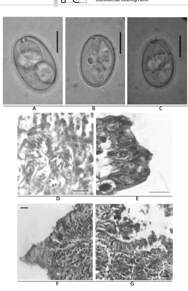
Three species of the genus Eimeria (Figure 1, A, B and C) previously reported affecting Japanese quails
(Teixeira & Lopes, 2002) were identified in the present study, as follows:
a. Eimeria tsunodai Tsutsumi, 1972: Sporulated oocysts were ovoid, measuring 20.2 ± 1.5 by 14.88 ± 0.79 µm and 1.36 shape index. Oocyst wall was smooth, double layered, with brownish inner layer and colorless outer layer, and measured 0.99 ± 0.1 µm. Despite usually only one polar granule was present, it could appear in pairs and refractive. Micropyle and residual body of the oocyst were absent. The sporocysts varied from ovoid to ellipsoid and measured 10.41 ± 0.6 by 5.39 ± 0.3 µm. They had a finer end where a small and fainted Stieda body projected. The residual body of the sporocyst was present and dispersed among the sporozoites, which were in pairs with globules visible at the enlarged extremity.
b. Eimeria uzura Tsunoda & Muraki, 1971: Sporulated oocysts were ovoid, measuring 22.2 ± 6.76 by 16.16 ± 1.13 µm, and 1.32 shape index. Oocyst wall was smooth, double layered, with brownish inner layer and colorless outer layer, measuring 1.08 ± 0.12 µm. Two to five polar granules were observed in the oocysts, sometimes with a massive aspect, but not refractive). The micropyle and residual body of the oocysts were absent. The sporocysts were ovoid measuring 11.76 ± 0.74 by 5.94 ± 0.45 µm, with a finer end, where a piriform Stieda body projected. The residual body of the sporocysts was present and had concentric granules between the sporozoites, which had refractive globules at the enlarged extremity.
c. Eimeria bateri Bathia, Pandey & Pande, 1965: Sporulated oocysts were subspherical, ovoid or ellipsoid, measuring 21.50 ± 1.84 by 16.36 ± 1.32 µm and shape index 1.32. Oocyst wall was smooth, double layered, with brownish inner layer and colorless outer layer, measuring 0.99 ± 0.1 µm. A single and refractive polar granule was present, but micropyle and the residual body of the oocyst were absent. Sporocysts were ovoid and measured 10.35 ± 0.73 by 6.66 ± 0.58 µm, with a prominent knob-like Stieda body. The residual body of the sporocyst was dispersed among the sporozoites, which had refractive globules at the enlarged extremity.
been developed (Kucera & Reznicky, 1991; Daugschies
et al., 1999; Gruber & Fernandez, 1999; Pereira et al.,
2001). However, morphological differentiation associated to specificity factors is still useful. Duszynski & Wilber (1997) emphasized and encouraged precision in the description of species, and established basic characteristics for an appropriate description of the oocysts. However, accuracy and caution were already recommended by Bandoni & Duszynski (1988). In the present study, species previously described were found, and the final identification was carried out by comparison with the original descriptions of the
parasites. Some Eimeria species with similar
morphology were initially excluded because of the differences in the size of the oocysts. The measurements, in the beginning of the investigation, the measurements allowed to distinguish from closely related species. Initially, E. coturnicis (Chakravarty &
Kar, 1947), E. tahamensis (Amoudi, 1987) and E.
fluminensis (Teixeira & Lopes, 2002) were excluded. Despite the similar measurements, E. taldykurganica
(Svambaev & Utebaeva, 1973) did not fit with the descriptions because it has a very different form, essentially ellipsoid with shape index 1.86. Lastly, E. dispersa (Tyzzer, 1929) may infect C. coturnix but is not infective to C. japonica (Tsunoda & Muraki, 1971) and therefore was also dismissed. On the other hand,
E. tsunodai, E. uzura and E. bateri were identified in the present study based on the measurements and other features of oocysts, such as polar granules, Stieda body and refractive globules, which were essential to confirm identification.
Clinical signs and Gross lesions
During the first week of life, few young quails presented diarrhea, weakness and small blood spots in the upper small intestine (jenunum and ileum). Later, softening of feces at the 14th day and an increased
cecum were seen in quails necropsied at the 21st days.
From the 35th to the 42nd days, a significant number of
quails had diarrhea, which disappeared soon.
There are few reports about the pathogenicity of
Eimeria in quails. Mazurkiewiewicz et al. (1967) reported clinical signs such as lack of appetite, ruffled feathers, uncoordinated movements, inhibition of laying and loss of weight in naturally infected young and mature quails reared at the laboratory. Norton & Pierce (1971) infected young Japanese quails experimentally with E. bateri and observed mild loss of weight and, although anorexia and softening of feces were observed at the third day of infection, the disease was
considered mild and easy to overcome. Tsunoda & Muraki (1971) also reported low pathogenicity in Japanese quails experimentally infected with 1 x 10⁵
oocysts of E. uzura, observing diarrhea and anemia from the 5th to 8th day of infection. No mortality was
not reported in these researches, and disease was considered similar to coccidiosis caused by E. acervulina
in chickens. Later, Ruff & Fagan (1984) used pure and
mixed cultures of E. uzura to infect quails. They
reported mortality, lower weight gain and poor reproductive performance. Concerning weight gain,
E. tsunodai was considered more pathogenic than
E. bateri. Finally, w h en pathogenicity w as investigated, the authors suggested that young quails are more susceptible to the coccidiosis effects. The results in the present work corroborate such opinion, since clinical signs were seen only in young quails. Finally, coccidiosis was assumed to be characteristically subclinical in this rearing farm with nonspecific and very mild clinical signs when the whole period of study was considered.
Histological Observations
Endogenous stages of the parasites were found in the small intestine. These were usually located in the villi, mainly above the nucleus of apical epithelial cells, or in the medium portion close to the glands (Figure 1, D and E). These observations resemble those described by Tsunoda & Muraki (1971), Norton & Pierce (1971) and Tsutsumi (1972) not only because of the site of infection, but also morphology was similar. Thus, endogenous stages observed in the small intestine were assumed to be developmental stages of E. bateri
and E. uzura, while the species found in the cecae might be E. tsunodai.
Pathological changes were also observed in the mucosa of the small intestine. Villous erosion (Figure 1, F and G), frequently concomitant with hyperplasia of the crypts of Lieberkühn, was often observed, as well as inflamatory infiltrate characterized by the presence of granulocytes and mononuclear cells, usually associated with edema.
disturbances and functional changes are usually related to the intensity of the parasitic infection.
CONCLUSION
Three species of the genus Eimeria were found and identified as E. bateri, E. tsunodai and E. uzura; and
the natural infection was characterized as subclinical. Despite of causing mild and nonspecific clinical signs, coccidiosis was considered an important disease because endogenous stages of the parasites and a high number of oocysts in feces were associated with intestinal lesions. The results strongly suggest that such infection might represent a limiting factor for quail production.
Table 1- Characteristics of species of the genus Eimeria described from Coturnix quails.
REFERENCES
Amoudi MA. Eimeria tahamensis n. sp. (Apicomplexa: Eimeriidae) from the Arabian quail (Coturnix delegorguey arabica). Journal of Parasitology 1987; 34:455-456.
Bandoni SM, Duszynsnki DW. A plea for improved presentation of type material for coccidia. Journal of Parasitology 1988; 74:519-523.
Bathia BB, Pandey TP, Pande BP. Eimeria bateri n. sp. from Indian common quail (Coturnix coturnix). Indian Journal of Microbiology 1965; 5:61-64.
Behmer AO, Tolosa EMC, Neto AGF. Manual de Técnicas para histologia normal e patológica. São Paulo: EDART, Editora da Universidade de São Paulo; 1976.
Bigland CH, Da Massa AJ, Woodard AE. Diseases of Japanese quail (Coturnix coturnix japonica) A flock survey and experimental transmission of selected avian pathogens. Poultry Science 1964;13:212-219.
Chakravarty M, Kar AB. A study on the coccidian of Indian birds. Proceedings of the Royal Society of Edinburgh 1947; 62:225-233.
Daugschies A, Imarom S, Bollwahn, W. Differentiation of porcine Eimeria spp. by morphologic algorithms. Veterinary Parasitology 1999; 81:201-210.
Duszynski DW, Wilber PG. A guideline for the preparation of species descriptions in the Eimeriidae. Journal of Parasitology 1997; 83: 333-336.
Garcia EA, Mendes AA, Pizzolante CC, Veiga N, Mattos TK. Alimentação de codornas com milho moído e ração de postura no
período pós-jejum durante a muda forçada e seus efeitos sobre o desempenho. Revista Brasileira de Ciência Avícola 2002; 4:119-126.
Gruber A, Fernandez S. Recentes avanços na biologia molecular de protozoários do gênero Eimeria. In: II Simpósio Internacional sobre coccidiose aviária; 1999; Foz do Iguaçu, PR. Brasil. p. 9-21.
Kucera J, Reznicky M. Diferentiation of species of Eimeria from the fowl using a computerized image analysis system. Folia Parasitologica 1991; 38:107-113.
Levine ND. Protozoan Parasites of Domestic animals and of man. 2 ed. Minneapolis: Burgess Pub. Co; 1982.
Long PL, Joyner LP. Problems in the identification of species of Eimeria. Journal of Protozoology 1984; 31:535-541.
Mazurkiewicz M, Podlewska D, Wachnik Z. Kokcydioza u przepiórek japonskich. Medycyna Weterynaryjna 1967; 23:536-537.
Menezes RCA, Lopes CWG. Epizootiologia da Eimeria arloingi em caprinos na microrregião Serrana Fluminense, Rio de Janeiro, Brasil. Revista da Universidade Federal Rural do Rio de Janeiro 1995; 17: 5-12.
Norton CC, Pierce MA. The life cycle of Eimeria bateri (Protozoa: Eimeriidae) in the Japanese quail Coturnix japonica. Journal of Protozoology 1971; 18:57-62.
Pereira MJS, Fonseca AH, Lopes CWG. Regressão linear na caracterização de variações morfométricas em coccidia. Revista Brasileira de Parasitologia Veterinária 2001; 10:75-78.
Ruff MD; Fagan JM. Pathogenicity of Japanese Quail (Coturnix japonica). Poultry Science, 1984, 63:55-60.
O o c y s t S p o r o c y s t S p e c i e s A u t h o r H o s t
s h a p e s iz e ( µ m ) S .I .
a W a ll P o l a r
G r a n u l e s h a p e s iz e ( µ m )
S t i e d a B o d y
E . u z u r a T s u n o d a M u r a k i, 1 9 7 1 C . ja p o n ic a e llip s o i d a l- o v o id
o v o id *
1 8 . 7 5 - 2 9 . 0 x 1 5 . 0 - 2 2 . 7 5 1 8 . 8 6 - 2 2 . 7 6 x 1 5 .0 4 - 1 7 . 4 2 *
1 .3 1 1 .3 2 *
d o u b le d o u b le *
0 - 4 2 - 4 *
? o v o id *
? 1 1 . 0 2 - 1 2 . 5 x 5 . 4 9 - 6 . 3 9 *
? p ir i f o r m
E . t s u n o d a i T s u ts u m i, 1 9 7 2 C . ja p o n ic a o v o id o v o id *
1 5 . 5 - 2 2 . 5 x 1 6 . 5 - 1 8 . 5 1 8 . 7 - 2 1 . 7 x 1 4 . 0 9 - 1 5 . 6 7 *
1 .3 0 1 .3 6 * d o u b le
0 - 5 1 - 2 *
? o v o id *
1 0 . 3 - 1 1 . 5 x 5 . 0 - 6 . 1 9 .8 1 - 1 1 . 0 1 x 5 . 0 9 - 5 . 6 9 *
f a in t e d f a in t e d *
E . b a t e r i B a t h ia e t a l .,1 9 6 5
C . c o t u r n ix
C . ja p o n ic a*
e llip s o i d a l, o v o i d o r s u b s p h e r ic a l e llip s o i d a l, o v o i d o r s u b s p h e r ic a l
1 5 . 0 - 2 8 . 0 x 1 4 . 0 - 2 3 . 0
1 9 . 6 6 - 2 3 . 3 4 x 1 5 .0 4 - 1 7 . 6 8 * 1 .2 6
1 .3 2 * d o u b le *
1
1 * o v o id
o v o id *
9 .0 - 1 3 .0 x 5 . 0 - 8 . 0
9 .6 6 - 1 1 . 8 x 6 . 0 8 - 7 .2 4 *
p r o m i n e n t p r o m i n e n t n ip p le - lik e *
E . f lu m i n e n s is T e i x e ir a e L o p e s , 2 0 0 2 C . ja p o n ic a s u b s p h e ric a l 1 5 . 4 4 - 1 9 . 1 8 x 1 4 .7 3 - 1 8 . 1 7 1 .0 5 d o u b le ? o v o id 9 .3 2 - 1 1 . 5 3 x 5 . 3 4 - 6 . 5 p ir i f o r m
E . d is p e r s a T y z z e r , 1 9 2 9 C . c o t u r n ix o v o id 1 7 . 2 - 2 6 . 4 x 1 5 . 4 - 2 2 . 4 ? s in g le ? o v o id ? ?
E . c o t u r n ic is C h a k r a v a r t y K a r , 1 9 6 7 C . c o t u r n ix o v o id 2 6 . 4 - 3 8 . 8 x 1 9 . 8 - 2 6 . 4 ? d o u b le 1 o v o id 1 3 . 2 - 1 7 . 2 x 8 . 8 - 1 1 .0 k n o b - lik e
E . t a ld y k u r g a n ic a S v a m b a e v U t e b a e v a , 1 9 7 3 C . c o t u r n ix o v o id 2 1 . 8 8 - 2 5 . 4 0 x 1 1 .9 - 1 3 . 1 1 .8 6 d o u b le 1 o v o id 8 .1 - 1 1 .6 - 3 . 5 9 - 4 . 8 ?
Soulsby EJL. Parasitologia y enfermedades parasitarias em los animales domésticos. 7 ed. México: Nueva Editorial Interamericana; 1987.
Svambaev SK, Utebaeva MK. Coccidial infections of Phasianus cochicus mongolicus and Coturnix coturnix in Kazakhstan. Izvestiia Akademii Nauk SSSR. Seriia biologicheskaia 1973; 6:62-68.
Teixeira M, Lopes CWG. Species of the genus Eimeria (Apicomplexa: Eimeriidae) from Japanese quails (Coturnix japonica) in Brazil and E. fluminensis for the preoccupied E. minima of this quail. Revista Brasileira de Ciências Veterinárias 2002; 9:53-56.
Tsunoda L, Muraki Y. A new coccidium of Japanese quails: Eimeria uzura n. sp. Japanese Journal of Veterinary Science 1971; 33: 227-235.
Tsutsumi Y. Eimeria tsunodai n.sp. (Protozoa:Eimeriidae). A cecael coccidium of Japanese quails (Coturnix japonica). Japanese Journal of Veterinary Science 1972; 34:1-9.
A B C
D E
F G
Figure 1 - Sporulated oocysts of (A) Eimeria bateri, (B) E. tsunodai and (C) E. uzura (in saturated sugar solution); Caeca of Japanese quail at the 42nd days, (D) Schizonts with merozoites and ileum at the 14th days, (E) macro and microgamonts, HE; Jejunum of Japanese
quail at the 14th days with (F) villous and (G) epithelial erosion, HE; (scale bar = 10µm).