Rev Odontol UNESP. 2018 July-Aug.; 47(4): 205-209 © 2018 - ISSN 1807-2577
ORIGINAL ARTICLE
Doi: https://doi.org/10.1590/1807-2577.06218
This is an Open Access article distributed under the terms of the Creative Commons Attribution License, which permits unrestricted use, distribution, and reproduction in any medium, provided the original work is properly cited.
Evaluation of calcium release and pH value of light-cured cavity
liners for pulp-capping materials
Avaliação da liberação de cálcio e alteração de pH de forradores cavitários fotoativados para capeamento pulpar
Kamila de Figueiredo PEREIRA
a*, Rosymere Freitas de Sousa CRUVINEL
b,
Andrea Abi Rached DANTAS
a, Milton Carlos KUGA
aaUNESP – Universidade Estadual Paulista, Faculdade de Odontologia de Araraquara, Araraquara, SP, Brasil
bUniRV – Universidade de Rio Verde, Rio Verde, GO, Brasil
Resumo
Introdução: Forradores cavitários à base de hidróxido de cálcio apresentam baixa resistência e alta solubilidade. Para resolver este problema, foi desenvolvido forradores à base de hidróxido de cálcio fotoativáveis contendo resina para
melhorar suas propridades. Objetivo: A proposta deste estudo foi avaliar a alteração de pH e liberação de cálcio de
forradores fotoativáveis. Material e método: Foram preparadas amostras (n=10) com Ultra-Blend plus, Biocal
(cimentos fotoativáveis) e Hydro C (controle). As amostras foram armazenadas com 10 mL de água destilada e
mantidas em estufa à 37 °C. Depois de 24 horas, 7 e 14 dias, foram avaliados os níveis de liberação de cálcio e a alteração de pH. Os dados foram analisados estatisticamente pelo teste ANOVA, seguido do pós teste Tukey (α = 0.05).
Resultado: Os resultados de pH mostraram Hydro C > Ultra-Blend plus > Biocal. Biocal apresentou a pior liberação
de cálcio. Conclusão: Materiais fotoativados apresentam menor liberação de cálcio e alteração nos valores de pH.
Descritores: Hidróxido de cálcio; forrador cavitário dental; concentração de íon de hidrogênio.
Abstract
Introduction: Cavity liners based calcium hydroxide present low strength and high solubility that is consider a disadvantage. In order to enhance these properties it was developed a light-cured cavity liner based calcium hydroxide
containing resin. Objective: The purpose of this study was to evaluate the pH and calcium release of light-cured cavity
liners. Material and method: There were prepared specimens (n=10) with the Ultra-Blend plus, Biocal (light cured
cements) and Hydro C (control). The samples were stored in 10 mL of distilled water and maintained at 37 °C. After
24 hours, 7, and 14 days, there were analyzed pH and the release of calcium levels. The data were statistically analyzed
by ANOVA and Tukey test (α = 0.05). Result: The results of pH showed Hydro C > Ultra-Blend plus > Biocal. Biocal
presented worst calcium release. Conclusion: Light-cured materials present lower calcium release and alteration
in pH values.
Descriptors: Calcium hydroxide; dental cavity lining; dydrogen-ion concentration.
INTRODUCTION
The use of cavity liners has been recommended in order to prevent the diffusion of residual and/or leached components through the dentin tubules. These materials are also used to overcome the problems related to the potential toxicity of dentin
bonding agents when applied in deep cavities1-3. Cavity liner
based calcium hydroxide has been largely used as a powerful cavity liner4-6.
Calcium hydroxide in cavity liners dissociate into ions calcium and hydroxyl, which are responsible for the antimicrobial properties
and induction of mineralization7,8. The calcium ions release is
essential for the mineralization process, subsequently it promotes
cell migration and differentiation9,10 while the hydroxyl ions
provide high pH (about 12), and promotes enzymatic inhibition
of microorganisms6,11. Those hydroxyl ions also release alkaline
phosphatase, which participates in the mineralization process10,11.
Therefore, the use of calcium hydroxide on the pulp tissue promotes repair12 and subsequent formation of dentin bridge4,13.
Cavity liners must be resistant to solubility in water, to organic solvents of acid etching, exhibit resistance to support the restoration, and resist to occlusal forces during mastication14 in order to protect
the pulp15. Currently, conventional calcium hydroxide liners have
and restorative materials16,17, in addition presents difficulty in
manipulation and application18,19.
With the purpose of improving the properties of conventional calcium hydroxide cavity liners, were developed resin-based cavity liners containing calcium hydroxide. These materials are light-cured20,
highly resistant to etchants21, present superior physical properties,
and handling characteristics14. Currently, there are no studies
showing if resin-based cavity liners with calcium hydroxide present alteration in pH and release of calcium ions when compared to conventional cavity liners. Thus, the aim of the present investigation was to evaluate pH and calcium release of resin-based cavity liners light-cured with calcium hydroxide. The null hypothesis was that all cements showed similar calcium release and pH change.
MATERIAL AND METHOD
The materials evaluated were Hydro C (Dentsply Ind. and Com.
Ltd., Petrópolis, RJ, BR), Ultra-Blend plus (Ultradent Products,
Inc, South Jordan, UT) and Biocal (Biodinâmica, Ibiporã, PR, BR).
Analysis of pH and Calcium Release
Ten samples of each material were prepared using polyethylene tubes (n=10). The tubes with dimensions of 10 mm in height and 1 mm in diameter were filled with the cavity liner based in calcium hydroxide (Table 1). Hydro C was evaluated after cured and Ultra-Blend
plus and Biocal were evaluated light-cured and not light-cured.
The samples of Ultra-Blendplus and Biocal were light-cured
with a light-curing unit (1200 mW/cm2) during 20 seconds, as
recommended by the manufacturer. The tubes filled with materials
were immersed in 10 mL of distilled water and stored in individual flasks at 37 °C at a relative humidity 100% in 24 hours, 7 and 14 days. After each period the tubes were removed and transferred to new flasks with 10 mL of distilled water. The solutions contained in the flask were used after each experimental period to determine pH
and release of calcium (Ca2+), based on the methodology used by
Santos et al.22. The measurement of pH was performed by digital
pHmeter (Q-400 Quimis instrument, São Paulo, SP, BR), which was previously calibrated with buffer solutions (pH 4.0 and 7.0). To evaluate the release of calcium was used atomic absorption spectrophotometry (Spectraa 55B - Varian, Inc., Palo Alto, CA, EUA). Lanthanum oxide was added to all samples to eliminate ionic interference specific for phosphate ions. Solutions containing calcium concentrations of 0, 1, 2, 3, 4 and 5 ppm were used for calibration. The data of pH and calcium release were subjected to parametric
tests ANOVA and Tukey post-hoc, with 5% of significance level.
RESULT
Table 2 shows the results of the pH test. Hydro C obtained the highest pH values in all experimental periods (p<0.05). Ultra-Blend plus showed higher pH than Biocal (p<0.05). After 24 hours, Ultra-Blend plus which was not light-cured showed higher pH values than the same material photo polymerized (p<0.05). On 7 and 14 days, Ultra-Blend plus light-cured and not light-cured showed no significantly difference (p<0.05). Biocal, which was light-cured, showed acid pH during all experimental periods.
Table 3 shows the results obtained in the evaluation of calcium release. Ultra-Blend plus not cured released more calcium when compared to Ultra-Blend plus cured, Hydro C and Biocal (cured and
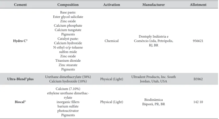
Table 1. Materials used in this study
Cement Composition Activation Manufacturer Allotment
Hydro C
Base paste: Ester glycol salicilate
Zinc oxide Calcium phosphate
Calcium tungstate Pigments Catalyst paste: Calcium hydroxide N-ethyl-o/p-toluene
sulfon-mide Zinc oxide Titanium dioxide
Zinc stearate Pigments
Chemical
Dentsply Indústria e Comércio Ltda, Petrópolis,
RJ, BR
956621
Ultra-Blendplus Urethane dimethacrylate (58%)
Calcium hydroxide (10%) Physical (Light)
Ultradent Products, Inc. South
Jordan, Utah, USA B5962
Biocal
Calcium (7.10%) ethylene urethane
dimethac-rylate inorganic fillers
barium sulfate photoactivator Pigments
Physical (Light) Biodinâmica
not cured) after 24 hours and 7 days (p<0.05). In 14 days, Hydro C showed higher release of calcium than the others cavity liners (p<0.05). Ultra-Blend plus not cured showed intermediate values when compared with the others. Biocal cured and not cured had the lowest calcium release, with no significant difference from Ultra-Blend plus cured in 14 days (p<0.05).
DISCUSSION
A conventional calcium hydroxide and two resin-based cements (cured and not cured) were used as lining materials and were evaluated in terms of calcium release and pH change. The null hypothesis was rejected, because all cements showed different calcium release and pH values during the evaluated periods.
Calcium hydroxide cements as Dycal and Hydro C are the most commonly conventional lining materials used in direct and indirect pulp capping, because they present alkaline pH, biocompatibility,
induce pulp-dentin mineralization, and reduce infection23,24.
Lining materials should remain on the interface between the tooth and the restoration, some of these conventional materials based with calcium hydroxide has shown high water sorption and
solubility14 the cavity liners containing resin components were
created to improve those mechanical properties.
In the present study, Hydro C, showed high pH values (p<0.05) and had an increase of calcium release throughout the evaluation. This data is similar with the literature25, where the principal font
of ions release is calcium hydroxide, but calcium silicate cements release more hydroxyl and calcium ions, which were not evaluated in this study.
Non-cured Ultra-Blend plus presented superior calcium release than Hydro C, cured Ultra-Blend plus and Biocal groups after 24 h and 7 days, despite the lower pH values compared to Hydro C. Currently, cavity liners containing resin components need to be cured and this causes a reduction in release of calcium
ions. Therefore, in comparison with other study25, Dycal present
lower calcium release than calcium silicate materials, in neutral pH. This could be compared with the superior calcium release of non-cured Ultra-Blend plus in lower pH value.
Cured Ultra-Blend plus presented lower calcium release than Hydro C during the first period (24h), but after 7 days both did not obtain statistically significant difference. Although Duarte et al.10
has analyzed calcium release of Ultra-Blend and Hydro C with spectrophotometry through atomic absorption and a calcium cathode lamp, Ultra-Blend presented higher value than Hydro C for 24 h and 7 days. Duarte et al.10 attributed this value to the
presence of hydroxyapatite in the material, which could be released and detected by spectrophotometer. The different methodology of this study might have shown distinct results.
Cured Ultra-Blend plus showed neutral pH (around 7.50) without significant difference after 7 and 14 days than the same material non-cured. However, Ultra-blend plus non-cured was better in the first period analyzed (24 h). In addition Ultra-blend plus cured and non-cured were better than Biocal throughout the experiment (p<0.05). Biocal cured had acid pH in this study.
The difference of ions release could be observed in vitro, however that may not occur in vivo. Further studies are being carried out to evaluate the interaction between the calcium release and pH change,
Table 2. pH values recorded at different time periods (mean and SD)
Hydro C Cured Ultra-Blend
plus
Not cured
Ultra-Blend plus Cured Biocal Not cured Biocal
Mean SD Mean SD Mean SD Mean SD Mean SD
24 hs 10.59a 0.28 7.35c 0.39 8.95b 0.38 5.57e 0.12 6.33d 0.33
7 d 10.76a 0.26 7.20b 0.22 7.10b 0.06 5.25d 0.13 5.72c 0.12
14 d 10.68 a 0.32 7.50b 0.75 7.40b 0.20 5.54d 0.43 6.35c 0.18
a,b,c,d,e The different letters in the same line indicate significant differences between groups (p<0.05).
Table 3. Calcium release recorded over different periods of time (mean and SD)
Hydro C Cured Ultra-Blend
plus
Not cured
Ultra-Blend plus Cured Biocal Not cured Biocal
Mean SD Mean SD Mean SD Mean SD Mean SD
24 hs 16.99 b 7.25 4.85c 3.08 41.82a 14.11 0.13d 0.03 0.16d 0.16
7 d 24.86b 11.21 25.72b 9.74 37.24a 6.03 0.15c 0.10 0.20c 0.26
14 d 30.63 a 16.85 4.84c 2.39 18.98b 5.10 0.07c 0.14 0.41c 1.12
also mechanical tests should be added for a better understand of dental cavity liners.
CONCLUSION
Within the limitations of this study, it is concluded that: 1- Hydro C had the highest pH rates; 2- Hydro C had an increase of calcium release during the experiment; 3- Ultra-Blend not cured had better calcium release than others materials for 24h and 7 days; 4- Ultra-Blend not cured had better pH and calcium release than
Ultra-Blend cured; 5- Biocal cured had the lowest pH value and the worst calcium release for all experimental period.
ACKNOWLEDGEMENTS
K.F.P. received a scholarship from the National Counsel of Technological and Scientific Development for her master’s dissertation. The authors are thankful to UNESP for providing the facilities for this study. The authors deny any conflicts of interest related to this study.
REFERENCES
1. Weiner RS. Liners, bases, and cements: a solid foundation. Gen Dent. 2002 Sep-Oct;50(5):442-6. PMid:12448897.
2. Duque C, Hebling J, Smith AJ, Giro EM, Oliveira MF, Souza Costa CA. Reactionary dentinogenesis after applying restorative materials and bioactive dentin matrix molecules as liners in deep cavities prepared in nonhuman primate teeth. J Oral Rehabil. 2006 Jun;33(6):452-61. http://dx.doi.org/10.1111/j.1365-2842.2005.01585.x. PMid:16671993.
3. Weiner R. Liners and bases in general dentistry. Aust Dent J. 2011 Jun;56(1 Suppl):11-22. http://dx.doi.org/10.1111/j.1834-7819.2010.01292.x. PMid:21564112.
4. Silva LA, Freitas AC, Carvalho FK, Queiroz AM, Nelson-Filho P, Porto-Neto ST. Direct pulp capping with a self-etching adhesive system: histopathologic evaluation in dogs’ teeth. Oral Surg Oral Med Oral Pathol Oral Radiol Endod. 2009 Jul;108(1):e34-40. http://dx.doi. org/10.1016/j.tripleo.2009.03.017. PMid:19540442.
5. Aguilar P, Linsuwanont P. Vital pulp therapy in vital permanent teeth with cariously exposed pulp: a systematic review. J Endod. 2011 May;37(5):581-7. http://dx.doi.org/10.1016/j.joen.2010.12.004. PMid:21496652.
6. An S, Gao Y, Ling J, Wei X, Xiao Y. Calcium ions promote osteogenic differentiation and mineralization of human dental pulp cells: implications for pulp capping materials. J Mater Sci Mater Med. 2012 Mar;23(3):789-95. http://dx.doi.org/10.1007/s10856-011-4531-0. PMid:22190198.
7. Schuurs AH, Gruythuysen RJ, Wesselink PR. Pulp capping with adhesive resin-based composite vs. calcium hydroxide: a review. Endod Dent Traumatol. 2000 Dec;16(6):240-50. http://dx.doi.org/10.1034/j.1600-9657.2000.016006240.x. PMid:11202889.
8. Modena KC, Casas-Apayco LC, Atta MT, Costa CA, Hebling J, Sipert CR, et al. Cytotoxicity and biocompatibility of direct and indirect pulp capping materials. J Appl Oral Sci. 2009 Nov-Dec;17(6):544-54. http://dx.doi.org/10.1590/S1678-77572009000600002. PMid:20027424.
9. Schröder U. Effects of calcium hydroxide-containing pulp-capping agents on pulp cell migration, proliferation, and differentiation. J Dent Res. 1985 Apr;64(Spec No):541-8. http://dx.doi.org/10.1177/002203458506400407. PMid:3857254.
10. Duarte MA, Martins CS, Oliveira Cardoso Demarchi AC, Godoy LF, Kuga MC, Yamashita JC. Calcium and hydroxide release from different pulp-capping materials. Oral Surg Oral Med Oral Pathol Oral Radiol Endod. 2007 Jul;104(1):e66-9. http://dx.doi.org/10.1016/j. tripleo.2007.01.024. PMid:17499530.
11. Estrela C, Sydney GB, Bammann LL, Felippe O Jr. Mechanism of action of calcium and hydroxyl ions of calcium hydroxide on tissue and bacteria. Braz Dent J. 1995;6(2):85-90. PMid:8688662.
12. Okabe T, Sakamoto M, Takeuchi H, Matsushima K. Effects of pH on mineralization ability of human dental pulp cells. J Endod. 2006 Mar;32(3):198-201. http://dx.doi.org/10.1016/j.joen.2005.10.041. PMid:16500225.
13. Fernandes AM, Silva GA, Lopes N Jr, Napimoga MH, Benatti BB, Alves JB. Direct capping of human pulps with a dentin bonding system and calcium hydroxide: an immunohistochemical analysis. Oral Surg Oral Med Oral Pathol Oral Radiol Endod. 2008 Mar;105(3):385-90. http://dx.doi.org/10.1016/j.tripleo.2007.08.031. PMid:18280971.
14. Francisconi LF, Freitas AP, Scaffa PMC, Mondelli RFL, Francisconi PAS. Water sorption and solubility of different calcium hydroxide cements. J Appl Oral Sci. 2009 Sep-Oct;17(5):427-31. http://dx.doi.org/10.1590/S1678-77572009000500014. PMid:19936520.
15. von Fraunhofer JA, Marshall KR, Holman BG. The effect of base/liner use on restoration leakage. Gen Dent. 2006 Mar-Apr;54(2):106-9. PMid:16689065.
16. Cox CF, Hafez AA, Akimoto N, Otsuki M, Mills JC. Biological basis for clinical success: pulp protection and the tooth-restoration interface. Pract Periodontics Aesthet Dent. 1999 Sep;11(7):819-26. PMid:10853583.
17. Mickenautsch S, Yengopal V, Banerjee A. Pulp response to resin-modified glass ionomer and calcium hydroxide cements in deep cavities: A quantitative systematic review. Dent Mater. 2010 Aug;26(8):761-70. http://dx.doi.org/10.1016/j.dental.2010.03.021. PMid:20452013.
18. Nair PN, Duncan HF, Pitt Ford TR, Luder HU. Histological, ultrastructural and quantitative investigations on the response of healthy human pulps to experimental capping with mineral trioxide aggregate: a randomized controlled trial. Int Endod J. 2008 Feb;41(2):128-50. http:// dx.doi.org/10.1111/j.1365-2591.2007.01329.x. PMid:17956562.
20. Kitasako Y, Shibata S, Tagami J. Migration and particle clearance from hard-setting Ca(OH)2 and self-etching adhesive resin following direct pulp capping. Am J Dent. 2006 Dec;19(6):370-5. PMid:17212080.
21. Burke FJ, Watts DC. Weight loss of three resin-based lining materials containing calcium following a phosphoric acid-etching and washing cycle. J Dent. 1989 Feb;17(1):38-40. http://dx.doi.org/10.1016/0300-5712(89)90008-0. PMid:2645332.
22. Santos AD, Moraes JC, Araújo EB, Yukimitu K, Valério WV Fo. Physico-chemical properties of MTA and a novel experimental cement. Int Endod J. 2005 Jul;38(7):443-7. http://dx.doi.org/10.1111/j.1365-2591.2005.00963.x. PMid:15946264.
23. Pinto AS, Araujo FB, Franzon R, Figueiredo MC, Henz S, García-Godoy F, et al. Clinical and microbiological effect of calcium hydroxide protection in indirect pulp capping in primary teeth. Am J Dent. 2006 Dec;19(6):382-6. PMid:17212082.
24. Corralo DJ, Maltz M. Clinical and ultrastructural effects of different liners/restorative materials on deep carious dentin: a randomized clinical trial. Caries Res. 2013;47(3):243-50. http://dx.doi.org/10.1159/000345648. PMid:23343804.
25. Natale LC, Rodrigues MC, Xavier TA, Simões A, Souza DN, Braga RR. Ion release and mechanical properties of calcium silicate and calcium hydroxide materials used for pulp capping. Int Endod J. 2015 Jan;48(1):89-94. http://dx.doi.org/10.1111/iej.12281. PMid:24646329.
CONFLICTS OF INTERESTS
The authors declare no conflicts of interest.
*CORRESPONDING AUTHOR
Kamila de Figueiredo Pereira, UNESP – Universidade Estadual Paulista, Faculdade de Odontologia de Araraquara, 14801-903 Araraquara - SP, Brasil, e-mail: [email protected]