ABSTRACT
http://dx.doi.org/10.1590/1678-775720150084
Bioactivity, physical and chemical properties of
MTA mixed with propylene glycol
Vaishali Prakash NATU, Nileshkumar DUBEY, Gerald Choon Leong LOKE, Teng Seng TAN, Wee Hsuan NG, Chee Weng YONG, Tong CAO, Vinicius ROSA
Faculty of Dentistry, National University of Singapore, Singapore.
Corresponding address: Vinicius Rosa - National University of Singapore, Faculty of Dentistry - 11 Lower Kent Ridge Road - Singapore - 119083 - Singapore - Phone: +65 6779 5555 ext 1650 - Fax: + 65 6778 5742 - e-mail: [email protected]
6XEPLWWHG0DUFK0RGL¿FDWLRQ0D\$FFHSWHG-XQH
O
and chemical (pH change, calcium release, crystallinity) properties and the biological outcomes (cell survival and differentiation) of mineral trioxide aggregate (MTA) mixed using different proportions of propylene glycol (PG) and water. Material and Methods: White MTA was mixed with different water/PG ratios (100/0, 80/20 and 50/50). Composition (XRD), ! "! #$%%&'* "+-3"4" 67&$999 Knoop hardness (100 g/10 s) and chemical characteristics (pH change and Ca2+ release for 7 days) were evaluated. Cell proliferation, osteo/odontoblastic gene expression and mineralization induced by MTA mixed with PG were evaluated. MTA discs (5 mm in diameter, 2 mm thick) were prepared and soaked in culture medium for 7 days. Next, the discs were removed and the medium used to culture dental pulp stem cells (DPSC) for 28 days. Cells survival was evaluated using MTS assay (24, 72 and 120 h) and differentiation with RT-PCR (ALP, OCN, Runx2, DSPP and MEPE) and alizarin red staining (7 and 14 days). Data were analysed using one-way ANOVA and Tukey’s post-hoc analysis (D=0.05). Results:@FIJ#2+ release, but it
compromised the hardness of the material. SEM showed that 50/50 group resulted porous material after setting due to the incomplete setting reaction, as shown by XRD analysis. The addition of PG (80/20 and 50/50) was not capable to improve cell proliferation or to enhance gene expression, and mineralized deposition of DPSC after 7 and 14 days '9939Q# U@@FI promote further improvements on the chemical and physical properties evaluated, and it was not capable of enhancing the bioactivity of the MTA.
Keywords: Dental pulp. Stem cells. Proliferation. Cell differentiation. Physical properties. Chemical properties. Hardness. Endodontics.
INTRODUCTION
Mineral trioxide aggregate (MTA) is a material with osteogenic, cementogenic and odontogenic potential that can be used for perforation repairs, pulp capping and pulpotomy17,29. It is also a retrograde J@[ surgery procedures7. It has positive effects on proliferation of dental pulp stem cells (DPSC) in a dose-dependent manner11,27,28. The exposure of DPSC to MTA promotes upregulation of dentin sialophosphoprotein (DSPP), bone sialoprotein, increase in alkaline and phosphatase (ALP) activity.
It may also induce biomineralization11,16,18.
Water is normally used to mix MTA. It is necessary for the cement hydration reaction to release Ca2+ that ultimately stimulate differentiation
of pulp cells13,26Q ^ U @J
manipulate, and the setting time of MTA is long4,21. Other vehicles have been proposed to alleviate these problems. Yet, the clinical effects are controversial. NaOCl, a common irrigating solution in endodontics, may improve handling properties and the setting time, but it also compromises the compressive strength of the compound14.
and odourless FDA-approved compound with antibacterial properties3,22. PG can be added to the mixing of MTA to improve handling properties. This strategy may also result in higher push-out bond strength25, increase in its sealing ability5 and higher pH and Ca2+ dissociation during the initial post-mixing periods10,12.
Different ratios of PG and water affect the physical and chemical properties of MTA, since crystal hydration is an important factor for the setting reaction of MTA6. The addition of high ratios @FIj69qU deemed too long to be clinically acceptable. This can translate into higher solubility and greater formation of pores, which compromises the mechanical strength of the material10. As the decrease in water content in the mixture changes physical and chemical characteristics of MTA, the question that arises is whether it could also affect its bioactivity. Thus, the aim of this study was to evaluate the effects of PG at different concentrations on physical and chemical properties, and the bioactivity of MTA.
MATERIAL AND METHODS
MTA preparation
0.1 g of White MTA (ProRoot MTA, Dentsply, |}~"U*9@ delivery vehicle prepared using different proportions of ultrapure water (UW) and PG (Table 1).
pH and Ca2+ release
Twenty-four polypropylene sterile micro pipette tips (length of 31.1 mm, inner diameter: 3.35 mm 9QJ MTA mixed with different proportions of UW and PG (Table 1) and allowed to set for 24 hours at 37°C (standard setting protocol used, except for
Q +U micro
pipette tips were placed independently in tubes containing 10 mL of UW. Half of the tubes were used to evaluate pH changes after 3, 24, 72 and 168 hours of incubation (n=3). The remaining tubes were used to assess Ca2+ release at the same time points (n=3) by inductively coupled plasma mass spectrometer (ICP-MS, Agilent 770, Santa Clara, #@ ~"Q $ @ J ~ !"J$q@Q @ microliters of this solution were added to 4.95 ml of UW in an ICP-MS vial (Autosampler vial, Nanonex Technology, Singapore) after calibration with standard solutions. UW alone was used as control agent in both tests.
6HWWLQJWLPHÀRZDELOLW\DQGKDUGQHVV Setting time (n=5) was measured with an ASTM C266-13 standard test method2. MTA mix was inserted into moulds, and the initial setting
time was evaluated using a Gillmore needle (113.4 g) every 60 seconds. Only the initial setting time was considered, since it governs the handling time available for the clinicians.
Flowability testing (n=5) was performed as recommended by ANSI/ADA 57-20001. Mixed MTA 9Q$ after spatulation. After 3 minutes from the time of spatulation, another glass slab was placed over the mixture. Additional weight (120 g) was placed on top of the overlying glass slab, and the set-up was kept in an incubator (37°C and 95% relative humidity) for 10 minutes. The set up was then carefully transferred onto a piece of graph paper and the additional weight was removed. The maximum and minimum diameter of the circular shape formed by the cement was measured (mm). Samples with a difference in minimum and maximum diameter of more than 1 mm were rejected. The average value of the two diameters was recorded as the @@Q
For microhardness (n=3), five indentations were made per specimen after setting using Knoop diamond indenter (FM-100, Futuretech, Kawasaki, Kanagawa Prefecture, Japan, load of 100 g and dwell time of 10 seconds) and Knoop hardness (HK), calculated according to equation 1 where Cp is the correction factor related to the shape of the indenter (0.070279), P is the test load (kgf) and L @Q
Equation 1
Scanning electron microscopy (SEM) and X-ray diffraction (XRD)
After setting, the MTA disks (5 mm in diameter, 2 mm in thickness) were fractured and the cross-sections observed under SEM (Quanta 650 FEGSEM, FEI, Hillsboro, Oregon, USA). Crystalline phase analysis of the unhydrated MTA powder and set MTA was obtained using an X-ray diffractometer (D8 Advance Powder X-ray Diffractometer, Bruker AXS, Karlsruhe, Baden-Württemberg, Germany) +J#'Q6$6" at 40kV and 40 mA (scan range: 10-80°, scanning rate of 0.02°/second). Crystalline formations were J#F4#F 4@@JQ!" used as control.
Stem cell culture and preparation of MTA conditioned media
The use of human dental pulp stem cells in this study was approved by the Institutional Review Board/NUS (Approval Number: NUS 2094). DPSCs between passages 3 and 6 (Allcells, Alameda, California, USA) were used to evaluate the cytotoxic and bioactivity of the groups. Cells
4!! 4 !@ Eagle Medium (Invitrogen, Carlsbad, California, USA)] supplemented with 10% fetal bovine serum (Invitrogen) and 1% penicillin/streptomycin (Invitrogen)24. Cells were passaged (TrypLE Select, -%9&79q@Q
To prepare the MTA conditioned media (extracts), molds (5 mm in diameter, 2 mm in thickness) were J!"U@@@ UW and PG (Table 1) and allowed to set. The discs obtained were immersed separately in 10 ml of the culture medium described above for 7 days. After this period, the disks were removed and the extract kept in 4°C for no longer than 2 weeks.
Cell viability assay
DPSCs (5x10³) were seeded in 96 well plates and treated with culture media (control) or MTA extracts (n=3). Cell viability was determined after 1, 3 and 5 days by using MTS assay (CellTiter 96 Aqueous |F!~" -J!$99!@ Meilen, Switzerland) at 490 nm.
Quantitative Real time polymerase chain reaction (qRT-PCR) and alizarin Red staining
Expression of osteo/odontoblastic-related genes was performed with qRT-PCR after exposing the DPSCs to the MTA extracts for 7 and 14 days (Figure 1). RNA extraction was carried out with the RNA extraction kit (PureLink™ RNA Mini Kit, LifeTech, Carlsbad, California. USA), followed by cDNA synthesis (iScript RT Supermix, Bio-Rad, Hercules, California, USA) and PCR (Biorad-CFX Connect™ Real time system, Hercules, California, USA). The primer sequences are listed in Figure 1. DPSC in culture medium alone served as control. Alizarin red staining (ARS) was performed to detect mineralization after 14 days. The cells were washed F JU @ *9 Q " solution (Sigma-Aldrich, Saint Louis, Missouri, USA) was added and incubated for 30 minutes at 37°C. Ten percent of cetylpyridinium chloride solution (Sigma-Aldrich) was added to each well, and the optical density was measured by a plate reader (wavelength 540 nm). The culture medium was the negative control, and the osteogenic induction medium was included as a positive control. Tests were performed in triplicates.
Gene Primer Sequence
Alkaline phosphatase (ALP) Forward: 5’-AAGTACTGGACAGACCAAGC-3’
Reverse: 5’-AGAGGGCCAAGAAGGGGAACT-3’
Osteocalcin (OCN) Forward: 5’-ATGAGAGCCCTCAGACTCCTC-3’
Reverse: 5'-CGGGCCGTAGAAGCGCCGATA-3'
Runt-related transcription factor 2 (Runx2) Forward: 5’-CACTGGCGCTGCAACAAGA-3’
Reverse: 5’-CATTCCGGAGCTCAGCAGAATAA-3’
Dentin sialophosphoprotein (DSPP) Forward: 5’-TGGCGATGCAGGTCACAAT-3’
Reverse: 5’-CCATTCCCACTAGGACTCCCA-3’
Matrix extracellular phosphoglycoprotein (MEPE) Forward: 5’-GGCCAGTGACTGCGATTAAAC-3’
Reverse: 5’-CCTTCGAGTGTGCTTTAGCAT-3’
b-Actin Forward: 5’-CAGGCTGTGCTATCCCTGTA-3’
Reverse: 5’-CATACCCCTCGTAGATGGGC-3’
Figure 1- Primer sequences
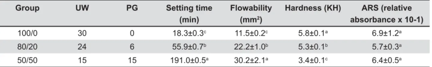
Group UW PG Setting time
(min)
Flowability (mm2)
Hardness (KH) ARS (relative
absorbance x 10-1)
100/0 30 0 18.3±0.3c 11.5±0.2c 5.8±0.1a 6.9±1.2a
80/20 24 6 55.9±0.7b 22.2±1.0b 5.3±0.1b 5.7±0.3a
50/50 15 15 191.0±0.5a 30.2±2.1a 3.4±0.1c 6.4±0.5a
Table 1- UW/PG ratio per 30 P
Statistical analyses
Statistical analyses were performed with either the one-way analysis of variance (ANOVA) and Tukey’s post-hoc analysis (D=0.05, SPSS V.22, IBM, USA).
RESULTS
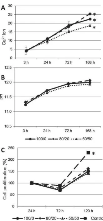
Figure 2 shows the Ca2+ release (A) and pH changes (B). After 168 hours, the amount of Ca2+ J @@ @ with the highest amount for 80/20. There was no J^@ Q "@ 7$ J decrease in cell viability for all groups, except for
$#Q " J increase in cell proliferation was observed after 120 hours for all groups.
and highest hardness was observed for 100/0 compared to 80/20 and 50/50 groups (Table 1).
SEM (Figure 3) showed a smooth and compact surface for 100/0. For 80/20, needle-like structures were present on the crystals. For 50/50 these structures were more frequent, and voids were also present. The XRD (Figure 3) shows that for 100/0 and 80/20 there was a decline of intensities of tricalcium silicate (Ca3SiO5, $$Q7
silicate (Ca2SiO4 $*$Q'
aluminate (Ca3Al2O6$**Q$9}
to the MTA powder. The peaks for 50/50 group were almost similar to the XRD of powder.
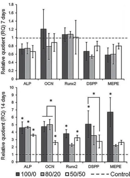
Figure 4 shows the relative gene expression after 7 and 14 days upon the exposure to different !"UQJ@@ for all genes tested after 7 days. In 14 days, OCN U J @ 69369 comparison to 100/0 and 80/20 groups, but higher than the control. For Runx2, only the 100/0 and 69369 J Q 4FF!FJ control only for 100/0. The MTA extracts failed to induce calcium deposition after 14 days (Table 1) as relative absorbances were similar to the control (culture medium only).
DISCUSSION
Mixing MTA with water and PG at different concentrations resulted in a smooth mix. Yet the addition of PG reduces the amount of water available for the hydration reaction, resulting in longer initial setting times (Table 1). The adverse effects include longer waiting time to restore the tooth and higher solubility, which compromises the sealing ability of the material15,30. PG also increased 'Q better adaptation to various irregularities present in the root canal system and improve the ability of the material to seep into perforations. Nevertheless, it @J material and insert the mix into the root canal in clinical practice.
Hardness can be used as an indicator of completeness of the setting reaction19. In our J increment of PG (Table 1). The higher hardness of 100/0 may be attributed to a well hydrated and J XRD and SEM as observed in Figure 3.
Although the initial Ca2+ dissociation was not affected by the addition of PG, after 168 hours, the amount released for 80/20 was higher (Figure 2A).
MTA sets by a hydration reaction to form a calcium silicate hydrate gel (CSH) and calcium hydroxide (CH). Ca2+ is produced in high proportions from CH and by the decomposition of CSH, which leads to an alkaline pH8. With a slower setting time, this process is probably sustained for a longer period. For 50/50, the available water will not be @J @ U resulting in lower availability of Ca2+. In fact, as the hydration reaction of 50/50 is incomplete, conform indicated by the XRD analysis (Figure 3), there is no consumption of the peaks of interest compared to the set material (100/0).
Formation of calcified barriers may involve differentiation of stem cells into cells capable to form mineralized tissues23,24. Addition of PG did not alter the relative expression of genes investigated J@Q} of increased gene expression may be explained by the proliferation curve given in Figure 2C, in which cell viability decreased by at least 30% for all UJQ
trends were also observed by treating odontoblast-like MDPC-23 cells with the MTA extracts27. However, one study observed that MTA can increase proliferation of human dental pulp cells28. This J@@ settings that resulted in a lower Ca2+ concentration in the medium28. The decrease in Ca2+ induced cell death observed after 5 days, which could be related to the homeostatic mechanisms of cells to regulate their intracellular Ca2+ levels20. Hence, it is possible that cells need to recover from the initially harmful process caused by the high levels of Ca2+ before starting differentiation processes.
After 14 days, 100/0 showed a consistent increase in expression of all genes tested while the addition of PG resulted in upregulation of the osteogenically related genes (ALP, OCN and Runx2). Although the amount of Ca2+ obtained for the 80/20 group was greater than 100/0, this did not translate into an increased gene expression probably due to the similar pH observed in both (Figure 2B). Except for the Runx2, gene expression in the 50/50
Figure 4-% & ; >?I &JQ&YZ !"#"$ #Z[ \
group was lower compared to that of the 100/0 and 80/20 groups. This can be credited to the lower Ca2+ release noted in the 50/50 group (Figure 2A). Hence, Ca2+ release and pH may act synergistically to induce cell differentiation. Despite the variation in gene expression, all the groups failed to induce mineralized matrix deposition (Table 1). This could be attributed to the short observation time of this study as DPSC are prone to differentiate towards a mineral-producing cell type9,23.
In summary, the addition of PG did not raise pH and calcium release over time, which resulted in deterioration of the cement’s hardness and increase in setting time. On the other hand, the addition of 20% of PG to the mix of MTA increases Ca2+ release, but it is not able to modify the pH J@Q" higher differentiation of dental pulp stem cells in vitro, it is unlikely to have major biological effects in complex clinical situations. Nonetheless, this strategy improves handling properties and favors @ Q be aware that the use of PG to the mix may be
considered to facilitate the penetration of MTA in curved root canals or to seal perforations, but the MTA bioactivity is not expected to be enhanced from the addition of this alternative vehicle.
ACKNOWLEDGEMENTS
@Q research was supported by grants from the National University of Singapore (R-221-000-061-133) and National Medical Research Council, Singapore (NMRC/CNIG/1107/2013). The authors want to thank Dr. Juan Alfredo Guevara Carrió (Universidade Presbiteriana Mackenzie) for the technical support provided for the XRD analysis.
REFERENCES
'&"4"Q"+-3"4"J67 Q# "4"$999Q
3- Ballal NV, Shavi GV, Kumar R, Kundabala M, Bhat KS. In vitro sustained release of calcium ions and ph maintenance from different vehicles containing calcium hydroxide. J Endod. $9'9*%6 %$&%Q
&^ IFQ#J@ mta to improve handling characteristics and decrease setting time. Q$997**'9 '$*'&Q
6& & ! " F 4 + " JA, Camilo CC, et al. Sealing ability of mta-angelus with propyleneglycol in furcal perforations. Acta Odontol Latinoam. $9'9$*$ '$&Q
6- Camilleri J. Hydration mechanisms of mineral trioxide Q-Q$9979% %$&79Q
7- Caron G, Azérad J, Faure MO, Machtou P, Boucher Y. Use of a J@ Q-|Q$9'% $69&*Q
8- Darvell BW, Wu RC. "MTA" - an hydraulic silicate cement: review Q4!Q$9''$76 97&$$Q 9- Davies OG, Cooper PR, Shelton RM, Smith AJ, Scheven BA. A comparison of the in vitro mineralisation and dentinogenic potential of mesenchymal stem cells derived from adipose tissue, bone marrow and dental pulp. J Bone Miner Metab. 2014. Epub ahead of print.
10- Duarte MA, Alves de Aguiar K, Zeferino MA, Vivan RR, Ordinola-Zapata R, Tanomaru-Filho M, et al. Evaluation of the propylene glycol association on some physical and chemical properties of UQ-Q$9'$6% 6%6&79Q 11- Hakki SS, Bozkurt SB, Hakki EE, Belli S. Effects of mineral trioxide aggregate on cell survival, gene expression associated with mineralized tissues, and biomineralization of cementoblasts. Q$99*6 6'*&Q
'$&^