INTRODUCTION
ZSM-5 zeolite is a heterogeneous catalyst important for application in fine chemical production, oil refinery, and petrochemistry [1, 2]. In addition, it has been used as a potential material as adsorbent [3] and catalytic support [4] for organic pollutant removal from aqueous solutions. However, due to the predominant presence of micropores in ZSM-5 structure, this limits the accessibility of large molecules on its active surface. Therefore, the creation of mesopores into the zeolite networks has been focus in recent years.
Several works have been reported in literature using different templates for obtaining the mesostructured ZSM-5 zeolites. Carbon particles [4, ZSM-5-7], ammonium-modified chitosan [8], amphiphilic compound [9], and monolithic nitrogen-doped carbon [10] have been used as mesopore templates. However, studies using chitin as template for the development of mesopores into the ZSM-5 zeolite
network has not been reported yet. Chitin is the second most abundant natural polysaccharide and it is mainly found in crustacean shells. The use of chitin as a template for making mesoporous material is justified since it is a low-cost, renewable and available material with interesting physicochemical characteristics [11, 12]. In addition, chitin can induce the formation of mesopores in nanometer scale [13]. Therefore, herein a new template for generation of mesopores on the ZSM-5 zeolite is investigated. Therefore, the aim of the present work was to generate mesoporosity into ZSM-5 zeolite using chitin as a new mesopore-generating agent. Influence of chitin amount used on the formation of mesopore into the ZSM-5 was investigated.
MATERIALS AND METHODS
Chemicals: sodium silicate (Na2SiO3; 53 wt% Na2O, 47 wt% SiO2), tetrapropylammonium hydroxide (TPAOH, 20 %v/v), aluminum sulfate [(Al2(SO4)3] were purchased from Sigma-Aldrich. All chemical reagents were of analytical grade and used as received without further purification.
Use of chitin as a template for the preparation of mesostructured ZSM-5
(Uso de quitina como um composto auxiliar na preparação
de ZSM-5 mesoestruturada)
F. C. Drumm1,J. S. de Oliveira1, M. S. P. Enders2,E. I.Müller2, E. A. Urquieta-González3, G. L. Dotto1,E. L. Foletto1*, S. L. Jahn1
1Federal University of Santa Maria, Department of Chemical Engineering, 97105-900, Santa Maria, RS, Brazil
2Federal University of Santa Maria, Department of Chemistry, Santa Maria, RS, Brazil
3Federal University of São Carlos, Research Centeron Advanced Materials and Energy, São Carlos, SP, Brazil
Abstract
In this work, mesostructured ZSM-5 zeolite was prepared using chitin as a new template to produce mesoporosity. Different
amounts of chitin were used for the synthesis of ZSM-5 in order to verify its influence on the formation of mesopores. All samples
were prepared by conventional hydrothermal process. For comparison purposes, ZSM-5 zeolite has also been synthesized without a
presence of chitin. The samples were characterized by X-ray diffraction, scanning electron microscopy and N2 adsorption techniques.
Experimental results showed that the mesopores volume of the obtained samples increased with increasing amount of chitin in the reaction mixture. Therefore, chitin showed to be a promising template for obtaining the ZSM-5 with mesoporous property.
Keywords: mesoporous ZSM-5, synthesis, chitin.
Resumo
Neste trabalho, zeólita ZSM-5 mesoestruturada foi preparada usando quitina como um novo auxiliar de síntese para produzir
mesoporosidade. Foram utilizadas diferentes quantidades de quitina para a síntese de ZSM-5, a fim de verificar a sua influência na formação de mesoporos. Todas as amostras foram preparadas por processo hidrotérmico convencional. Para fins de comparação,
a zeólita ZSM-5 também foi sintetizada sem a presença de quitina. As amostras foram caracterizadas por difração de raios X, microscopia eletrônica de varredura e isotermas de adsorção-dessorção de N2. Os resultados experimentais mostraram que o volume de mesoporos das amostras obtidas aumentou com a quantidade crescente de quitina na mistura reacional. Portanto, a quitina mostrou ser um auxiliar de síntese promissor para a obtenção da ZSM-5 com propriedade mesoporosa.
Palavras-chave: ZSM-5 mesoporosa, síntese, quitina.
Chitin (deacetylation degree of 45%, particle size of 625 μm, crystallinity index of 86±1%) was obtained from shrimp wastes (Penaeus brasiliensis) by demineralization, deproteinization, deodorization, drying and milling steps, whose physicochemical characteristics can be found in a previous work [14].
Synthesis procedure of mesoporous ZSM-5: in this work, synthesis procedure of mesoporous ZSM-5 was based on a previous work [15], which employs nucleating gel as structure-directing agent for the formation of the ZSM-5. Firstly, the nucleating gel was prepared containing the molar composition as follows: 1 SiO2:0.3 Na2O:0.05 (TPA)2O:24 H2O:0.3 OH-. After, the mixture was charged into a PTFE lined stainless-steel autoclave, and aged for 7 days at 60 °C resulting in a gel solution, i.e, the nucleating gel (solution A). Secondly, the precursor gel (solution B) for the synthesis of the Na-ZSM-5 zeolites was prepared using the molar composition as follows: 1 SiO2:0.033 Al2O3:0.6 Na2O:0.001 (TPA)2O:25 H2O:0.2 OH-. Then, an amount of 1 wt% of solution A was placed into the solution B under magnetic agitation for some minutes, resulting in a mixture with TPAOH/SiO2 molar ratio =0.001 [4]. The proportions of chemical compounds above mentioned were used in order to obtain a ZSM-5 zeolite containing SiO2/Al2O3 ratio of 30 [4, 16]. Posteriorly, chitin (particles smaller than ≤200 mesh) was added on the mixture at different amounts (Table I). The amount of chitin used ranged from 0 to 3.9 wt%, which corresponded to C/SiO2 molar ratios ranging from 0 to 1.0. The respective samples were named Z0.0, Z0.1, Z0.2, Z0.3, Z0.6, Z0.8 and Z1.0, where Z0.0 corresponded to sample synthesized without chitin. The resulting mixtures were homogenized during 30 min using ultrasound equipment. Then, the respective mixtures were charged into PTFE-lined stainless-steel autoclaves and submitted to a hydrothermal treatment at 170 °C for 24 h. After, the powders were separated using filtration, washed with distillated water and dried at 110 °C for 12 h. Then, the material was treated at 600 ºC by 5 h in muffle furnace under oxidizing atmosphere in order to remove the chitin by combustion reaction, generating larger cavities on the ZSM-5 structure.
Characterization methods: powder X-ray diffraction (XRD) patterns of the powered samples were performed on a Rigaku MiniFlex 300 diffractometer, with a Cu-Kα (λ= 1,5418 Å) radiation source, 30 kV, 10 mA, step size of 0.03° and a count time of 0.5 s per step. The morphology of the prepared samples was observed by field emission gun scanning electron microscopy (SEM-FEG, Carl Zeiss, Sigma 300 VP). Samples were covered with gold and a lens detector under high vacuum was used for the analysis. N2 adsorption-desorption isotherms were obtained on a Micromeritics ASAP 2020 instrument. Specific surface area (SBET) was determined by applying Brunauer-Emmet-Teller (BET) equation from adsorption branches in the relative pressure range of 0.05-0.3. The micropore area (Smic) and micropore volume (Vmic) were calculated by t-plot method. External surface area (SExt) was calculated as a difference between SBET and Smic. Total pore volume (VTotal) was obtained
at P/P0= 0.99. Mesopore volume (Vmes) was calculated as a difference between Vtotal and Vmic.
RESULTS AND DISCUSSION
X-ray diffraction patterns for the samples synthesized with different chitin contents are shown in Fig. 1, where it can be observed that the crystalline phase obtained for all samples corresponds to MFI-type structure, confirming the formation of the ZSM-5 zeolite [17, 18]. In addition, it can be observed that the intensity of XRD peaks decreases with the increasing of the chitin amount, resulting in a decrease of the relative crystallinity, whose quantitative values expressed in terms of percentage are shown in Table I. Similar results have been observed by other researchers [4], which used different amounts of carbon particles as template for the formation of mesoporous ZSM-5. Relative crystallinity was calculated according to the standard method described in the ASTM D3906-03 [19]. Therefore, in this work, the degree of crystallinity of the samples prepared with chitin was obtained by comparing the total area of the diffraction peaks in the 2θ range of 22-25° to that of the same peaks in the sample prepared without chitin (considered as reference sample). Therefore, an increase of the chitin content on the ZSM-5 synthesis can inhibit the crystallization rate, leading to the decrease of its crystallinity [20, 21].
N2 adsorption-desorption isotherms and corresponding pore size distribution curves of the ZSM-5 samples are shown in Figs. 2a and 2b, respectively, and the textural properties are summarized in Table II. The N2 adsorption-desorption isotherms (Fig. 2a) recorded on the sample prepared without
Figure 1: X-ray diffraction patterns of ZSM-5 samples prepared
with different amounts of chitin. Z0.0 corresponds to sample prepared without chitin.
[Figura 1: Difratogramas de raios X das amostras de ZSM-5 preparadas com diferentes quantidades de quitina. Z0.0 corresponde à amostra preparada sem quitina.]
10
2q (degree)
Intensity
30
chitin displays shape typical for microporous material [22]. It can be observed that the hysteresis loops of the samples increase with increasing the amount of chitin in the reaction mixture, suggesting an increase in mesoporosity. From Fig. 2b, it is possible to observe a broader pore size distribution for the samples prepared with amount of chitin above 2.3
wt%, with some peaks centered in the mesoporous region (between 20 and 50 nm). In addition, wider pore size distributions that continue into the macropores region (size pore above 50 nm) are observed for the samples prepared with chitin above 3.1 wt% (Z0.8 and Z1.0 samples), thus indicating the presence of some macropores [23].
As summarized in Table II, despite the decreasing of SBET with the increase in amount of chitin on the reaction mixture, a gain in SExt was observed. For the sample prepared without chitin (Z0.0 sample), the SExt was 13% of the SBET, whereas the sample prepared with 3.9 wt% of chitin (Z1.0 sample), a gain in SExt of 28% was obtained. This gain in terms of SExt was also observed by other researchers [24], which used polystyrene spheres as template for the preparation of ZSM-5 containing relatively large mesopores. In addition, it was also verified that the increase of chitin amount resulted in increasing VTotal as well as Vmes. Therefore, these results showed that the use of chitin was very effective in the generation of mesoporosity on the ZSM-5 zeolite. In order to better visualize the significant increase in the volume Table I - Relative crystallinity of ZSM-5 samples.
[Tabela I - Cristalinidade relativa das amostras ZSM-5.]
Sample C/SiO2 molar ratio
Amount of chitin (wt%)
Degree of crystallinity (%)
Z0.0 0.0 0.0 100
Z0.1 0.1 0.4 98
Z0.2 0.2 0.8 90
Z0.3 0.3 1.2 83
Z0.6 0.6 2.3 70
Z0.8 0.8 3.1 46
Z1.0 1.0 3.9 33
Figure 2: N2 adsorption-desorption isotherms (a) and pore size distribution (b) of ZSM-5 samples prepared with different amounts of chitin. Z0.0 corresponds to sample prepared without chitin.
[Figura 2: Isotermas de adsorção-dessorção de N2 (a) e distribuição do tamanho de poro (b) das amostras de ZSM-5 preparadas com diferentes quantidades de quitina. Z0.0 corresponde à amostra preparada sem quitina.]
0.0 0.2 0.4 0.6 0.8 1.0
Relative pressure (P/P0)
N2
adsorbed volume
0.024
0.012 0.020
0.008 0.016
0.004
0.000
0
Pore diameter (nm)
20 40 60 80 100 120 140 160 180
dV/dD pore volume (cm.g
-1.nm -1)
b)
Sample SBET (m2.g-1)
Smic
(m2.g-1)
SExt
(m2.g-1)
Gain in
SExt* (%)
VTotal
(cm3.g-1) Vmic
(cm3.g-1)
Vmes
(cm3.g-1)
Z0.0 330 282 39 13 0.158 0.131 0.027
Z0.1 298 259 48 14 0.145 0.120 0.025
Z0.2 286 237 50 17 0.151 0.110 0.042
Z0.3 279 230 49 17 0.151 0.107 0.045
Z0.6 287 228 58 20 0.180 0.106 0.074
Z0.8 192 147 45 23 0.175 0.068 0.107
Z1.0 164 118 46 28 0.183 0.055 0.129
* Gain in SExt (%) was defined as [SExt/SBET]x100.
Table II - Physical parameters of ZSM-5 samples. [Tabela II - Parâmetros físicos das amostras de ZSM-5.]
of mesopores as a consequence of increasing the amount of chitin employed on the ZSM-5 synthesis, Fig. 3 was constructed. It can be observed an increase in the volume of mesopores to the detriment of the significant reduction in the volume of micropores.
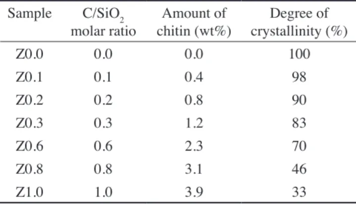
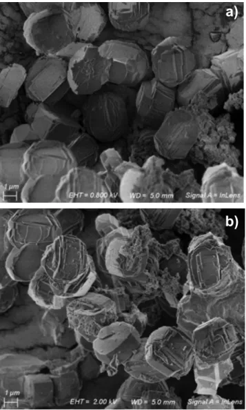
Fig. 4 shows SEM images of samples prepared with and without chitin. It can be seen that the size and shape of particles for both samples are similar, demonstrating that the use of chitin as mesopore template did not affect the morphological characteristics of ZSM-5 crystallites. This finding is consistent with a recently reported work, where carbon particles were used as mesopore template for preparation of ZSM-5 [4]. From Fig. 4, it can be also observed that the particle size for both samples was around 5 μm.
CONCLUSIONS
In this work, chitin was employed as a new template for generation of mesoporous ZSM-5. The addition of chitin retarded the crystallization rate, but did not modify the crystal morphology. Total and mesopore volumes of obtained materials increased with increasing amount of chitin in the reaction mixture. However, a prominence can be given to the large increase in the mesopores volume obtained by the use of chitin with template. In summary, chitin can be a very promising template for the synthesis of mesoporous zeolite crystals.
ACKNOWLEDGMENT
F.C.D. sincerely thanks CAPES for the scholarship.
REFERENCES
[1] P.A. Jacobs, J.A. Martens, Studies Surf. Sci. Catal. 58 (1991) 445.
[2] J. Cejka, H. van Bekkum, A. Corma, F. Schüth (Eds.), Introduction to zeolite science and practice, Studies Surf. Sci. Catal., 168, 3rd Ed., Elsevier, Amsterdam (2007).
[3] M. Lamia, D. Fatiha, B. Mohammed, D. Ayada, Oriental
J. Chem.32 (2016) 171.
[4] J.S. Oliveira, M.A. Mazutti, E.A. Urquieta-González, E.L. Foletto, S.L. Jahn, Mater. Res. 19 (2016) 1399.
[5] Y.H. Chou, C.S. Cundy, A.A. Garforth, V.L. Zholobenko, Micropor. Mesopor. Mater. 89 (2006) 78.
[6] Z. Pavlackova, G. Kosova, N. Zilkova, A. Zukal, J. Cejka, Studies Surf. Sci. Catal.162 (2006) 905.
[7] J.B. Koo, N. Jiang, S. Saravanamurugan, M. Bejblová, Z. Musilová, J. Cejka, S.E. Park, J. Catal. 276 (2010) 327. [8] J. Jin, X. Zhang, Y. Li, H. Li, W. Wu, Y. Cui, Q. Chen, L. Li, J. Gu, W. Zhao, J. Shi, Chem. Eur. J. 18 (2012) 16549. [9] B.K. Singh, D. Xu, L. Han, J. Ding, Y. Wang, S. Che, Chem. Mater. 26 (2014) 7183.
[10] R.J. White, A. Fischer, C. Goebel, A. Thomasa, J Am. Chem. Soc. 136 (2014) 2715.
[11] G.L. Dotto, G.S. Rosa, M.A. Moraes, R.F. Weska,
Figure 4: SEM images of samples prepared: (a) without chitin (Z0.0), and (b) with chitin (Z1.0).
[Figura 4: Micrografias obtidas por microscopia eletrônica de varredura das amostras preparadas: (a) sem quitina (Z0.0); e (b)
com quitina (Z1.0).]
Figure 3: Effect of C/SiO2 molar ratio added to the synthesis gel.
[Figura 3: Efeito da razão molar C/SiO2 adicionado ao gel de síntese.]
0.18
0.14
0.10
0.06
0.02 0.16
0.12
0.08
0.04
0.00 0.0
C/SiO2 molar ratio
Pore volume (cm
3.g -1)
0.4 0.8
0.2 0.6 1.0
a)
L.A.A. Pinto, J. Environm. Chem. Eng. 1 (2013) 50. [12] G.L. Dotto, J.M.N. Santos, I.L. Rodrigues, R. Rosa, F.A. Pavan, E.C. Lima, J. Coll. Interf. Sci. 446 (2015) 133. [13] A. Einbu, S.N. Naess, A. Elgsaeter, K.M. Vårum, Biomacromolecules5 (2004) 2048.
[14] G.L. Dotto, J.M.N. Santos, J.M. Moura, L.A.A. Pinto, e-Polymers 16 (2016) 49.
[15] D. Stamires, Y.L. Lam, J. Gorne, R. Wasserman, J.C.M. Ferreira, J. Silva, Patent Coop. Treaty WO/2006/087337, 2006 Aug. 24.
[16] J.S. Oliveira, F.C. Drumm, M.A. Mazutti, E.L. Foletto, S.L. Jahn, Cerâmica 62 (2016) 281.
[17] M.M.J. Treacy, J.B. Higgins, Collection of simulated XRD powder patterns for zeolites, 4th Ed., Struct. Comm. Int. Zeolite Ass., Elsevier, New York (2001).
[18] E.L. Foletto, N.C. Kuhnen, H.J. José, Cerâmica 46
(2000) 210.
[19] ASTM D3906-03, “Standard test method for determination of relative X-ray diffraction intensities of faujasite-type zeolite-containing materials” (2013).
[20] L. Chu, G. Liu, Q. Xiao, Mater. Res. Bull. 60 (2014) 746.
[21] L. Li, Q. Meng, J. Wen, J. Wang, G. Tu, C. Xu, F. Zhang, Y. Zhong, W. Zhu, Q. Xiao, Micropor. Mesopor. Mater. 227 (2016) 252.
[22] X. Wei, P.G. Smirniotis, Micropor. Mesopor. Mater. 97 (2006) 97.
[23] D. Liu, P. Yuan, D. Tan, H. Liu, T. Wang, M. Fan, J. Zhu, H. He, J. Colloid Interface Sci. 388 (2012) 176. [24] K.A. Sashkina, V.S. Labko, N.A. Rudina, V.N. Parmon, E.V. Parkhomchuk, J. Catal. 299 (2013) 44.
(Rec. 05/04/2017, Rev. 16/06/2017, Ac. 16/09/2017)