UNIVERSIDADE DA BEIRA INTERIOR
Sciences
Biosynthesis, solubilization and purification of human
membrane bound catechol-O-methyltransferase in
Brevibacillus choshinensis cells
Augusto Quaresma Henriques Pedro
Thesis for the obtention of a Master degree in
Biochemistry
(2
ndCycle of Studies)
Supervisor: Prof. Doctor Luís António Paulino Passarinha
Co-Supervisor: Prof. Doctor Cláudio Jorge Maia Baptista
iii
Para a minha Mãe e o meu Avô,
Com todo o carinho e amor…
v
A
A
c
c
k
k
n
n
o
o
w
w
l
l
e
e
d
d
g
g
m
m
e
e
n
n
t
t
s
s
Em primeiro lugar, quero deixar uma sincera palavra de agradecimento ao Professor Doutor Luís Passarinha e ao Professor Doutor Cláudio Maia, não só pela orientação deste trabalho, como pela confiança depositada em mim para a sua concretização. Nunca me esquecerei da vossa acessibilidade mas também do respeito e consideração com que sempre me trataram. Não posso deixar de referir que foi para mim um verdadeiro privilégio mas também um prazer poder trabalhar ao vosso lado.
Ao Professor Doutor João Queiroz, da Universidade da Beira Interior, quero expressar a minha sincera gratidão pela sua disponibilidade e contribuição no desenvolvimento deste projecto de investigação.
A todos os membros e colaboradores do CICS, particularmente do grupo “Biotechnology and Biomolecular Sciences”, quero agradecer pela salutar convivência bem como por todos os ensinamentos que me ministraram. Ao Vítor Gaspar, à Filomena Silva, bem como à Patrícia Pereira deixo um sentido obrigado, vocês foram, são e continuarão a ser a minha fonte de inspiração.
À Sofia e à Margarida agradeço a vossa contribuição para o bom desempenho do meu trabalho.
Aos meus verdadeiros amigos que desde sempre têm estado a meu lado, tanto nos bons como nos maus momentos, o meu muito obrigado.
À minha adorada família, especialmente os meus avós José, Alice e Júlia, os meus tios José e Isabel, o meu irmão José Miguel e ao meu querido Pai por todo o amor, sacrifício e apoio, quero agradecer-vos do fundo do meu coração. Todo o meu sucesso se deve a vocês.
vi
R
R
e
e
s
s
u
u
m
m
o
o
As proteínas membranares constituem cerca de 20 a 30 % de todas as proteínas codificadas pelo genoma de vários organismos. Elevadas quantidades de proteínas num estado de elevada pureza são necessárias quer para estudos farmacológicos, quer para estudos cristalográficos, daí a imperativa necessidade de desenvolver novos sistemas para a sobre-expressão heteróloga de proteínas membranares. Especificamente, nós testámos a aplicação de Brevibacillus choshinensis para a biosíntese da isoforma membranar da catecol-O-metiltransferase humana. No que diz respeito ao processo de produção, obteve-se uma moderada a elevada expressão num meio complexo com um valor de 45 nmol/h/mg para a actividade biológica da hMBCOMT, atingida às 20 horas de cultura a 37 ºC e 250 rpm. No que diz respeito à solubilização da proteína alvo, a eficiência de reconstituição para a hMBCOMT é nula na presença de detergentes iónicos tais como o SDS. No entanto, a aplicação de baixas concentrações de detergentes não-iónicos parece ser ideal para solubilizar a fracção membranar visto que a hMBCOMT recombinante retém elevados valores para a actividade biológica. Dos detergentes testados, a digitonina a 0.5 % (m/v) parece ser o mais adequado. De facto, o método descrito nesta tese é simples e poder-se-á tornar muito útil se aplicado num diagrama global para o isolamento da MBCOMT tendo em vista a sua caracterização bioquímica ou biofísica, onde se destaca a determinação da sua estrutura por cristalografia de raios-X ou estudos de interacção da hMCOMT com inibidores.
P
P
a
a
l
l
a
a
v
v
r
r
a
a
s
s
-
-
c
c
h
h
a
a
v
v
e
e
Proteínas membranares; MBCOMT humana; Produção heteróloga de proteínas; Detergentes; Doença de Parkinson
viii
A
A
b
b
s
s
t
t
r
r
a
a
c
c
t
t
Membrane proteins constitute 20-30% of all proteins encoded by the genome of various organisms. While large amounts of purified proteins are required for pharmaceutical and crystallization attempts, there is an unmet need for the development of novel heterologous membrane protein overexpression systems. Specifically, we tested the application of Brevibacillus choshinensis cells for the biosynthesis of human membrane bound catechol-O-methyltransferase (hMBCOMT). In terms of the upstream stage moderate to high expression was obtained for complex media formulation with a value near 45 nmol/h/mg for hMBCOMT specific activity achieved at 20 hours culture at 37 ºC and 250 rpm. Subsequently, the efficiency for reconstitution of hMBCOMT is markedly null in the presence of ionic detergents, such as SDS. However, the application of lower concentrations of non ionic detergents seems to be ideal to solubilize the membrane fraction since recombinant hMBCOMT retains more biological activity. From the detergents tested, digitonin at 0.5 % (w/v) appears to be the most suitable. Indeed, the straightforward method describe in this paper can be very useful and apply in a global flow sheet of MBCOMT isolation for biochemical or biophysical characterization including X-ray crystal structure determination or crosslinking interaction inhibitors studies.
K
K
e
e
y
y
w
w
o
o
r
r
d
d
s
s
Membrane proteins; Human MBCOMT; Heterologous protein production; Detergents; Parkinson Disease
x
T
T
a
a
b
b
l
l
e
e
o
o
f
f
C
C
o
o
n
n
t
t
e
e
n
n
t
t
s
s
PageChapter I
Introduction 1Section I – Brevibacillus Expression System 1
Subsection I – An Overview 1
Subsection II – Brevibacillus choshinensis Cells 1
Section II – The COMT Enzyme 3
Subsection I – COMT Multiple Functions 3
Subsection II – COMT Gene 3
Subsection III – COMT Genetic Polymorphisms 4
Subsection IV – COMT and its implication in several human disorders 5
Section III – The COMT Isoforms 8
Subsection I – Intracellular localization of COMT isoforms 8 Subsection II – Relative Distributions of S and MBCOMT 8 Subsection III – Analytical Methods in COMT Assays 10
Section IV – MBCOMT 11
Subsection I – Recombinant hMBCOMT Production 12
Subsection II – Recombinant hMBCOMT Purification 13
Section V – The Mimics of the Lipid Bilayer: A mandatory Tool for Membrane Protein
Production 14
Subsection I - Detergents 15
Subsection II – Non micellar membrane-mimicking systems 16
Chapter II
Materials and Methods 19
Section I – Materials 19
Section II – Methods 20
Subsection I – Strains, plasmids and media 20
Subsection II – Construction of Expression Vector pNCMO2-hMBCOMT 20
Subsection III – Agarose Gel Electrophoresis 21
Subsection IV – Recombinant hMBCOMT Production 21
xi
Subsection VI – MBCOMT Solubilization 22
Subsection VII – Total Protein Quantification 22
Subsection VIII – MBCOMT Enzymatic Assay 23
Subsection IX – SDS-PAGE and Western-Blot 23
Subsection X – Determination of Cell Density and Dry Brevibacillus choshinensis
Weight 24
Subsection XI – Hydrophobic Interaction Chromatography 24
Chapter III
Results and discussion 25
Section I – Construction of Expression Vector pNCMO2-hMBCOMT 25
Section II – Recombinant hMBCOMT Production 28
Subsection I – Dry Brevibacillus choshinensis Weight 28 Subsection II – Analysis of the different fractions obtained in hMBCOMT Production 29 Subsection III – Time Course Profile of hMBCOMT Production 30
Subsection IV – Detergent Screening 34
Subsection V – Kinetic Characterization of hMBCOMT 36
Section III – hMBCOMT Purification Initial Trials - HIC 40
Chapter IV
Conclusions 44Chapter V
Future Perspectives 45Chapter VI
References 46Chapter VII
Appendices 49xiii
L
L
i
i
s
s
t
t
o
o
f
f
F
F
i
i
g
g
u
u
r
r
e
e
s
s
Page
Chapter I - Introduction
Figure 1 – Electron Microscopy Image of Brevibacillus choshinensis cells ... 2
Figure 2 –pNCMO2 DNA vector map. ... 2
Figure 3 – The O-methylation of the catechol substrate catalysed by COMT. ... 3
Figure 4 - Structure of human COMT gene ... 4
Figure 5 – Pathways for the oxidative metabolism, redox cycling and inactivation of estradiol and estrone in mammalian cells and tissues ... 5
Figure 6 – Proposed mechanism of homocysteine pathophysiology and pathogenesis based on accumulation of intracellular SAH ... 6
Figure 7 - Representative structures and examples of the three major classes of COMT inhibitors ... 7
Figure 8 – Analytical methods applied to COMT activity analysis. ... 11
Figure 9 - Elution of MBCOMT from a Resource Q column at pH 7.8 by a NaCl gradient ... 14
Figure 10 - Schematic representation of different solubilization methods for integral membrane proteins.. ... 15
Figure 11 – Structure of the different types of detergents ... 16
Chapter II – Materials and Methods Figure 1 – BSA (bial buffer with Triton X-100 1% (v/v)) calibration curve ... 22
Chapter III – Results and Discussion Figure 1 – Agarose gel electrophoresis pictures of the several stages during the construction of the expression vector pNCMO2-hMBCOMT. ... 27
Figure 2 – Relationship between the OD (660 nm) and the dry B. choshinensis weight. ... 29
Figure 3 – Western-blot and SDS-PAGE analysis of recombinant human MBCOMT in the three different fractions obtained during the process of recombinant human MBCOMT production 30 Figure 4 - Growth profile of B. choshinensis and recombinant hMBCOMT specific activity profile at different incubation periods and different culture conditions ... 32
Figure 5 – Growth profile of B. choshinensis harboring the plasmid pNCMO2-BLA at different incubation periods (ranging from 0 to 72 hours) at 120 rpm and 30 ºC ... 33
Figure 6 - Western-blot and SDS-PAGE analysis of recombinant hMBCOMT at different times of incubation period on 2SYNm medium ... 34
Figure 7 – Densitometric quantification based on figure 17 of the bands obtained at different times of incubation period on 2SYNm comparatively to the control ... 34
xiv
Figure 8 – Effect of concentration and type of detergent used to solubilize the membrane fraction on the human recombinant MBCOMT biological activity (nmol/h/mg of protein) .... 35 Figure 9 – Percentage of increase on hMBCOMT biological activity relatively to the control of the membrane fractions solubilized with different types and concentrations of detergents . 36 Figure 10 – Relationship between the total protein concentration and the hMBCOMT specific activity (nmol/h/mg) ... 37 Figure 11 – Relationship between the incubation time and the hMBCOMT specific activity (nmol/h/mg) ... 37 Figure 12 – Saturation curve of recombinant hMBCOMT ([SAM] = 250.0 µM) and Lineweaver-burk plot of the saturation curve of recombination hMBCOMT. ... 38 Figure 13 – Saturation curve of recombinant hMBCOMT ([SAM] = 100.0 µM) and Lineweaver-burk plot of the saturation curve of recombination hMBCOMT ... 39 Figure 14 – Hydrophobic interaction Chromatography on butyl-sepharose 4FF with 0.6 M, 0.4 M and 0.2 M ammonium sulphate, respectively for A, B and C in 10 mM Tris-Cl (pH 7.8). ... 41 Figure 15 – SDS-PAGE and Western-blot analysis of recombinant hMBCOMT eluted at different concentrations of ammonium sulphate on the butyl-sepharose resin. ... 43xvi
L
L
i
i
s
s
t
t
o
o
f
f
T
T
a
a
b
b
l
l
e
e
s
s
Page
Chapter I - Introduction
Table 1 – Relative quantification of SCOMT and MBCOMT proteins in rat tissues expressed as % of total COMT in the immunoblot. ... 9 Table 2 – Relative quantification of SCOMT and MBCOMT proteins in human tissues and cells expressed as % of total COMT in the immunoblot. ... 9
Chapter III – Results and Discussion
xviii
L
L
i
i
s
s
t
t
o
o
f
f
A
A
c
c
r
r
o
o
n
n
y
y
m
m
s
s
AADC Aromatic amino acid descarboxylase B. chosinensis Brevibacillus choshinensis
BSA Bovine serum albumin
cDNA Complementary deoxyribonucleic acid
CHAPS 3 – [(3-Cholamidopropyl) dimethylammonio] – 1 – propanesulfonate CMC Critic micellar concentration
COMT Catechol-O-Methyltransferase CTAB Methylammonium bromide DNA Deoxyribonucleic acid
DTT Dithiotreitol
E. coli Escherichia coli
EDTA Ethylenediamine tetraacetic acid
HIC Hydrophobic Interaction Chromatography
hMBCOMT Human Membrane bound Catechol-O-Methyltransferase HPLC High Performance Liquid Chromatography
IPTG Isopropylthiogalactosidase
MBCOMT Membrane bound Catechol-O-Methyltransferase MgCl2 Magnesium chloride
NaCl Sodium chloride
NLP Nanolipoprotein
OD660 Cell density at 660 nm
PCR Polymerase chain reaction
PD Parkinson disease
PVDF Polyvinyl difluoride
rHDL Reconstituted high density lipoproteins SAH S-adenosyl-l-homocysteine
SAM S-adenosyl-l-methionine
SCOMT Soluble Catechol-O-Methyltransferase SDS Sodium dodecyl sulphate
1
Chapter I
Introduction
Section I - Brevibacillus Expression System:
Subsection I - An Overview:
The Brevibacillus Expression System based on Brevibacillus choshinensis (B. choshinensis) cells is well-suited for secretory production of heterologous proteins with high efficiency [1]. This system presents several advantages such as the fact that the host bacterium secretes proteins very efficiently and produces low levels of extracellular proteases so that the products remained unscatched in the culture medium [1]. In addition, the proteins are produced as active forms; the host bacterium is a safe organism and is amenable to genetic engineering [1]. Many proteins from eukaryotic and prokaryotic organisms have been successfully produced in this system at high levels and with native biological activity [2 - 6]. In particular, for eukaryotic proteins with cystein residues that make intramolecular bonds and, consequently, its disulfide bonds must be formed at an exact location, it is very difficult to achieve an efficient production at traditional bacterial systems based on intracellular expression [1]. However, this can be overcome using extracellular expression strategies based on B. choshinensis.
Subsection II - Brevibacillus choshinensis cells:
B. choshinensis (figure 1) is a gram-positive bacterium that has excellent ability to produce many kinds of proteins in an extracellular model [1]. These cells exhibit high transformation efficiency by electroporation, which makes the construction of expression clones a straightforward process [1].
Figure 1
In general, there are several expression vectors that can be used in the expression system [1]. Some of them like the pNI or the pN
intracellular protein production and others such as the pNCMO2 or the pNY326 extracellular protein biosynthesis
From the mentioned expression vectors, the one chosen for this work was the pNCMO2 (figure 2) which is a shuttle vector between
to construct an expression plasmid using this vector, first the vector is transferred into
The pNCMO2 presents several unique features. Among others, this vector has a neomycin and ampicilin resistant genes as
Also possesses a secretory sign into the medium culture [1
of the five promoters driving the transcription
Figure 1 – Electron Microscopy Image of B. choshinensis cells [7
here are several expression vectors that can be used in the ]. Some of them like the pNI or the pNI-His DNA allow intracellular protein production and others such as the pNCMO2 or the pNY326
biosynthesis [1].
From the mentioned expression vectors, the one chosen for this work was the pNCMO2 (figure which is a shuttle vector between B. choshinensis and Escherichia coli
to construct an expression plasmid using this vector, first E. coli is used as the host and then the vector is transferred into B. choshinensis cells [1].
Figure 2 – pNCMO2 DNA vector map [1].
The pNCMO2 presents several unique features. Among others, this vector has a neomycin and as a selection marker in B. choshinensis and E. coli
secretory signal segment that is responsible by the protein secretion dire into the medium culture [1]. Also, the pNCMO2 vector contains the P2 promoter (which is one of the five promoters driving the transcription of cell wall protein) that does not work in
2
cells [7].
here are several expression vectors that can be used in the Brevibacillus His DNA allows preferentially an intracellular protein production and others such as the pNCMO2 or the pNY326 lead to an
From the mentioned expression vectors, the one chosen for this work was the pNCMO2 (figure (E. coli) [1]. In order is used as the host and then
The pNCMO2 presents several unique features. Among others, this vector has a neomycin and E. coli, respectively [1]. that is responsible by the protein secretion directly ]. Also, the pNCMO2 vector contains the P2 promoter (which is one of cell wall protein) that does not work in E.
3
coli. but has a strong promoter activity in B. choshinensis, enabling efficient protein production and making pNCMO2 suitable for cloning genes of which expressions are stressful for E. coli [1].Section II - The COMT Enzyme:
Subsection I - COMT multiple functions:
Catechol-O-methyltransferase (COMT; EC 2.1.1.6) is a magnesium-dependent enzyme that catalyzes the methylation of catechol substrates using S-adenosyl-l-methionine (SAM) as a methyl donor and yielding, as reaction products, the O-methylated catechol and S-adenosyl-l-homocysteine (SAH) (see figure 3) [8].
Figure 3 – The O-methylation of the catechol substrate catalysed by COMT (Adapted from [9, 10]).
Typically, the substrates of COMT in mammals include catecholamines with hormonal and neurotransmission activities such as dopamine, norepinephrine, epinephrine, catecholestrogens and their metabolites, ascorbic acid, some indolic intermediates of melanin metabolism and xenobiotic catechols, like carcinogenic catechol-containing flavonoids [9].
Subsection II - COMT Gene:
The protein COMT is present in prokaryotes and eukaryotes [8]. In mammals, it appears in two molecular forms, a soluble form (SCOMT) and in membrane form bound to the rough endoplasmatic reticulum membrane (MBCOMT) [8]. Both the isoforms are encoded by a single gene that, in humans, is located on chromosome 22 band q11.21 and is composed by six exons (see figure 4) [9]. The first two exons are non-coding and the translation initiation codons for
both the isoforms are located on the th
direct the synthesis of two partially overlapped transcripts (see figure 4), one (P2) of 1.5 Kb that is constitutively expressed and another (P1) of 1.3 Kb is subject
regulation [9]. The short transcript translates SCOMT and the longer transcript translates MBCOMT but also the soluble form by the leaky scanning mechanis
[8].
Subsection III - COMT Genetic Polymorp
COMT activity is ubiquitous in animal tissues but the COMT levels vary among different species, in individuals of the same specie as well as in tiss
The highest enzymatic activity, within individuals, is present in the gastrointestinal tract while the lowest enzymatic activity is r
[8]. In humans, COMT activity can be distributed in three groups, one with high activity (COMTH/H), one with intermediate activi
[8]. The difference in activity is correlated with a functional COMT polymorphism at codon 108/158 (respectively for
valine in the polypeptide chain [
activity and decreased thermal stability, while the Val 108/158 is activity [8]. Several others polymorphisms have been reported to COMT but physiological significance [8
are located on the third exon [8]. In humans, two separate promoters direct the synthesis of two partially overlapped transcripts (see figure 4), one (P2) of 1.5 Kb that is constitutively expressed and another (P1) of 1.3 Kb is subject
ort transcript translates SCOMT and the longer transcript translates MBCOMT but also the soluble form by the leaky scanning mechanism of translational initiation
Figure 4 – Structure of human COMT gene [11].
COMT Genetic Polymorphisms:
COMT activity is ubiquitous in animal tissues but the COMT levels vary among different species, in individuals of the same specie as well as in tissues from the same individuals [8 The highest enzymatic activity, within individuals, is present in liver, followed by kidneys and the gastrointestinal tract while the lowest enzymatic activity is reported to the cardiac tissue ]. In humans, COMT activity can be distributed in three groups, one with high activity
), one with intermediate activity (COMTH/L) and other with low activity (COMT ]. The difference in activity is correlated with a functional COMT polymorphism at codon
respectively for SCOMT and MBCOMT) involving a substitution of a methione to a line in the polypeptide chain [9]. The Met 108/158 variant is associated with low enzymatic activity and decreased thermal stability, while the Val 108/158 is associated with high
]. Several others polymorphisms have been reported to COMT but physiological significance [8].
4
]. In humans, two separate promoters direct the synthesis of two partially overlapped transcripts (see figure 4), one (P2) of 1.5 Kb that is constitutively expressed and another (P1) of 1.3 Kb is subject to tissue-specific ort transcript translates SCOMT and the longer transcript translates m of translational initiationCOMT activity is ubiquitous in animal tissues but the COMT levels vary among different ues from the same individuals [8]. liver, followed by kidneys and eported to the cardiac tissue ]. In humans, COMT activity can be distributed in three groups, one with high activity ) and other with low activity (COMTL/L) ]. The difference in activity is correlated with a functional COMT polymorphism at codon MBCOMT) involving a substitution of a methione to a 8/158 variant is associated with low enzymatic associated with high ]. Several others polymorphisms have been reported to COMT but with irrelevant
5
Subsection IV
COMT and its implication in several human disorders:
During the last decades, the COMT enzyme has been implicated in several human disorders such as cardiovascular diseases [12], neurologic disorders [13 - 15] and estrogen-induced cancers [16, 17].
In particular, the metabolism of steroidal estrogens are depicted in figure 5 where the oxidation of estradiol (E2) occurs primarily at C-2 (and C-4) to form the 2-3 and 3-4 catechols, at C-17 to form estrone (E1) and C-16 to form the 16α-hydroxy (OH) E1 [17]. The E2 and the E1 catechols are intermediates in the generation of more reactive semiquinones and quinones which can serve as substrates for redox cycling and, consequently, generation of reactive oxygen species. Beyond the glucoronidation and sulfatation, the catechols are inactivated by O-methylation mediated by COMT [17].
The E2 catechols 2-OH and 4-OH are O-methylated by COMT with the metabolic clearance of 4-OH E2 being slower than that of 2-OH E2 due to the fact that probably the 2-OH E2 inhibits the O-methylation of 4-OH E2 that persists and participates in redox cycling to generate increased oxidative DNA damage [17]. Some studies with hamsters revealed that the inhibition of COMT potentiates tumorigenesis but it is still unknown whether the susceptibility to steroid-hormone cancers in humans is associated with the levels of COMT activity [17].
Figure 5 – Pathways for the oxidative metabolism, redox cycling and inactivation of estradiol and estrone in mammalian cells and tissues [17].
6
The implication of COMT in cardiovascular diseases is related to SAH (see figure 6), one of the products formed from the O-methylation of catechol substrates by COMT, commonly described as an endogenous inhibitor of COMT-mediated O-methylation of endogenous as well as exogenous catechols [12]. In the case of catecholamines, the inhibition of its methylation by SAH, results in accumulation and, subsequently, in the overstimulation of the adrenoreceptor-mediated functions of the cardiovascular system [12]. Also, the constant exposure of the vascular endothelial cells to high levels of circulating catecholamines would lead to their chronic cumulative damage due to the large amounts of the oxidative products (catechol quinones or semiquinones and oxyradicals) formed from the endogenous catecholamines. [12].Figure 6 – Proposed mechanism of homocysteine pathophysiology and pathogenesis based on accumulation of intracellular SAH [12].
Parkinson’s disease (PD) is a neurological disorder characterized by the degeneration of dopaminergic neurons, with consequent reduction in striatal dopamine levels leading to characteristic motor symptoms [8]. The most appropriated treatment for this disease is the dopamine replacement therapy with levodopa together with an inhibitor of aromatic amino acid descarboxylase (AADC) [8]. It has been stated that the use of COMT inhibitors as adjuvants to levodopa/AADC inhibitor therapy significantly improves the clinical benefits of this treatment [8]. The first-generation COMT inhibitors (figure 7) comprise several compounds such as Pyrogallol and tropolone that contain a catechol substrate or some related
7
bioisosteric moiety [8]. Tipically, these compounds are competitive substrates of COMT leading, in some cases to lowers efficacies in vivo and extremely toxic [8]. The second-generation COMT inhibitors (figure 7) (tolcapone, nebicapone, nitecapone, among others) constituted a new class of di-substituted catechols whose enhanced potency is assigned to the substitution with electron-withrawing groups at a position ortho to a hydroxyl group of the catechol moiety [8]. For these compounds, the necessary concentration to inhibit 50 % of COMT activity is in the nanomolar range (increased potency when compared with the first-generation inhibitors whose concentration is at a micromolar range) [8]. It has been discussed whether COMT inhibition should be restricted to the peripheral tissues (like the Nitecapone) or, alternatively, a COMT inhibitor should present broad tissue selectivity (like tolcapone) [8]. Since is attributed to SAM an antidepressive effect and it is well known that the COMT inhibition both central and peripherally leads to the depletion of SAM in the striatum, it is accepted that the COMT inhibitors should present a limited access to the brain in order to minimize the side effects of the PD adjuvant therapy [8].Nowadays, there is a third class of COMT inhibitors – late atypical inhibitors (figure 7) – that comprises both the bisubstrate inhibitors and the bifunctional inhibitors. In spite of the first ones were designed to target simultaneously the catechol and the SAM binding sites, the bifunctional inhibitors were made by including two catecholic pharmacofores in the same inhibitor molecule [8]. In what concerns to this last class of compounds, it seems that they have more potency in vitro than the compounds previously described but more studies are necessary to address several issues like its efficacy in vivo or its safety [8].
Figure 7 – Representative structures and examples of the three major classes of COMT inhibitors (Tropolone, Pyrogallol refers to the first-generation inhibitors, Tolcapone and Nebicapone to the second generation and the Bisubstrate and bifunctional inhibitors represents the late atypical inhibitors) [8].
8
Section III – The COMT isoforms:
In mammals, COMT is present in two molecular forms: a soluble form (SCOMT) that contains 221 amino acid residues and a molecular weight of 24.7 KDa (humans) and in another form associated with the membrane (MBCOMT) [8]. The last one, beyond the 221 amino acids from the soluble isoform, has an additional peptide in its amino terminal of 50 amino acid residues corresponding to a molecular weight of 30 KDa (humans) [8]. This extra peptide contains a stretch of 21 (humans) hydrophobic amino acid residues that constitute the membrane anchor region [8]. MBCOMT is an integral membrane protein with the carboxy-terminal portion of the polypeptide (the region catalytically active) exposed to the cytoplasmic side of the membrane [18].
Subsection I - Intracellular localization of COMT isoforms:
In general, the enzyme SCOMT is present in the cytoplasm but it also has been reported in the nucleus of rat transfected COS-7 cells [9] as well as in mammary epithelial cells under certain circumstances such as increased levels of catecholestrogens [19]. On the other hand, MBCOMT, previously assigned to the outer mitochondrial membrane [20] and plasma membrane [21], actually is attributed to the rough endoplasmic reticulum membrane [22].
Subsection II - Relative distributions of S and MBCOMT:
In most human and rat tissues, the levels of SCOMT greatly exceed the levels of MBCOMT (see tables 1 and 2), except for the human brain where it only represents 30 % of the total COMT [8]. This difference in the soluble to MBCOMT ratios (where mainly the long transcript is found) between the rat and human brains is probably due to the fact that in humans the MB-AUG initiation codon is located in a more favorable context than the S-MB-AUG initiation codon [8]. Therefore, most of the translation is initiated from the MB-AUG site [18].
9
Table 1 – Relative quantification of SCOMT and MBCOMT proteins in rat tissues expressed as % of total COMT in the immunoblot [9].
Table 2 – Relative quantification of SCOMT and MBCOMT proteins in human tissues and cells expressed as % of total COMT in the immunoblot [9].
T Tiissssuuee SSCCOOMMTT MMBBCCOOMMTT L Liivveerr 93 7 K Kiiddnneeyy 75 25 H Heeaarrtt 79 21 C Ceerreebbeelllluumm 86 14 T Teelleenncceepphhaalloonn 69 31 T Tiissssuuee SSCCOOMMTT MMBBCCOOMMTT L Liivveerr 85 15 K Kiiddnneeyy 77 23 A Addrreennaall 74 26 D Duuooddeennuumm 89 11 B Brraaiinn 30 70 H HeeLLaa--CCeellllss 35 65 M MCCFF77--CCeellllss 92 8
10
Subsection III - Analytical methods in COMT assays:
Tipically, a COMT activity trial usually consists on the handling of the sample and incubation followed by separation and detection of the reaction products [23]. The several analytical methods described for COMT activity analysis are depicted in figure 8 [23}. Some of these methods involve the direct quantification of the substrate or the product and don’t include a separation step between the O-methylated products and the substrate or co-substrate [23]. During the last decades, more reliable and sensitive methods have been developed, involving liquid-liquid extraction or chromatographic techniques with different types of detection [23]. The first methods to separate an O-methylated metabolite from the parent compound included liquid-liquid extraction with organic solvents [23]. After extraction, the organic phase was usually evaporated and the products assess by fluorometric or radiochemical detection [23]. Paper and thin layer chromatography has been used to identify the O-methylated products, extracted from COMT incubation mixtures, by comparing the Rf values with standard reference compounds [23]. Also, gas chromatography have been applied to separate regioisomeric O-methylated products from each other [23] This kind of methods usually require derivatisation of the products and the use of an internal standard [ 23]. In spite of the methods described above, High Performance Liquid chromatography (HPLC) is the most common technique used for COMT activity measurements. Normally, HPLC with UV detection shows poor selectivity and moderate sensitivity [23]. On the other hand, HPLC with fluorescence detection can be very sensitive and allows the analysis of Omethylated compounds if they exhibit native fluorescence themselves or if they can be derivatised with fluorogenic reagents [23]. The sensitivity of HPLC with radiochemical detection is comparable with other types of detection but when compared with other systems, are restricted to special cases, in order to avoid working with radioactive material [23].
The catechols and phenolic hydroxils of the O-methylated products are easily oxidized, which allows electrochemical detection in COMT activity assays [23]. This technique can be used in a wide range of substrates (catecholamines, L-dopa, among others) and it has been applied to determination of the inhibitory effect of COMT inhibitors [23].
Figure 8 –
Section IV – MBCOMT:
Many kinetic and biochemical properties of MBCOMT are similar from those SCOMT isoform. Specifically,
requirement and similar Km va
several differences between the two enzymes. MBCOMT is an integral membrane protein [ 24] with pI values higher from those of SC
the COMT isoforms is that with exception of estrogen catechols, which possess similar K values for both isoforms of COMT, all other catechol substrates including catecholamines have lower KM values for MBCOMT [
influence the kinetic parameters of both isoforms since at low concentrations of catecholamines, O-methylation by the low K
predominate, and only when this enzyme becomes saturated with substrate, does the contribution of the hig
catecholamine substrates have a higher affinity but lower reaction velocity with MBCOMT
Analytical methods applied to COMT activity analysis [23
MBCOMT:
Many kinetic and biochemical properties of MBCOMT are similar from those
isoform. Specifically, the mechanism reaction, Ca2+ inhibition, pH optimum, Mg requirement and similar Km values for the methyl donor AdoMet [24]. However, there are several differences between the two enzymes. MBCOMT is an integral membrane protein [
] with pI values higher from those of SCOMT [20]. Another important difference between that with exception of estrogen catechols, which possess similar K values for both isoforms of COMT, all other catechol substrates including catecholamines have
values for MBCOMT [24]. Interestingly, the substrate concentration seems to e the kinetic parameters of both isoforms since at low concentrations of methylation by the low KM membrane-bound form of COMT would
predominate, and only when this enzyme becomes saturated with substrate, does the contribution of the high KM, soluble form of COMT becomes significant [
catecholamine substrates have a higher affinity but lower reaction velocity with MBCOMT
11
to COMT activity analysis [23].
Many kinetic and biochemical properties of MBCOMT are similar from those described to inhibition, pH optimum, Mg2+
4]. However, there are several differences between the two enzymes. MBCOMT is an integral membrane protein [18, ]. Another important difference between that with exception of estrogen catechols, which possess similar KM
values for both isoforms of COMT, all other catechol substrates including catecholamines have 4]. Interestingly, the substrate concentration seems to e the kinetic parameters of both isoforms since at low concentrations of bound form of COMT would predominate, and only when this enzyme becomes saturated with substrate, does the , soluble form of COMT becomes significant [24]. The catecholamine substrates have a higher affinity but lower reaction velocity with MBCOMT
12
than SCOMT, suggesting that MBCOMT is more important for the metabolism of catecholamines in vivo [24, 25].Subsection I - Recombinant hMBCOMT Production:
Membrane proteins and COMT, in particular, are implicated in several disorders (previously described). However, studies of these disorders are hampered by a lack of information of the proteins involved which makes the knowledge of the structure of hMBCOMT an essential prerequisite for understanding its function and, further, how its function can be modified by small molecules, one of the major goals of pharmaceutical industry [26]. As the majority of medically and pharmaceutically relevant membrane proteins, hMBCOMT is present in tissues at low concentration, which makes heterologous expression in large-scale production-adapted cells a prerequisite for structural studies [26].
In the last years, recombinant hMBCOMT was produced in several eukaryotic systems such as Sf9 insect cells [21], in transfected human embryonic kidney fibroblast cell lines [27] and in human HeLa and hamster BHK-cells [28].
The system reported by Ulmanen et al. (1992) based on Sf9 insect cells for recombinant hMBCOMT production yielded great amounts of hMBCOMT proteins whose polypeptide wasn’t processed in the lower-molecular mass SCOMT form [21]. Moreover, they also evaluated the effects of Triton X-100 treatment on substrate methylation of hMBCOMT containing fractions and found that the meta/para methylation ratio increases upon treatment with Triton X-100 [21].
In what concerns to human kidney cell lines, its transfection with the plasmid containing the MBCOMT cDNA, yielded a value of 206 U/mg of protein (using 2.5 mM pyrocatechol as substrate), comparatively to the 9,98 U/mg of protein in the untransfected cells [27]. Also in this work, the inhibitory effect of Ro 40-7592 and tropolone on MBCOMT activity was assessed. Indeed, it was found that these two compounds inhibited hMBCOMT in a dose-dependent manner with an IC50 (700 nM and 1mM for Ro 40-7592 and tropolone respectively)
similar from those obtained for the native enzyme [27].
Lundstrom et al. (1997) described a system for hMBCOMT production in human HeLa and hamster BHK-cells from which they assessed the intracellular localizations of COMT isoforms [28]. Indeed, they found that hSCOMT is predominantly found in the cytoplasm but it also could be detected in nuclei [28]. On the other hand, hMBCOMT was attributed to rough endoplasmic reticulum membrane [28].
A common characteristic from almost all works published concerning recombinant hMBCOMT production were largely based on fractionations of cell or tissue homogenates since in that time there weren’t available any hMBCOMT-specific antibodies [28].
13
Several prokaryotic systems have been described for recombinant hMBCOMT production with one major common characteristic, the employment of E. coli as the host. In one of these systems, it wasn’t able to express the recombinant protein and the authors supposed that the hydrophobic sequence that anchors MBCOMT to the membrane was toxic for the bacteria [9]. In spite of these results, recombinant hMBCOMT in a catalytical active form was produced at relatively high levels in E. coli SG 13009 [29, 30] and BL21 strains [11].The experiments for hMBCOMT production using E. coli SG 13009 under the IPTG inducible T7 promoter were carried out with two complementary Deoxyribonucleic acid (cDNA), one coding for the MBCOMT and the other lacking the 24 hydrophobic N-terminal amino acids of MBCOMT [29, 30]. These two cDNAs were introduced into the expression vector pDS56/RBSII and relatively high levels of catalytically active MBCOMT were obtained while the levels of the truncated COMT protein were much lower [29, 30].
A recently work from Zhu et al. (2007) presented a system for hMBCOMT production, from which the authors performed a wide set of kinetic analyses using several substrates, computational modeling studies in order to understand the molecular mechanisms underlying the catalytic behavior of hMBCOMT with respect to cofactor SAM, various substrates and the regioselectivity for the formation of mono-methyl ether products [11]. In fact, the enzyme kinetic analyses indicated that the produced MBCOMT was functionally active and the homology models developed for MBCOMT showed that the binding energy values calculated for most substrates agreed well with measured kinetic parameters [11]. Also, the model developed predicted precisely the regioselectivity for the O-methylation of several substrates at different hydroxyl groups since the predicted values matched the experimental data [11].
Subsection II - Recombinant MBCOMT Purification:
Actual scientific literature fails in terms of relevant information concerning purification trials for MBCOMT isolation. However, there is a report [31] where the authors described a partial purification by anion-exchange chromatography of a fraction containing rat MBCOMT previously isolated from rat liver homogenates and solubilized with Triton X-100 0,5 %. In this strategy, the authors used a Resource Q column, prepacked with Source 15Q, combining high speed, elevated capacity with high resolution [31]. The figure 9 represents a typical chromatogram obtained with a Resource Q column for the purification of MBCOMT [31].
Figure 9 – Elution of MBCOMT from a Resource Q column at pH 7.8 by a NaCl gradient. The full line represents the absorbance at 280 nm and the dashed line repre
grey bar indicates the fractions positive for COMT activity. The inset represents an analysis by western blot of the MBCOMT microssomal fraction (1), the solubilized MBCOMT fraction (2) and the Resource Q fractions positive for COMT activity (3
Section V – The mimics of the lipid bilayer: a mandatory tool for
membrane protein production:
There are several difficulties concerning the study of membrane proteins [ membrane proteins are natu
simplest organism is a complex, heterogenous and dynamic environment limits (but does not preclude) the use of many standard biophysical techniques to determine
and function such as NMR, X previous protein extraction environment in vitro [32].
In addition, membrane proteins are not generally soluble in aque
reside in surroundings that satisfy their high hydrophobicity. This can be accomplished using special synthetic systems for
solubilization techniques used for functional proteins [22].
Elution of MBCOMT from a Resource Q column at pH 7.8 by a NaCl gradient. The full line represents the absorbance at 280 nm and the dashed line represents the conductivity read on
grey bar indicates the fractions positive for COMT activity. The inset represents an analysis by western blot of the MBCOMT microssomal fraction (1), the solubilized MBCOMT fraction (2) and the Resource Q
ve for COMT activity (3 – 7) [31].
The mimics of the lipid bilayer: a mandatory tool for
membrane protein production:
There are several difficulties concerning the study of membrane proteins [
membrane proteins are naturally embedded in a mosaic lipid bilayer, which in even the simplest organism is a complex, heterogenous and dynamic environment limits (but does not preclude) the use of many standard biophysical techniques to determine
as NMR, X-ray christallography, among others [32]. Such methods require extraction from its native membrane and studied in a detergent or a lipid
].
In addition, membrane proteins are not generally soluble in aqueous solution, so they need to reside in surroundings that satisfy their high hydrophobicity. This can be accomplished using special synthetic systems for in vitro trials [32]. In figure 10 are depicted several solubilization techniques used for functional and structural characterization of membrane
14
Elution of MBCOMT from a Resource Q column at pH 7.8 by a NaCl gradient. The full line sents the conductivity read on-line. The grey bar indicates the fractions positive for COMT activity. The inset represents an analysis by western-blot of the MBCOMT microssomal fraction (1), the solubilized MBCOMT fraction (2) and the Resource Q
The mimics of the lipid bilayer: a mandatory tool for
There are several difficulties concerning the study of membrane proteins [32]. The fact that rally embedded in a mosaic lipid bilayer, which in even the simplest organism is a complex, heterogenous and dynamic environment limits (but does not preclude) the use of many standard biophysical techniques to determine protein structure ]. Such methods require from its native membrane and studied in a detergent or a lipid
ous solution, so they need to reside in surroundings that satisfy their high hydrophobicity. This can be accomplished using are depicted several rization of membrane
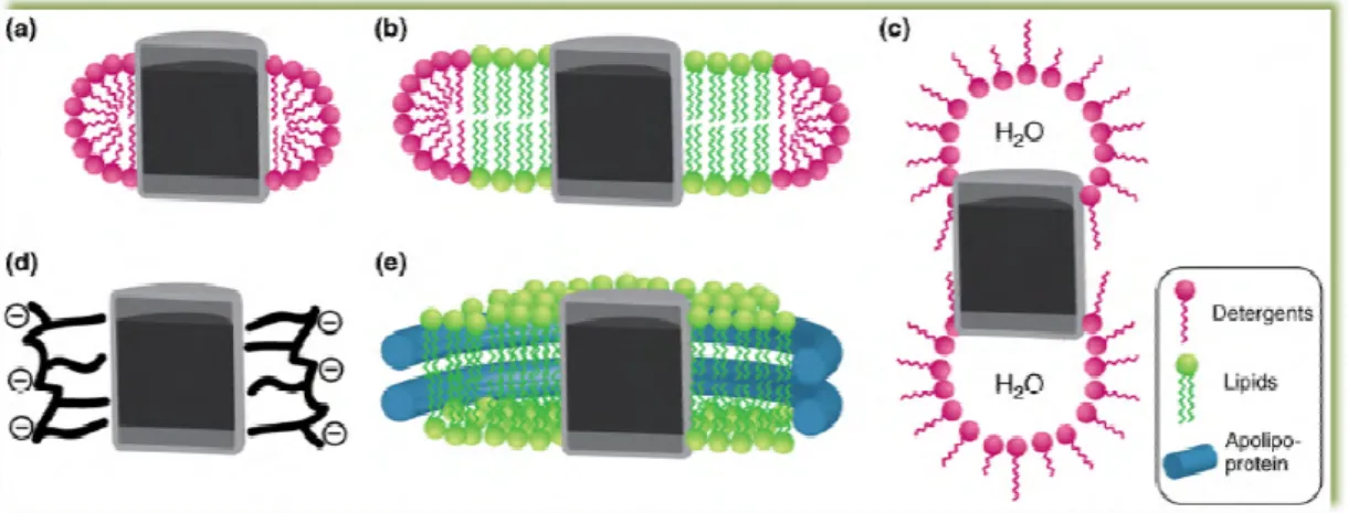
Figure 10 – Schematic representation of different solubilization methods for integral membrane proteins. Micelle (a), bicelle (b), reverse micelle (c), amphipol (d) and nanolipoprotein particle ( membrane protein is depicted in grey with its hydrophilic and hydrophobic r
[22].
Subsection I - Detergents:
The importance of detergents as tools for the study of membrane proteins cannot be underestimated since they
applied in the primary solubili
molecules, consisting of a polar head group and a hydrophobic chain that exhibit unique properties in aqueous solution by forming spherical micellar structures in which membrane proteins are usually soluble
Detergents are classified in four major classes of detergents (whose st figure 11), the ionic, non
detergents contain a head group with a net charge that can be anionic or cationic and a hydrophobic hydrocarbon chain or steroidal back
sodium dodecyl sulphate (
proteins but it can promote irreversible denaturation detergents which differ from SDS in their backbone [32]. As a result, they have a polar and apolar face inste the capacity to form small kidney
the traditional ionic detergents [32 head groups of either polyo
mild and non-denaturing as they break lipid protein-protein contacts [32
biological active form [32
Schematic representation of different solubilization methods for integral membrane proteins. Micelle (a), bicelle (b), reverse micelle (c), amphipol (d) and nanolipoprotein particle ( membrane protein is depicted in grey with its hydrophilic and hydrophobic regions in light and dark grey
Detergents:
The importance of detergents as tools for the study of membrane proteins cannot be underestimated since they are usually vital in the isolation and purification of the protein and in the primary solubilization step of reconstitution [32]. Detergents are amphipatic molecules, consisting of a polar head group and a hydrophobic chain that exhibit unique erties in aqueous solution by forming spherical micellar structures in which membrane proteins are usually soluble (see figure 10 a) [32].
Detergents are classified in four major classes of detergents (whose structure is depicted in non-ionic, zwiterionic and bile acid salts detergents [32
detergents contain a head group with a net charge that can be anionic or cationic and a hydrophobic hydrocarbon chain or steroidal backbone [32]. This type of detergents comprises dodecyl sulphate (SDS) which is extremely effective in the solubilization of membrane it can promote irreversible denaturation [32]. The bile acid salts are ionic detergents which differ from SDS in their backbone, which consists on a rigid s
]. As a result, they have a polar and apolar face instead of a well-defined head group, with form small kidney-shaped aggregates unlike the spherical micelles formed by raditional ionic detergents [32]. Non-ionic detergents contain uncharged hydrophilic head groups of either polyoxyethilene or glycosidic groups. Typically, are considered to be
denaturing as they break lipid-lipid and lipid-protein interactions
[32]. This allows membrane protein solubilization and isolation in a biological active form [32]. Finally, zwiterionic detergents combine the properties of ionic
15
Schematic representation of different solubilization methods for integral membrane proteins. Micelle (a), bicelle (b), reverse micelle (c), amphipol (d) and nanolipoprotein particle (e). The egions in light and dark grey
The importance of detergents as tools for the study of membrane proteins cannot be are usually vital in the isolation and purification of the protein and ]. Detergents are amphipatic molecules, consisting of a polar head group and a hydrophobic chain that exhibit unique erties in aqueous solution by forming spherical micellar structures in which membrane
ructure is depicted in nd bile acid salts detergents [32]. The ionic detergents contain a head group with a net charge that can be anionic or cationic and a of detergents comprises is extremely effective in the solubilization of membrane ]. The bile acid salts are ionic a rigid steroidal group defined head group, with shaped aggregates unlike the spherical micelles formed by rgents contain uncharged hydrophilic are considered to be protein interactions rather than s allows membrane protein solubilization and isolation in a zwiterionic detergents combine the properties of ionic
16
and ionic detergents and, in general have more deactivating characteristics than non-ionic detergents [32].The critic micellar concentration (CMC) can be defined as the minimum concentration of detergent for individual detergent molecules to cluster and form micelles promoting a sudden change in surface tension and other pertinent physical properties [32].
Figure 11 – Structure of the different types of detergents [32].
Subsection II - Non-micellar membrane-mimicking systems:
The appropriated function of membrane proteins is likely to depend strongly on the chemical and physical properties of the membrane [22]. A major limitation for their functional and structural characterization is thus the requirement for an artificial environment that mimics the native membrane to preserve the integrity and stability of the membrane protein [22]. The most commonly employed methods are detergent micelles, which can be detrimental to membrane protein activity and stability [22]. Therefore, there is an imperative need for the development of alternative nonmicellar solubilization techniques.
17
The amphipols (see figure 10 d) were designed to minimize the micellar phase in order to limit the membrane protein instability in detergents [33].The amphipols are composed of a hydrophilic polymeric chain onto which hydrophobic alkyl chains are randomly grafted [33]. Thus, these molecules possess a high affinity for the surfaces of the protein (multipoint attachment) and even very low concentrations of free surfactant allow the protein to remain soluble [33]. Furthermore, the diminished protein-protein interaction and the reduced dynamics of conformational transitions in the helix bundle may limit protein aggregation and unfolding phenomena [33].The amphipols, as well as detergent micelles, are able to stabilize membrane proteins in aqueous solution, but they don’t closely resemble the native lipid bilayer [22]. Therefore, a membrane mimic comprising a lipid bilayer is preferable, as it may better maintain the structural integrity of a membrane protein [22]. This requirement can be achieve applying four groups of membrane mimics, the lipossomes, reverse bicelles, bicelles and nanolipoproteins [22].
Specifically, coupling of proteins to the liposome surface offers the possibility of cell-specific targeting with important therapeutic implications [34]. Several methods have been described for the insertion of membrane proteins into artificial membranes and it usually involves the use of detergents [34]. Recently, a three steps method for transmembrane proteins has been described, focus the solubilization of liposomes by a detergent, addition of the protein and the detergent removal [34]. However, liposomes can limit solubility, improve the formation of multilamellar vesicles and the inaccessibility of the vesicle interior may interfere with functional investigations [22].
Reverse or inverted micelles (see figure 10 c) contain hundreds of surfactant molecules with geometric and charge properties that drive the spontaneous organization of a typically spherical particle with an inner water core [35]. In these systems, integral membrane proteins should be transferred from an aqueous detergent solubilized state to the reverse micelle system maintaining the structural integrity of the protein [35]. This can be accomplished by employing a special kind of surfactant (such as methylammonium bromide – CTAB), one acting as an aqueous detergent and as a reverse micelle surfactant [35]. These two characteristics showed by the surfactants are sometimes conferred by the use of co-surfactants such as hexanol [35].
The bicelles are binary, water-soluble assemblies of lipids and detergents [22]. In a bicelle (see figure 10, the lipids form the central part and the detergents form the edge of a disc-shaped assembly (see figure 10 b) [22]. For NMR experiments, micelles offer an advantage over bicelles since its large molecular weight leads to broad resonance lines [22]. However, the use of bicelles is preferable for the study of interactions that are not retained in micelles [22].
The nanolipoprotein (NLPs) or reconstituted High Density Lipoproteins (rHDLs) particles provided a novel tool for studying membrane proteins in a native-like membrane environment
18
[22]. NLPs consist of a noncovalent assembly of phospholipids arranged as a discoidal bilayer, surrounded by amphipatic apolipoproteins (see figure 10 e). In their native context, apolipoproteins assemble into roughly spherical HDL particles [22]. The in vitro reconstitution of HDLs has served as a base for the development of disc-shaped rHDLs for membrane protein solubilization [22]. In general, the native-like bilayer architecture provided by NLPs is likely to support both protein stability and functionality of an incorporated membrane protein [22]. The in vitro reconstitution of NLPs from purified lipids or membrane extracts offers the unique possibility to precisely mimic the native composition of a particular membrane and to probe the effect of selected lipids on membrane-protein function [22]. The major advantage of these systems is render membrane proteins water-soluble in the complete absence of detergent molecules [22].19
Chapter II
Materials and Methods
Section I - Materials:
High fidelity PCR enzyme mix, Nco I and EcoR I restriction enzymes were purchased from Fermentas International Inc (Thermo Scientific). Taq DNA polymerase, Wizard Plus SV Minipreps DNA Purification System and pGEM-T easy vector system were obtained from Promega (Madison, USA). PCR clean-up Gel extraction was obtained from Macherey-Nagel (Duren, Germany). Escherichia coli competent cells XL1B and Top 10 were obtained from Invitrogen (USA). The pNCMO2 expression vector, pNCMO2-BLA control vector and B. choshinensis Electro-Cells were purchased from Takara Bio Inc. Ultrapure reagent-grade water was obtained with a Mili-Q system (Milipore/Waters). Ampicilin (sodium salt), neomycin (trisulfate salt hydrate), isopropylthiogalactosidase (IPTG), glucose, calcium chloride dihydrate, yeast extract, ferrous sulfate heptahydrate, manganese sulfate monohydrate, zinc sulfate heptahydrate, magnesium chloride anhydrous, lysozyme, dithiotreitol (DTT), S-(5’-Adenosyl)-L-methionine chloride, CAPS, DNase, RNase, epinephrine (bitartrate salt), disodium Ethylenediamine tetraacetic acid (EDTA), sodium octil sulfate, dibutylamine, Bovine serum albumin (BSA), LB-Agar and Agar were obtained from Sigma Chemical Co (St Louis, MO, USA). Potassium acetate (anhydrous), potassium chloride and sodium chloride were supplied by Fluka (Buchs, Switzerland). Bacto Soytone and Polypeptone were purchased from Becton Dickinson (New Jersey, USA). Bis-Acrylamide 30 % was obtained from Bio-RAD, Hercules, CA. The High-Range Rainbow molecular weight markers used for estimation of subunit molecular weight and the anti-rabbit IgG alkaline phosphatase secondary antibody were purchased from GE Healthcare Biosciences (Uppsalla, Sweden). Polyclonal rabbit anti-COMT antibody was produced in Bial using purified recombinant rat COMT [36]. All other chemicals were of analytical grade and used without further purification.
20
Section II – Methods:
Subsection I - Strains, plasmids and media:
The B. choshinensis cells were used in this study for protein recombinant production. In addition, E. coli TOP 10 and XL1B cells were applied for deoxyribonucleic acid (DNA) manipulations. The vector pGEM-T easy was applied for DNA manipulations and the plasmid pNCMO2, the E. coli - B. choshinensis shuttle vector, was used as the expression vector. Usually, a LB formulation medium was essential to allow the growth of E. coli TOP 10 cells. Alternatively, for B. choshinensis cells, we used the 2SYNm medium (20.0 g/L glucose, 40.0 g/L Bacto Soytone, 5.0 g/L Bacto Yeast Extract, 0.15 g/L CaCl2.2H2O and 50.0 µg/ml
Neomycin), the MTNm liquid medium (10.0 g/L glucose, 10.0 g/L Polypeptone, 5.0 g/L Bacto Yeast extract, 10.0 mg/L FeSO4.7H2O, 10.0 mg/L MnSO4.4H2O, 1.0 mg/L ZnSO4.7H2O, 4.1 g/L
MgCl2 and 50.0 µg/ml Neomycin) and MTNm plates (MT Liquid medium, 3.75 g/L Agar and 10.0
µg/ml Neomycin).
Subsection II - Construction of pNCMO2-hMBCOMT expression
vector:
Brevibacillus Expression system (Takara Bio Inc) was used for the expression of human MBCOMT in its native form and all the process was carried out according to manufacturer’s instructions. Briefly, a 813 bp DNA fragment coding for MBCOMT was obtained using normal human liver cDNA as template. Polymerase chain reactions (PCR) were carried out using specific primers (forward primer, 5’ TACCATGGCTTTCGCTATGCCGGAGGC 3’; reverse primer, 3’ TAGAATTCTCAGGGCCCTGCTTC 5’) with Nco I and EcoR I restriction sites for directional cloning. PCR was conducted as follows: denaturation at 95 ºC for 5 minutes, followed by 30 cycles at 95 ºC for 30 seconds, 64 ºC for 30 seconds and 72 ºC for one minute, and a final elongation step at 72ºC for 5 minutes. The amplified cDNA was purified by low melting agarose gel electrophoresis and was cloned into pGEM-T easy vector. The construct was transformed into E. coli Top 10 cells and grown overnight at 37 ºC in plates with LB-Agar medium containing ampicilin (50.0 µg/ml), IPTG (0.1 mM) and X-GAL (40.0 µg/ml). Next, some white positive colonies were inoculated in 2.0 ml of LB medium and grown at 37 ºC and 250 rpm overnight. From these cultures, highly purified plasmids were prepared using Wizard SV Plus Minipreps (Promega) and were sequenced (Stabvida, Oeiras, Portugal) to confirm the identity of the amplicon [37].
The MBCOMT DNA cloned into pGEM-T easy was digested with Nco I and EcoR I, purified by low melting agarose gel electrophoresis and was then cloned into the expression vector
21
pNCMO2 (previously digested with the same restriction enzymes) by T4 DNA ligase. This construct was transformed into E. coli Top 10 cells, grown overnight at 37 ºC in plates with LB-Agar medium containing ampicilin (50.0 µg/ml) and colonies were screened for the presence of the construct pNCMO2-hMBCOMT. Therefore, some colonies were inoculated in 2.0 ml of LB medium and grown at 37 ºC and 250 rpm overnight. From these cultures, highly purified plasmids were prepared using Wizard SV Plus SV Minipreps and digested with Nco I and EcoR I restriction enzymes to confirm the insert. Next, pNCMO2-hMBCOMT vector was sequenced to confirm the identity of the amplicon, orientation and frame. Since the sequence was confirmed to correspond to human MBCOMT [37], the target plasmid was introduced into B. choshinensis by electroporation, according to the manufacturer’s instructions and grown overnight at 37 ºC in MTNm plates containing neomycin (50.0 µg/ml). After, some colonies were inoculated in 2.0 ml of MT medium and grown overnight at 30 ºC and 120 rpm for screening the presence of the construct pNCMO2-hMBCOMT.Subsection III – Agarose gel electrophoresis:
The DNA electrophoresis was performed on a gel containing 1% agarose (Hoefer, San Francisco, CA, USA). The electrophoresis was carried out in Tris-acetic acid (TAE) buffer (40 mM Tris base, 20 mM acetic acid and 1 mM EDTA, pH 8.0) and runed at 120 V for 30 min. The bands corresponding to DNA was visualized under ultra violet light after staining the gels with 0.01% ethidium bromide. The gels were visualized under UV light in a Vilber Lourmat system at 320 nm (ILC Lda, Lisbon, Portugal).
Subsection IV - Recombinant hMBCOMT Production:
Unless otherwise stated, recombinant hMBCOMT was carried out according to the following protocol. Cells containing the expression construct were grown overnight at 37 ºC in MTNm plates. A single colony was inoculated in 62.5 ml of 2SYNm medium in 250 ml shake flasks. Cells were grown at 30 ºC and 120 rpm until the cell density at 660 nm (OD660) reached 2.6.
Subsequently, an aliquot was added in 62.5 ml of 2SYNm medium on 250.0 ml shake flasks, since the inoculation volume was fixed to achieve an initial OD660 of 0.2 units. After a 48 h
growth at 30 ºC and 120 rpm, cells were harvested by centrifugation (5000 x g, 25 min, 4 ºC) and stored frozen at – 20.0 ºC until use.
22
Subsection V - Cell Lysis:
The bacterial cell pellet (62,5 ml or 125,0 ml) was ressuspended in 10,0 ml of an appropriate Buffer (150 mM NaCl, 10 mM DTT, 50 mM Tris, MgCl2 1 mM, pH 8,0) with protease inhibitors
(5,0 µg/ml leupeptin and 0,7 µg/ml pepstatin), disrupted by lysozyme treatment (10,0 mg/ml) for 15 minutes at room temperature and followed by six freeze (- 196 ºC in liquid nitrogen)/thaw (42 ºC) cycles. Then, deoxyrribonuclease (1,0 mg/ml) was added to the lysate and the soluble material removed by centrifugation (16000 x g, 20 min, 4 ºC).
Subsection VI - MBCOMT Solubilization:
Unless otherwise stated, solubilization was carried out by incubating the pellet (containing the total membrane) obtained after freeze thaw/lysis with 1.0 % (v/v) Triton X-100 in an appropriate buffer (150 mM NaCl, 10 mM dithiotreitol, 5 µg/ml leupeptin, 50 mM Tris pH 8.0) at4 ºC until full solubilization of the pellet.
Subsection VII - Total protein quantification:
Protein content in samples was measured by the Pierce BCA Protein Assay Kit (Thermo Scientific, USA), using BSA as the standards (0.025 – 2.0 mg/ml), according to manufacturer’s instructions.
23
Subsection VIII - MBCOMT Enzymatic assay:
The methylating efficiency of recombinant MBCOMT was evaluated by measuring the amount of metanephrine formed from epinephrine as substrate as previously described [38]. To determine the recombinant MBCOMT kinetics parameters KM and Vmax, aliquots of the
solubilised membrane (previously optimized for 2.0 mg of total protein per ml) were added to increasing concentrations of epinephrine (20 – 400 µM) (maintaining cofactor SAM concentration on 250 µM) during 15 min (previously optimized) at 37 ºC. The reaction was stopped with 2 M of percloric acid and the samples were processed as described elsewhere [38].Finally, the incubation samples were injected and analyzed in a HPLC with an electrochemical amperometric detection system.
Subsection IX - SDS-PAGE and Western-blot:
Reducing sodium dodecyl sulphate-polyacrylamide gel electrophoresis (SDS-PAGE) was performed according to the method of Laemmli [39] and as previously described [40]. Samples were boiled in a loading buffer containing 500 mM Tris-CL (pH 6,8), 10 % (w/v) SDS, 0,02 % bromophenol blue (w/v), 0,2 % glycerol (v/v), 0,02 % β-mercaptoethanol (v/v) for 5 min and then run on 4 % stacking and 12,5 resolving gels containing 0,1 % SDS, with a running buffer containing Tris (25 mM), glycine (192 mM), SDS (0,1 % w/v) at 150 V for 95 min. Then, one gel was stained by Coomassie brilliant blue and the other gel was transferred to a polyvinylidene difluoride (PVDF) membrane, in to perform the western blots experiments. The proteins were transferred over a 44 min period at 750 mM at 4 ºC in a buffer containing 10 mM CAPS and 10 % (v/v) of methanol. After the blotting, the membranes were blocked with TBS-T (pH 7,4) containing 5 % (w/v) non-fat milk for 65 min at room temperature and exposed overnight at 4 ºC to a rabbit anti-rat MBCOMT polyclonal antibody, that cross reacts with the human protein, at 1:2500 dilution in TBS-T 1 %. The filters were washed three times (15 min each) with TBS-T and adherent antibody was detected by incubation for 1 h with an anti-rabbit IgG alkaline phosphatase secondary antibody at a 1:10000 dilution in TBS-T 1 %. The PVDF membranes were air dried, incubated with 200 ml of ECL substrate for 5 min and enhanced by exposure to chemiluminescence’s detection.

![Figure 3 – The O-methylation of the catechol substrate catalysed by COMT (Adapted from [9, 10])](https://thumb-eu.123doks.com/thumbv2/123dok_br/18068303.864278/22.892.159.777.518.617/figure-o-methylation-catechol-substrate-catalysed-comt-adapted.webp)
![Figure 4 – Structure of human COMT gene [11].](https://thumb-eu.123doks.com/thumbv2/123dok_br/18068303.864278/23.892.151.793.325.470/figure-structure-human-comt-gene.webp)
![Figure 5 – Pathways for the oxidative metabolism, redox cycling and inactivation of estradiol and estrone in mammalian cells and tissues [17]](https://thumb-eu.123doks.com/thumbv2/123dok_br/18068303.864278/24.892.161.773.683.1029/figure-pathways-oxidative-metabolism-cycling-inactivation-estradiol-mammalian.webp)
![Figure 6 – Proposed mechanism of homocysteine pathophysiology and pathogenesis based on accumulation of intracellular SAH [12]](https://thumb-eu.123doks.com/thumbv2/123dok_br/18068303.864278/25.892.227.709.425.763/figure-proposed-mechanism-homocysteine-pathophysiology-pathogenesis-accumulation-intracellular.webp)

![Table 1 – Relative quantification of SCOMT and MBCOMT proteins in rat tissues expressed as % of total COMT in the immunoblot [9]](https://thumb-eu.123doks.com/thumbv2/123dok_br/18068303.864278/28.892.291.644.237.505/table-relative-quantification-mbcomt-proteins-tissues-expressed-immunoblot.webp)

![Figure 11 – Structure of the different types of detergents [32].](https://thumb-eu.123doks.com/thumbv2/123dok_br/18068303.864278/35.892.150.785.217.722/figure-structure-different-types-detergents.webp)
