Clinical Study
A shared biomechanical environment for bone and posture development
in children
Fábio A. Araújo, MSc
a,b,*
, Ana Martins, MSc
a,b, Nuno Alegrete, MD
c,d, Laura D. Howe, PhD
e,f,
Raquel Lucas, PhD
a,baISPUP-EPIUnit, Universidade do Porto, Rua das Taipas 135, Porto 4050-600, Portugal
bDepartamento de Ciências da Saúde Pública e Forenses e Educação Médica, Faculdade de Medicina, Universidade do Porto, Alameda Prof. Hernâni Monteiro, Porto 4200-319, Portugal
cCentro Hospitalar São João, Alameda Prof. Hernâni Monteiro, Porto 4200-319, Portugal
dDepartamento de Cirurgia, Faculdade de Medicina, Universidade do Porto, Alameda Prof. Hernâni Monteiro, Porto 4200-319, Portugal eMRC Integrative Epidemiology Unit, Oakfield House, Oakfield Grove, Bristol BS8 2BN, United Kingdom
fSchool of Social and Community Medicine, University of Bristol, Senate House, Tyndall Avenue, Bristol BS8 1TH, United Kingdom
Received 16 August 2016; revised 13 March 2017; accepted 24 April 2017
Abstract BACKGROUND CONTEXT: In each specific habitual standing posture, gravitational forces
de-termine the mechanical setting provided to skeletal structures. Bone quality and resistance to physical stress is highly determined by habitual mechanical stimulation. However, the relationship between bone properties and sagittal posture has never been studied in children.
PURPOSE: This study aimed to investigate the association between bone physical properties and
sagittal standing postural patterns in 7-year-old children. We also analyzed the relationship between fat or fat-free mass and postural patterns.
STUDY DESIGN: Cross-sectional evaluation.
PATIENT SAMPLE: This study was performed in a sample of 1,138 girls and 1,260 boys at 7
years of age participating in the Generation XXI study, a population-based cohort of children fol-lowed since birth (2005–2006) and recruited in Porto, Portugal.
OUTCOME MEASURES: Sagittal standing posture was measured through photographs of the
sag-ittal right view of children in the standing position. Three angles were considered to quantify the magnitude of major curves of the spine and an overall balance measure (trunk, lumbar, and sway angles). Postural patterns were identified using latent profile analysis in Mplus.
METHODS: Weight and height were measured. Total body less head fat or fat-free mass and bone
properties were estimated from whole-body dual-energy X-ray absorptiometry scans. The associa-tions of fat or fat-free mass and bone physical properties with postural patterns were jointly estimated in latent profile analysis using multinomial logistic regressions.
FDA device/drug status: Not applicable.
Author disclosures: FAA: Grant: FCT – Fundação para a Ciência e a Tecnologia (POCI-01-0145-FEDER-016837; PTDC/DTP-EPI/1687/2014; PTDC/DTP-EPI/3306/2014) (Paid directly to institution/employer), FCT – Fundação para a Ciência e a Tecnologia (SFRH/BD/85398/2012), Administração Regional de Saúde Norte (Ministry of Health) (Paid direct-ly to institution/employer), Fundação Calouste Gulbenkian (Paid directdirect-ly to institution/employer), Unidade de Investigação em Epidemiologia – Instituto de Saúde Pública da Universidade do Porto (EPIUnit) (POCI-01-0145-FEDER-006862; UID/DTP/04750/2013 (Paid directly to institution/ employer), pertaining to the submitted manuscript. AM: Grant: FCT – Fundação para a Ciência e a Tecnologia (POCI-01-0145-FEDER-016837; PTDC/DTP-EPI/1687/2014; PTDC/DTP-EPI/3306/2014) (Paid directly to institution/employer), Administração Regional de Saúde Norte (Ministry of Health) (Paid directly to institution/employer), Fundação Calouste Gulbenkian (Paid directly to institution/employer), Unidade Investigação em Epidemiologia – Instituto de Saúde Pública da Universidade do Porto EPIUnit)
(POCI-01-0145-FEDER-006862; UID/DTP/04750/2013 (Paid directly to institution/ employer), pertaining to the submitted manuscript. NA: Nothing to disclose.
LDH: Nothing to disclose. RL: FCT – Fundação para a Ciência e a Tecnologia
(POCI-01-0145-FEDER-016837; EPI/1687/2014; PTDC/DTP-EPI/3306/2014) (Paid directly to institution/employer), FCT – Fundação para a Ciência e a Tecnologia (SFRH/BPD/88729/2012), Administração Region-al de Saúde Norte (Ministry of HeRegion-alth) (Paid directly to institution/ employer), Fundação Calouste Gulbenkian (Paid directly to institution/ employer), Unidade de Investigação em Epidemiologia – Instituto de Saúde Pública da Universidade do Porto (EPIUnit) (POCI-01-0145-FEDER-006862; UID/DTP/04750/2013 (Paid directly to institution/employer), pertaining to the submitted manuscript.
The disclosure key can be found on the Table of Contents and at www.TheSpineJournalOnline.com.
* Corresponding author. ISPUP-EPIUnit, Universidade do Porto, Rua das Taipas, 135-139, Porto 4050-600, Portugal. Tel.:+351 22206 1820.
E-mail address:[email protected](F.A. Araújo)
http://dx.doi.org/10.1016/j.spinee.2017.04.024 1529-9430/© 2017 Elsevier Inc. All rights reserved.
RESULTS: The identified patterns were labeled as Sway, Flat, and “Neutral to Hyperlordotic”
(in girls), and “Sway to Neutral,” Flat, and Hyperlordotic (in boys). In both genders, children in the Flat pattern showed the lowest body mass index, and children with a rounded posture presented the highest: mean differences varying from−0.86 kg/m2to 0.60 kg/m2in girls and from−0.70 kg/m2to
0.62 kg/m2in boys (vs. Sway or “Sway to Neutral”). Fat and fat-free mass were inversely associated
with a Flat pattern and positively associated with a rounded posture: odds ratio (OR) of 0.23 per standard deviation (SD) fat and 0.70 per SD fat-free mass for the Flat pattern, and 1.85 (fat) and 1.43 (fat-free) for the Hyperlordotic pattern in boys, with similar findings in girls. The same direc-tion of reladirec-tionships was observed between bone physical properties and postural patterns. A positive association between bone (especially bone mineral density) and a rounded posture was robust to ad-justment for age, height, and body composition (girls: OR=1.79, p=.006 fat-adjusted, OR=2.00, p=.014 fat-free mass adjusted; boys: OR=2.02, p=.002 fat-adjusted, OR=2.42, p<.001 fat-free mass adjusted).
CONCLUSIONS: In this population-based pediatric setting, there was an inverse association between
bone physical properties and a Flat posture. Bone and posture were more strongly positively linked in a rounded posture. Our results support that both bone properties and posture mature in a shared and interrelated mechanical environment, probably modulated by pattern-specific anthropometrics and body composition. © 2017 Elsevier Inc. All rights reserved.
Keywords: Body composition; Body size; Bone density; Child; Sagittal standing posture; Spine
Introduction
Sagittal standing posture refers to the way that people stand upright. Sagittal posture is commonly described by thoracic kyphosis and lumbar lordosis (outward and inward curves of the spine in the sagittal plane, respectively), complemented with sagittal balance, that is, the positioning of the center of mass in the upright position[1]. In each specific habitual stand-ing posture, gravitational forces determine the mechanical setting provided to skeletal structures. An anteriorly dis-placed center of mass, as frequently observed in patients with osteoporosis due to thoracic hyperkyphosis, results in an in-creased forward bending moment of the upper body, leading to higher compressive moments in thoracolumbar and lumbar
regions[2]. This probably explains hyperkyphosis as a risk
factor for new vertebral fractures independently of bone
mineral density (BMD) or previous fracture[3,4]. Stronger
extensor trunk muscles are then needed to compensate for hyperkyphosis while keeping a stable upright posture, which
increases spinal compressive[2,5–7]and shear[5–7]loading
that may promote spinal disorders and additional vertebral fractures in the long run. Thus, sagittal standing posture seems to be a key macrostructural factor in defining the amount of physical stimuli imposed on spinopelvic bone tissue.
At the microstructural level, bone quality and resistance to physical stress is highly determined by habitual mechan-ical stimulation: the network of bone trabeculae is modeled to resist the specific stress to which it is usually exposed[8,9]. Prepubertal years are a particularly sensitive stage for the at-tainment of optimal bone strength because the skeleton is especially responsive to mechanical loading and shows greater plasticity[10–13]. But despite the direct relationship between bone structure (size and architecture) and vertebrae shape and
local alignment[7,14,15], the relationship between bone
prop-erties and sagittal posture has never been studied in children.
In addition, body size and composition contribute to the mechanical environment of spinopelvic structures, with fat and fat-free mass operating as extraskeletal modulators of bone morphology. The skeleton has to support and deal with loading
moments resulting from weight bearing[2,7], and adiposity
and muscles can also directly affect posture by changing the orientation of vertebral bodies toward increased lumbar
lor-dosis[2,16–19]. Thus, mechanical loading of the pediatric
skeleton by extraskeletal tissues seems to be a crucial factor
to increase bone mineral accrual[10]and promote postural
health[16,20,21]. Because both bone and posture are
con-tinuously matured in a shared and mutually interrelated mechanical environment, the influences of body size and composition need to be taken into account in evaluating the biological link between bone and posture.
Our primary goal was to investigate the association between bone physical properties and sagittal postural patterns among a large sample of 7-year-old children selected from the Gen-eration XXI birth cohort. We also explored the roles of fat and fat-free mass in this association.
Materials and methods
Subjects
This study was conducted within Generation XXI, a population-based birth cohort of 8,647 live born infants and their mothers[22,23]. Participants were recruited between 2005 and 2006 at five public maternity units serving the six mu-nicipalities of the metropolitan area of Porto, North of Portugal. Initially, 91.4% of invited mothers agreed to participate. Seven years after birth, all Generation XXI children were invited to a follow-up evaluation based on their date of birth, and 80% of the cohort’s children were reevaluated. The present study was based on an additional wave of assessment held for 2,998
eligible children consecutively attending the 7-year-old follow-up (December 2012 to August 2013), and without a diagnosis of severe neurologic impairment (n=7). This additional eval-uation occurred between March 2013 and February 2014. Ethical approval was obtained from the Ethics Committee of São João Hospital/University of Porto Medical School. Anthropometric variables
Weight was measured in light indoor clothing to the nearest 0.1 kg using a digital scale (Xinyu Electronic Company, Limited [Zhongshan, Guangdong, China]) and height was mea-sured to the nearest 0.1 cm using a wall stadiometer (SECA). Body mass index was defined as weight in kg divided by height squared in m2.
Dual-energy X-ray absorptiometry
Whole-body dual-energy X-ray absorptiometry (DXA) scans were performed (Hologic Discovery QDR 4500W, Bedford, MA, USA). Total body less head fat and fat-free mass was used. Fat and fat-free mass indices were then calcu-lated by dividing fat mass and fat-free mass (kg) by height
squared (m2)[24]. Total body less head bone mineral content
(BMC) was obtained, and BMD was expressed as BMC (in g)
per projected bone area (in cm2). Area-adjusted BMC (aBMC)
was derived as a measure of volumetric BMD by a regres-sion of BMC on bone area and adding the residuals of the
linear regression to mean BMC[25]. As recommended, total
body less head rather than total body measurements was used because the head is less responsive to environmental stimuli
[26]. The standard quality assurance tests using the
calibra-tion block were performed daily, and also each month using the spine phantom. Dual-energy X-ray absorptiometry scans
were removed from analysis because of anomalies caused by movement, artifacts, or other logistic issues. Nine trained ra-diology technicians were involved in DXA evaluations. Two of the examiners performed 84% of all the scans.
Pediatric sagittal postural patterns
Sagittal standing posture evaluation was performed by quan-titative assessment of photographs of the sagittal right view of children, which is documented as the safest method for
epidemiological studies among children[27,28]. Posture was
assessed before DXA scans to facilitate the attainment of the usual upright position.
Spherical retro-reflective markers were placed over ana-tomical landmarks on the right side of the child’s body by one of two qualified health professionals. Children assumed their habitual standing position with feet slightly apart and
looking straight ahead[27,29]. Full-body flash photographs
of the sagittal right view of children were then acquired. Angular measures formed by the lines drawn from the ana-tomical landmarks were obtained using the postural assessment
software PAS/SAPO (Ferreira, EAG)[30]. Three angles were
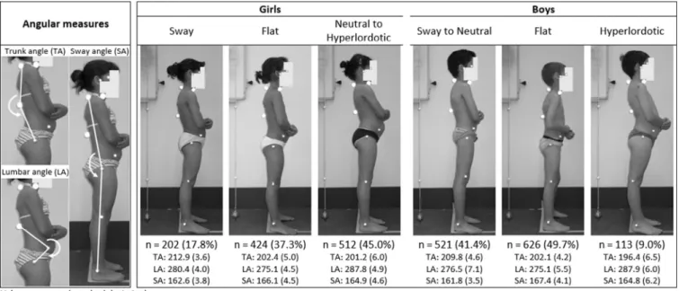
considered to quantify the magnitude of major curves of the spine (thoracic kyphosis and lumbar lordosis), and also an overall balance measure assessing body sway (as exempli-fied in the left panel ofFig. 1).
Children were ranked regarding their distance to the average postural values within each examiner’s distribution to elim-inate potential systematic differences between examiners (ie,
individual residuals of mixed effects models)[31], and
re-siduals were used to define postural patterns through the R
package Mclust[32]. Based on the interpretability of the
pat-terns among the models with the smallest Bayesian information
Fig. 1. Individual angular measures used to identify pediatric sagittal postural patterns using Mplus latent profile analysis (left panel), and a typical member of each pattern shown separately for girls and boys (right panel).
criterion[33], a final three-pattern solution was obtained separately for girls and boys. The methods and results of pattern identification are thoroughly described elsewhere[34]. Statistical analysis
Because we wished to estimate the postural patterns and their associations with the exposures in a single step to min-imize bias[35], we used the pediatric patterns described above as the basis for reestimation of postural patterns within Mplus (version 6.12, Muthén & Muthén, Los Angeles, CA, USA). Latent profile analysis was performed with multinomial lo-gistic regressions simultaneously computed in the same models (ie, including predictors of estimated postural latent pro-files). Initially, five different parameterizations of variance-covariance matrices were tested, with a fixed three-class solution for each gender based on our previous work on pediatric pat-terns[34]. Then, we selected the type of parameterization that optimized observed agreement of pattern assignment (based on the most likely class). Finally, latent postural classes were reestimated using information provided by each different set of predictors included in the models, and their associations were jointly quantified. This last step was used to account for uncertainty in the assignment of patterns and consequent-ly to obtain unbiased estimates of associations[35]. Sensitivity analysis was carried out by considering regional measures of body composition (trunk, upper, and lower limbs) derived from total body DXA scans.
Results
Pediatric postural patterns
A total of 1,138 girls and 1,260 boys accepted to partic-ipate and were included in the analysis (79% and 81% of eligible children, respectively). Compared with previous work on pattern
assignment of Generation XXI children[34], final postural
models obtained 68.5% concordance in girls and 79.7% in
boys (detailed information provided in Supplementary
Table S1). The average latent class assignment probabilities (for the most likely latent class membership) varied between 0.72 and 0.86 in girls and between 0.68 and 0.69 in boys.
Fig. 1(right panel) displays the average features of the three postural patterns and a typical child in each posture:
(1) Sway (girls) and “Sway to Neutral” (boys): increased trunk angle with backward tilt of the spine over the hips;
(2) Flat pattern in both genders: straight spine with forward trunk lean; and
(3) “Neutral to Hyperlordotic” (girls) and Hyperlordotic (boys): relatively increased lumbar angle and inter-mediate body sway.
Gender-specific aggregation of the neutral labeling was based on a different pattern prevalence between genders (see
Fig. 1).
Associations between DXA-derived parameters and postural patterns
Table 1shows descriptive analyses of anthropometric vari-ables and DXA-derived parameters according to participants’ most likely class assignment. Associations between DXA
pa-rameters and postural patterns are presented inTable 2 and
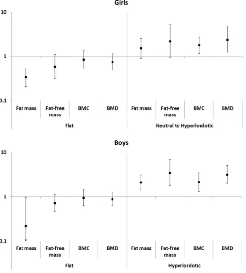
Fig. 2, using the Sway or “Sway to Neutral” pattern as refer-ence given their intermediate overall profile of anthropometrics and parameters of body composition.
Flat posture
In both genders, children in the Flat pattern showed the
lowest body mass index with mean differences of 0.86 kg/m2
in girls and 0.70 kg/m2
in boys, compared with the Sway or “Sway to Neutral” pattern. Fat and fat-free mass were in-versely associated with a Flat pattern: odds ratio (OR) of 0.36 per fat standard deviation (SD) and 0.60 per fat-free SD in girls (both p<.01), and in boys, ORs were 0.23 per fat SD (p=.001)
and 0.70 per fat-free SD (p=.023). However, when adjusted
for age and height (model 1), these associations remained statistically significant only for fat mass, and were
indepen-dent of fat-free mass (model 2b: girls OR=0.34, 95% confidence
interval [CI]: 0.21–0.55; boys OR=0.22, 95% CI: .06–0.81). Even though children in the Flat pattern had lower bone properties in crude analysis (especially in girls: ORs ranging from 0.66, 95% CI: 0.46–0.94 per BMD SD to 0.72, 95% CI: 0.53–0.99 per aBMC SD), those associations did not remain in adjusted models. Moreover, when adjusting for fat mass (model 2a), the Flat pattern was associated with increased bone properties, especially in girls (ORs varying from 1.17, 95% CI: 0.86–1.61 per aBMC SD to 1.60, 95% CI: 1.03–2.49 per BMC SD).
“Neutral to Hyperlordotic” or Hyperlordotic posture Children with a rounded posture presented the highest
body mass index: mean differences of 0.60 kg/m2in girls and
0.62 kg/m2in boys (vs. Sway or “Sway to Neutral”). The
like-lihood of having a “Neutral to Hyperlordotic” or a Hyperlordotic pattern increased per SD of fat mass and fat-free mass, and the relationships of posture with each component of body size were independent of each other (model 2b and 2a), although not significantly in girls. When adjusted for fat-free mass, ORs for fat mass were increased by 29% in girls (p=.370) and 77% in boys (p=.012), and when adjusted for fat mass, ORs for fat-free mass were increased by 50% in girls (p=.113) and 118% in boys (p=.016). Additionally, a rounded posture was associated with higher BMD and content (stronger for BMD than BMC) independently of fat or fat-free mass. In girls, across models 2a and 2b, ORs varied from 1.35 (95% CI: 0.85–2.17) per BMC SD to 2.00 (95% CI: 1.15–3.46) per BMD SD, and in boys, from 1.23 (95% CI: 0.73–2.08) per BMC SD to 2.42 (95% CI: 1.48–3.95) per BMD SD.
Sensitivity analysis
Similar results to the main analysis were obtained in the analysis of regional parameters from trunk, upper limb, and lower limb regions. No clear differences were observed between regions in respect to associations of fat or fat-free mass and bone parameters with postural patterns.
Discussion
In both genders, lower adiposity was associated with a flat-tened spine, and concordantly, higher fat and fat-free mass with a rounded posture type. There was an inverse association between bone physical properties and a Flat posture, but this relationship did not remain after accounting for differences in body composition across postural morphotypes. However, in a rounded posture, bone and posture were more strongly linked, possibly as the result of a shared accumulation of increased mechanical forces: children in the postural pattern character-ized by higher lumbar angle (“Neutral to Hyperlordotic” in girls and Hyperlordotic in boys) presented higher BMD, indepen-dently of anthropometrics and body composition.
Sagittal postural patterns in this work showed a plausi-ble association with body size and composition among the
pediatric population. For instance, both girls and boys with a hypercurved spine were heavier, taller, exhibited higher fat and fat-free mass, and had increased bone mass and density. Because compressive forces on spinopelvic structures are the sum of superincumbent fat and muscle loads acting on ver-tebral bodies in the axial plane, this implies specific weight-loading profiles for each postural morphotype even if not attributable to its sagittal configuration. In a rounded spine, higher forces are applied to bone structures because the skel-eton has more weight to support and needs higher muscle moments to regulate amplified oscillations of the upper body
over the hips [2,7]. Our characterization of postural
pat-terns reinforces that children with a flattened spine can keep an anteriorly displaced balance (increased sway angle) because they are lighter, whereas children with a rounded spine need to activate stronger extensor back muscles to avoid falling an-teriorly and to reestablish balance in an intermediate range. Numerous studies showed that low body mass index or weight is associated with a flattened posture and that in-creased body size is associated with a hypercurved spine
[16,21]. However, we showed, for the first time, that both fat mass and fat-free mass contribute to the associations of body size with sagittal posture. After adjustment for height, only
Table 1
Descriptive data for anthropometric variables and DXA parameters according to pediatric sagittal postural patterns, shown separately for girls and boys
All Sway Flat
Neutral to Hyperlordotic
Mean SD Mean SD Mean SD Mean SD
Girls, n=1,138 Age, y 7.4 0.4 7.4 0.4 7.4 0.4 7.4 0.4 Weight, kg 27.3 5.8 27.7 6.4 25.9 5.0 28.4 6.1 Height, cm 124.1 5.5 124.6 5.7 123.8 5.4 124.2 5.6 BMI, kg/m2 17.61 2.82 17.66 2.98 16.80 2.43 18.26 2.88 Fat mass, kg 8.5 3.6 8.8 3.8 7.5 3.0 9.2 3.8 Fat-free mass, kg 14.7 2.3 14.7 2.5 14.3 2.1 14.9 2.4
Fat mass index, kg/m2 5.45 2.04 5.56 2.1 4.84 1.7 5.90 2.1
Fat-free mass index, kg/m2 9.48 0.92 9.42 1.02 9.32 0.85 9.63 0.92
Area, cm2 962.9 62.7 965.1 63.4 962.0 63.8 962.7 61.6
BMC, g 591.6 85.5 592.9 88.4 582.0 83.5 599.1 85.4
BMD, g/cm2 0.61 .06 0.61 .06 0.60 .05 0.62 .06
aBMC, g 591.6 42.3 590.2 43.5 583.0 39.0 599.3 43.2
All Sway to Neutral Flat Hyperlordotic
Mean SD Mean SD Mean SD Mean SD
Boys, n=1,260 Age, y 7.4 0.4 7.4 0.4 7.5 0.4 7.4 0.4 Weight, kg 27.2 5.2 27.8 5.5 26.4 4.7 28.7 5.8 Height, cm 125.2 5.5 125.4 5.7 124.9 5.5 125.4 5.0 BMI, kg/m2 17.25 2.42 17.55 2.53 16.85 2.12 18.17 3.01 Fat mass, kg 7.0 3.1 7.4 3.3 6.5 2.7 8.0 3.9 Fat-free mass, kg 15.9 2.3 16.0 2.4 15.7 2.2 16.3 2.1
Fat mass index, kg/m2 4.42 1.79 4.66 1.86 4.12 1.56 5.03 2.28
Fat-free mass index, kg/m2 10.08 0.89 10.13 0.93 10.00 0.83 10.32 0.92
Area, cm2 959.7 65.3 961.4 65.7 958.8 66.5 956.8 57.1
BMC, g 601.0 85.4 603.4 88.4 597.5 85.1 609.6 71.9
BMD, g/cm2 0.62 .05 0.62 .06 0.62 .05 0.64 .05
aBMC, g 601.0 36.4 601.4 38.5 598.5 33.4 613.0 39.8
aBMC, area-adjusted BMC; BMC, bone mineral content; BMD, bone mineral density; BMI, body mass index; DXA, dual-energy X-ray absorptiometry; SD, standard deviation.
fat mass was inversely associated with a Flat posture, whereas both components of nonskeletal body mass were positively and independently related to a rounded posture type. The effect of adiposity on spinopelvic alignment is mainly derived from biomechanical constraints during posture development, po-tentially causing plastic deformation of bones and intervertebral
discs [16,20,21]. Adiposity also displaces balance
for-wardly, which increases lumbar lordosis as the most efficient
compensation to restore a stable basis of support[2,16,17].
On the other hand, stronger back extensor muscles lead to
an increase in lumbar lordosis[18,19], and a hyperlordotic
posture, through its sagittal organization alone, also re-quires higher muscle moments than all other postures,
especially during more demanding tasks [36]. These
me-chanical pathways are congruent with our findings, but the robust and exclusive association (after adiposity adjust-ment) between fat-free mass and a hyperlordotic posture probably implies a biological threshold for adiposity, above which muscles predominantly control upright balance. This threshold is likely related to balance instability caused by ad-iposity, which would explain why fat-free mass was not associated with a Flat pattern.
Table 2
Associations between standardized DXA parameters and pediatric sagittal postural patterns, shown separately for girls and boys
Flat Neutral to Hyperlordotic
OR 95% LCI 95% UCI p OR 95% LCI 95% UCI p
Girls, n=1,138 (reference pattern=Sway)
Fat mass Model 0 0.36 0.24 0.55 <.001 1.32 0.81 2.17 .268
Model 1 0.34 0.21 0.55 <.001 1.51 0.89 2.55 .126
Model 2b 0.34 0.21 0.55 <.001 1.29 0.74 2.25 .370
Fat-free mass Model 0 0.60 0.43 0.84 .003 1.33 0.71 2.51 .375
Model 1 0.59 0.31 1.10 .094 2.21 0.96 5.11 .064 Model 2a 1.02 0.57 1.84 .150 1.50 0.91 2.49 .113 BMC Model 0 0.69 0.53 0.90 .007 1.14 0.75 1.74 .530 Model 1 0.84 0.54 1.32 .460 1.79 1.17 2.74 .008 Model 2a 1.60 1.03 2.49 .038 1.48 0.97 2.24 .067 Model 2b 1.26 0.77 2.07 .360 1.35 0.85 2.17 .208 BMD Model 0 0.66 0.46 0.94 .023 1.49 0.68 3.25 .317 Model 1 0.75 0.49 1.15 .182 2.39 1.23 4.64 .010 Model 2a 1.55 1.01 2.38 .046 1.79 1.18 2.72 .006 Model 2b 0.96 0.57 1.60 .861 2.00 1.15 3.46 .014 aBMC Model 0 0.72 0.53 0.99 .040 2.27 1.43 3.60 .001 Model 1 0.75 0.56 1.02 .068 2.28 1.45 3.60 <.001 Model 2a 1.17 0.86 1.61 .322 1.49 1.07 2.07 .019 Model 2b 0.78 0.56 1.08 .139 1.91 1.18 3.10 .009 Flat Hyperlordotic
OR 95% LCI 95% UCI p OR 95% LCI 95% UCI p
Boys, n=1,260 (reference pattern=Sway to Neutral)
Fat mass Model 0 0.23 .07 0.71 .001 1.85 1.31 2.60 <.001
Model 1 0.22 .05 0.95 .043 2.08 1.41 3.08 <.001
Model 2b 0.22 .06 0.81 .023 1.77 1.14 2.77 .012
Fat-free mass Model 0 0.70 0.52 0.95 .023 1.43 0.99 2.08 .057
Model 1 0.72 0.45 1.13 .150 3.45 1.76 6.76 <.001 Model 2a 1.00 0.58 1.74 .991 2.18 1.16 4.11 .016 BMC Model 0 0.83 0.59 1.16 .270 1.30 0.89 1.89 .180 Model 1 0.94 0.62 1.43 .778 2.11 1.30 3.44 .003 Model 2a 1.22 0.73 2.02 .446 1.40 0.89 2.18 .143 Model 2b 1.10 0.70 1.74 .672 1.23 0.73 2.08 .430 BMD Model 0 0.80 0.58 1.10 .168 1.78 1.28 2.46 .001 Model 1 0.88 0.61 1.26 .484 3.15 1.98 4.99 <.001 Model 2a 1.25 0.79 1.98 .349 2.02 1.31 3.13 .002 Model 2b 1.02 0.63 1.63 .949 2.42 1.48 3.95 <.001 aBMC Model 0 0.82 0.60 1.13 .225 2.21 1.49 3.27 <.001 Model 1 0.90 0.67 1.20 .463 2.69 1.72 4.21 <.001 Model 2a 1.23 0.85 1.79 .269 1.90 1.31 2.77 .001 Model 2b 0.94 0.67 1.33 .728 2.32 1.50 3.59 <.001
aBMC, area-adjusted BMC; BMC, bone mineral content; BMD, bone mineral density; DXA, dual-energy X-ray absorptiometry; LCI, lower confidence interval; OR, odds ratio; UCI, upper confidence interval.
Model 0=crude associations; Model 1=adjusted for age and height; Model 2a=additionally adjusted for fat mass; Model 2b=as model 1 plus adjustment for fat-free mass.
The inverse relation between bone physical properties and a Flat pattern observed in our work may be explained by the profile of anthropometric and body composition character-istics featured by this typology. However, in the case of a rounded posture, an association between bone quality and posture remains after those adjustments. A specific shape and design of the spine establishes the morphologic configura-tion for the acconfigura-tion of gravity and muscles. A Sway posture minimizes muscle work and stresses in the resting standing position[36]. As lumbar lordosis increases up to a hyperlordotic posture, mechanical loads also increase. Flattened or neutral spines are better suited to minimize muscle work and stress
in weight-bearing activities[36], and the extremely
pro-nounced thoracic kyphosis in the Sway type can be expected to contribute to higher mechanical stress compared with the
Flat pattern[2,5,6]. Conversely, fat and lean mass positively
affect bone structure through mechanical and endocrine effects
[37,38], with a more important contribution of lean than fat
mass during childhood[38,39]. Therefore, both adiposity and
muscles can lead to changes in the morphology of vertebral bodies, namely by changing their anteroposterior height ratios[40]. These changes modify vertebral tilt and define local
alignment[7,14,15], and consequently overall postural
pat-terns due to adaptation of adjacent anatomical regions[1].
As examples, longitudinal vertebral growth in children may
increase lumbar lordosis[15], and both higher thoracic
ky-phosis[14,41,42] and higher lumbar lordosis[42]seem to
have an osteoporotic origin (decreased BMD) at more ad-vanced ages.
Our population-based finding of bone-posture potentia-tion in a hypercurved spine suggests that a bidirecpotentia-tional mechanism exists—that is, hyperlordosis promotes mechan-ical stress but bone growth changes vertebrae tilting—in a pattern-specific dynamic environment also defined by anthropometrics and body composition. This was also sup-ported by regional analysis of body composition, with no clear
Fig. 2. Graphs showing associations (odds ratio and 95% confidence interval) between standardized DXA parameters and pediatric sagittal postural pattern, shown separately for girls (reference= Sway) and boys (reference = Sway to Neutral). Odds ratios are per standard deviation higher DXA parameter, adjusted for age and height. DXA, dual-energy X-ray absorptiometry.
differences of bone-posture associations between load-bearing regions and the upper limbs. Increased loads and endocrine adaptations in the hyperlordotic posture probably culminate in stronger biological relations of fat, muscle, and bone with sagittal alignment. Probably because of weaker me-chanical forces, maturational processes of bone and posture do not seem more closely linked in a Flat than in a Sway posture. Furthermore, associations between bone and posture were stron-ger for boys than for girls. This may result from different aggregations of postural morphologies in the present classi-fications (“Neutral to Hyperlordotic” in girls and “Sway to Neutral” in boys), or represent a true gender-specific bone
response to mechanical stimuli[43,44].
One of the limitations of this work is the lack of direct mea-surement of mechanical stimuli imposed by anthropometrics, adiposity, and muscle contractions. Our analyses assumed that mechanical influences are captured by lean mass and re-flected in bone physical properties, both quantifiable by DXA measurements. The population-based nature of our work con-strained these assessments, but it ensured a wide representation of naturally occurring anthropometrics, body composition, bone physical properties, and postural angles in the pediatric pop-ulation. Furthermore, it has been previously shown that children participating in this wave of assessment (posture and DXA mea-surements) were similar to the general Generation XXI cohort at birth regarding anthropometrics, although maternal educa-tion was higher for included children[34]. The external validity of our findings is a key advantage because previous evidence had relied mainly on biomechanical model simulations without
any empirical measurements of bone quality[2,5,6,36].
Fur-thermore, it is essential to study posture morphotypes instead of isolated parameters because patterns add the effect of dif-ferent combinations of regional alignment on health, and consequently, allow a more comprehensive mechanical char-acterization of the upright posture[2,16,29,36]. Our associations between DXA-derived parameters and postural patterns may be biased because of the use of posture classification not com-pletely overlapping with the initial Mclust grouping, especially
in the Flat pattern (Supplementary Table S1). However, given
the direction of differences between classifications, this would bias results toward the null hypothesis and not create spuri-ous associations. Further, latent profile analysis in Mplus enables using information provided from model predictors (ie, DXA-derived parameters) to reestimate patterns and jointly quantify
unbiased associations[35], which probably surpasses
limita-tions resulting from the use of two different software. Because variables considered in this study have high physiological cor-relation, differentiating effects of posture from body size or composition on bone may be unrealistic. Moreover, given the observational nature of our study, the causal nature of rela-tionships between body composition, bone, and posture should be seen in the context of homeostatic feedback mechanisms rather than as a set of unidirectional effects.
This study evaluated for the first time the relations between bone physical properties and sagittal posture in 7-year-old chil-dren recruited from a population-based birth cohort. There
was an inverse association between bone physical proper-ties and a Flat posture, and bone and posture were more strongly positively linked in a rounded posture. As initially hypothesized, our results support that both bone and posture mature in a shared and interrelated mechanical environment modulated by pattern-specific anthropometrics and body composition.
Acknowledgment
Generation XXI was funded by FEDER through the Op-erational Programme Competitiveness and Internationalization and by national funds through the FCT – Fundação para a Ciência e a Tecnologia via grants POCI-01-0145-FEDER-016838 and POCI-01-0145-FEDER-016837, under the projects PTDC/DTP-EPI/1687/2014 and PTDC/DTP-EPI/3306/ 2014. Support by Administração Regional de Saúde Norte (Ministry of Health), Fundação Calouste Gulbenkian and Unidade de Investigação em Epidemiologia - Instituto de Saúde Pública da Universidade do Porto (EPIUnit) (POCI-01-0145-FEDER-006862; UID/DTP/04750/2013) is also acknowledged. Araújo FA and Lucas R were supported by grants SFRH/ BD/85398/2012 and SFRH/BPD/88729/2012, co-funded by FCT and the POPH/FSE.
The authors also gratefully acknowledge the families en-rolled in Generation XXI, and the contribution of the members of the research team and staff.
Supplementary material
Supplementary material related to this article can be at
http://dx.doi.org/10.1016/j.spinee.2017.04.024.
References
[1] Mac-Thiong J, Labelle H, Roussouly P. Pediatric sagittal alignment. Eur Spine J 2011;20(Suppl. 5):S586–90.
[2] Bruno A, Anderson D, D’Agostino J, et al. The effect of thoracic kyphosis and sagittal plane alignment on vertebral compressive loading. J Bone Miner Res 2012;27:2144–51.
[3] Huang M, Barrett-Connor E, Greendale G, et al. Hyperkyphotic posture and risk of future osteoporotic fractures: the Rancho Bernardo study. J Bone Miner Res 2006;21:419–23.
[4] Roux C, Fechtenbaum J, Kolta S, et al. Prospective assessment of thoracic kyphosis in postmenopausal women with osteoporosis. J Bone Miner Res 2010;25:362–8.
[5] Briggs A, van Dieën J, Wrigley T, et al. Thoracic kyphosis affects spinal loads and trunk muscle force. Phys Ther 2007;87:595–607.
[6] Keller TS, Colloca CJ, Harrison DE, et al. Influence of spine morphology on intervertebral disc loads and stresses in asymptomatic adults: implications for the ideal spine. Spine J 2005;5:297–309.
[7] Roussouly P, Pinheiro-Franco JL. Biomechanical analysis of the spino-pelvic organization and adaptation in pathology. Eur Spine J 2011;20(Suppl. 5):609–18.
[8] Frost H. Wolff’s law and bone’s structural adaptations to mechanical usage: an overview for clinicians. Angle Orthod 1994;64:175–88.
[9] Frost HM. Bone’s mechanostat: a 2003 update. Anat Rec A Discov Mol Cell Evol Biol 2003;275:1081–101.
[10] Behringer M, Gruetzner S, McCourt M, et al. Effects of weight-bearing activities on bone mineral content and density in children and adolescents: a meta-analysis. J Bone Miner Res 2014;29:467–78.
[11]Farr JN, Laddu DR, Going SB. Exercise, hormones and skeletal adaptations during childhood and adolescence. Pediatr Exerc Sci 2014;26:384–91.
[12] Janz KF, Letuchy EM, Eichenberger Gilmore JM, et al. Early physical activity provides sustained bone health benefits later in childhood. Med Sci Sports Exerc 2010;42:1072–8.
[13] Ducher G, Bass SL, Saxon L, et al. Effects of repetitive loading on the growth-induced changes in bone mass and cortical bone geometry: a 12-month study in pre/peri- and postmenarcheal tennis players. J Bone Miner Res 2011;26:1321–9.
[14] Stone M, Osei-Bordom D, Inman R, et al. Heritability of spinal curvature and its relationship to disc degeneration and bone mineral density in female adult twins. Eur Spine J 2015;24:2387–94.
[15] Mac-Thiong J, Berthonnaud E, Dimar JR 2nd, et al. Sagittal alignment of the spine and pelvis during growth. Spine 2004;29:1642–7.
[16] Araújo F, Lucas R. What do we know about the determinants of sagittal standing posture? OA Musculoskelet Med 2014;2:15.
[17] Whitcome KK, Shapiro LJ, Lieberman DE. Fetal load and the evolution of lumbar lordosis in bipedal hominins. Nature 2007;450:1075–8.
[18] Sinaki M, Itoi E, Rogers JW, et al. Correlation of back extensor strength with thoracic kyphosis and lumbar lordosis in estrogen-deficient women. Am J Phys Med Rehabil 1996;75:370–4.
[19] Hongo M, Miyakoshi N, Shimada Y, et al. Association of spinal curve deformity and back extensor strength in elderly women with osteoporosis in Japan and the United States. Osteoporos Int 2012; 23:1029–34.
[20] Boulay C, Tardieu C, Hecquet J, et al. Sagittal alignment of spine and pelvis regulated by pelvic incidence: standard values and prediction of lordosis. Eur Spine J 2006;15:415–22.
[21] Smith A, O’Sullivan P, Beales D, et al. Trajectories of childhood body mass index are associated with adolescent sagittal standing posture. Int J Pediatr Obes 2011;6:e97–106.
[22] Correia S, Rodrigues T, Barros H. Socioeconomic variations in female fertility impairment: a study in a cohort of Portuguese mothers. BMJ Open 2014;4:e003985.
[23] Larsen P, Kamper-Jørgensen M, Adamson A, et al. Pregnancy and birth cohort resources in Europe: a large opportunity for aetiological child health research. Paediatr Perinat Epidemiol 2013;27:393– 414.
[24] Wells J, Cole T. Adjustment of fat-free mass and fat mass for height in children aged 8 y. Int J Obes Relat Metab Disord 2002;26:947– 52.
[25] Macdonald-Wallis C, Tobias J, Smith G, et al. Relation of maternal prepregnancy body mass index with offspring bone mass in childhood: is there evidence for an intrauterine effect? Am J Clin Nutr 2010; 92:872–80.
[26] Bianchi M, Baim S, Bishop N, et al. Official positions of the International Society for Clinical Densitometry (ISCD) on DXA evaluation in children and adolescents. Pediatr Nephrol 2010;25:37– 47.
[27] Perry M, Smith A, Straker L, et al. Reliability of sagittal photographic spinal posture assessment in adolescents. Adv Physiother 2008;10:66– 75.
[28] Fortin C, Feldman D, Cheriet F, et al. Clinical methods for quantifying body segment posture: a literature review. Disabil Rehabil 2011;33:367– 83.
[29] Smith A, O’Sullivan P, Straker L. Classification of sagittal thoraco-lumbo-pelvic alignment of the adolescent spine in standing and its relationship to low back pain. Spine 2008;33:2101–7.
[30] Ferreira E, Duarte M, Maldonado E, et al. Postural assessment software (PAS/SAPO): validation and reliability. Clinics (Sao Paulo, Brazil) 2010;65:675–81.
[31] Pinheiro J, Bates D. Mixed-effects models in S and S-plus. Springer Verlag; 2000.
[32] Fraley C, Raftery AE. Model-based clustering, discriminant analysis, and density estimation. J Am Stat Assoc 2002;97:611–31.
[33] Schwarz G. Estimating dimension of a model. Ann Stat 1978;6:461–4.
[34] Araújo FA, Severo M, Alegrete N, et al. Defining patterns of sagittal standing posture in girls and boys of school age. Phys Ther 2017; 97:258–67.
[35] Bray B, Lanza S, Tan X. Eliminating bias in classify-analyze approaches for latent class analysis. Struct Equ Modeling 2015;22:1–11.
[36] Galbusera F, Brayda-Bruno M, Costa F, et al. Numerical evaluation of the correlation between the normal variation in the sagittal alignment of the lumbar spine and the spinal loads. J Orthop Res 2014;32:537–44.
[37] Reid IR. Relationships between fat and bone. Osteoporos Int 2008;19:595–606.
[38] Farr JN, Amin S, LeBrasseur NK, et al. Body composition during childhood and adolescence: relations to bone strength and microstructure. J Clin Endocrinol Metab 2014;99:4641–8.
[39] Medina-Gomez C, Heppe D, Yin J, et al. Bone mass and strength in school age children exhibit sexual dimorphism related to differences in lean mass: the generation R study. J Bone Miner Res 2015;31:1099– 106.
[40] Goh S, Price RI, Leedman PJ, et al. The relative influence of vertebral body and intervertebral disc shape on thoracic kyphosis. Clin Biomech (Bristol, Avon) 1999;14:439–48.
[41] Edmondston SJ, Singer KP, Price RI, et al. The relationship between bone mineral density, vertebral body shape and spinal curvature in the elderly thoracolumbar spine: an in vitro study. Br J Radiol 1994;67: 969–75.
[42] Pavlovic A, Nichols D, Sanborn C, et al. Relationship of thoracic kyphosis and lumbar lordosis to bone mineral density in women. Osteoporos Int 2013;24:2269–73.
[43] Kriemler S, Zahner L, Puder JJ, et al. Weight-bearing bones are more sensitive to physical exercise in boys than in girls during pre- and early puberty: a cross-sectional study. Osteoporos Int 2008;19:1749–58.
[44] Cardadeiro G, Baptista F, Ornelas R, et al. Sex specific association of physical activity on proximal femur BMD in 9 to 10 year-old children. PLoS ONE 2012;7:e50657.