In vivo evaluation of antiseptics and disinfectants on
control of Caseous Lymphadenitis: clinical, haematological,
serological and microbiological monitoring
Avaliação
in vivo
de antissépticos e desinfetantes no controle da Linfadenite
Caseosa: acompanhamento clínico, hematológico, sorológico e microbiológico
Lauana Borges Santiago1*, Raymundo Rizaldo Pinheiro1, Francisco Selmo Fernandes Alves1,
Vanderlan Warlington Souza dos Santos2, Apoliana de Sousa Rodrigues2 Ana Milena César Lima2,
Eduardo Luiz de Oliveira1, Fernando Henrique Melo Andrade Rodrigues de Albuquerque1
ABSTRACT: The objective of this experiment was to evaluate the efficacy of iodine tincture at 10% and sodium hypochlorite at 2.5% applied into the abscess of animals affected by Caseous Lymphadenitis (CL). Eighteen ewes were used, assorted into three groups: one treated with iodine tincture at 10% (IT), another one with sodium hypochlorite at 2.5% (SH) and the last group underwent the conventional treatment (CT). Conventional treatment was based on surgical drainage and chemical cauterization of the lesion with iodine tincture at 10%. Natural rupture of six abscesses from IT group was observed and in five of them the viability of Corynebacterium pseudotuberculosis
was confirmed on the lesion place, after rupture of lymph node. As for the SH group, spontaneous rupture was observed in five out of six abscesses treated, and the microorganism was identified on the lesion of five animals, after rupture. In the sixth animal of this group, abscess involution was noticed. A severe swelling was identified in the region of lymph node treated, resulting in wide lesion in animals from groups IT and SH. No difference (p > 0,05) was found in blood parameters due to treatments. As for the serological monitoring of animals, comparative analysis between months within each group showed that months 1, 2, 3 and 4 were different (p < 0,05) from month 0, for groups IT and SH. For CT group, there was no difference (p > 0,05) between months 1 to 5 and 0. Then, the application of iodine tincture at 10% or sodium hypochlorite at 2.5% into the abscess of animals affected by CL, at stage in which lesions are detected through inspection, is not effective for its control.
KEYWORDS: abscess; Corynebacterium pseudotuberculosis; sheep; treatment.
RESUMO: Objetivou-se com este experimento avaliar a eficácia da tintura de iodo a 10% e do hipoclorito de sódio a 2,5% aplicados no interior do abscesso de animais acometidos pela Linfadenite Caseosa (LC). Foram utilizadas 18 fêmeas ovinas, divididas em 3 grupos: o primeiro foi tratado com tintura de iodo a 10% (TI), o segundo com hipoclorito de sódio a 2,5% (HS) e o último grupo foi submetido ao tratamento convencional (CT). O tratamento convencional da doença consistia na drenagem e cauterização química dos abscessos, utilizando-se solução de iodo a 10%. Foi observada a ruptura natural dos seis abscessos do grupo TI, sendo que em cinco deles a viabilida-de do Corynebacterium pseudotuberculosis foi confirmada no local da lesão, após a ruptura do linfonodo. Quanto ao grupo HS, a ruptura espontânea foi observada em cinco dos seis abscessos tratados, sendo o microrganismo identificado na lesão dos cinco animais após o rom-pimento. No sexto animal desse grupo, foi constatada a involução do abscesso. Foi verificado um edema intenso na região do linfonodo tratado com formação de uma lesão de grande extensão nos animais dos grupos TI e HS. Não foi detectada diferença (p > 0,05) nos pa-râmetros hematológicos em decorrência dos tratamentos avaliados. Quanto ao acompanhamento sorológico dos animais, a análise es-tatística comparativa entre os meses, dentro de cada grupo, mostrou que os meses 1, 2, 3 e 4 foram diferentes (p < 0,05) do mês 0 para os grupos TI e HS. No grupo CT, não houve diferença (p > 0,05) entre os meses de 1 a 5 e o mês 0. Conclui-se que a aplicação de tintura de iodo a 10% ou hipoclorito de sódio a 2,5% no interior do abscesso de animais acometidos pela LC, em estágio no qual as lesões são detectadas por meio da inspeção, não é eficaz para o seu controle.
PALAVRAS-CHAVE: abscesso; Corynebacterium pseudotuberculosis; ovino; tratamento.
1Embrapa Caprinos e Ovinos – Sobral (CE), Brazil. 2Universidade do Vale do Acaraú – Sobral (CE), Brazil.
INTRODUCTION
In recent years, the production of goats and sheep has been representing an important part in the context of Brazilian agribusiness. Currently, it is characterised as a very important cultural, social and economical activity for the Northeast of Brazil, playing a crucial role in the development of this re-gion. The production of high biological value food (milk, meat and offal) and skin of excellent quality, besides the adaptability of animals to local ecosystems, allow the goat and sheep industry to fit as a good alternative for work and
income (Moraes Neto et al., 2003). However, there is still
a low productivity in the region due to inadequate han-dling and to frequent occurrence of diseases, like Caseous Lymphadenitis (CL).
CL is a chronic and debilitating disorder which affects small ruminants, characterised by the formation of abscesses
in one or more lymph nodes, caused by Corynebacterium
pseudotuberculosis, a positive Gram bacillus, short and
irregu-lar, with approximately 0.5 to 0.6 μm x 1.0 to 3.0 μm. It is
an infectious disease, which is a worldwide problem, respon-sible for causing severe economic losses for goat and sheep
industry (Alves et al., 2007). The depreciation of skins value,
carcass condemnation, falling in production of meat, milk, wool and reproductive efficiency are some of the main
dam-ages caused by CL (Krishna et al., 1977; Figueiredo et al.,
1982; Paton et al., 1994; Paton et al., 2003), in addition to
public health risk posed by the zoonotic potential described for C. pseudotuberculosis (Mills et al., 1997; Peel et al.,
1997; Join-Lambert et al., 2006).
The occurrence of injuries on the skin of animals is the main gateway for the microorganism, which is in the environ-ment due to rupture of abscesses of animals infected by the disease (Nairn; Robertson, 1974). The purulent exudate of a CL lesion is extremely rich in viable bacterial cells, with
an approximate concentration of 1 x 10⁶ to 5 x 10⁷ cells per
gram of material (Brown; Olander, 1987). Therefore, the main aspect related to the control of this illness is the im-mediate isolation of affected animals and surgical drainage of abscess before its natural rupture, once the microorganism is able to survive and persist in the environment for a long period of time (Williams, 1980). Such characteristic of the microorganism associated with inefficiency of the treatment based on antimicrobial agents and the difficult detection of infected animals make the eradication of CL an extremely arduous task to be performed (Williamson, 2001).
The currently recommended treatment for CL is drain-age and chemical cauterization of abscesses, using iodine solution at 10%, when they are in advanced stage of devel-opment. However, this technique represents high risks of environmental contamination, whether through unexpect-ed spontaneous rupture of abscess or by simple exposure to external environment of a highly contaminated material.
An experiment evaluating the application of formaldehyde solution at 10% into the lymph node has already been
ac-complished, which found the death of C. pseudotuberculosis
at the injury place. However, the use of this product inside the abscesses is responsible for the onset of various disor-ders as the occurrence of fibrosis, necrosis, destruction of epithelial and adjacent muscle tissues, and possible inva-sion of bone tissue (Alves; Pinheiro, 2003). Moreover, it is known that formaldehyde has teratogenic, mutagenic and carcinogenic effects on laboratory animals, and it is also responsible for causing birth defects and other adverse reactions (UFRGS, 2003).
Recently carried out in vitro studies aiming to evaluate
the efficacy of antiseptics and disinfectants of low toxicity
to the animal organism against C. pseudotuberculosis
report-ed the high sensitivity of this microorganism face to solution of iodine tincture at 10% and sodium hypochlorite at 2.5%
(Santiago et al., 2010). Therefore, the aim of this
experi-ment was to evaluate the efficacy of these substances inside the abscess of animals affected by CL in early stages of de-velopment, focusing on treating and controlling the disease.
MATERIAL AND METHODS
Experiment location and period
The experiment was conducted at Embrapa Goats and Sheep (CNPC), located in Sobral, in a semi-arid hinterland of Ceará (Brazil), at 3° 41’ 32” South latitude and 40° 20’ 53” West longitude, at an altitude of 75 m. The experiment was con-ducted between July and December of 2009, in accordance with ethical principles adopted by the National Council for Control of Animal Experimentation (CONCEA), Law no.
11.794, October 8th 2008.
Experimental groups
Eighteen crossbred Santa Ines ewes naturally infected by CL were used, belonging to CNPC experimental herd. The
animals were kept in a paddock of native pasture, had ad
three groups, containing six animals each. The first group of animals was treated with iodine tincture at 10% (IT), the second group was treated with sodium hypochlorite at 2.5% (SH) and the third group was used as control for the comple-tion of convencomple-tional treatment of this disease, through sur-gical drainage and chemical cauterization of the lesion with iodine tincture at 10% (CT). For the preparation of the solution of iodine tincture at 10%, 100g of iodine ressubli-mate, 60g of potassium iodide, 50 mL of distilled water and 950 mL of absolute alcohol were used. As for the solution of sodium hypochlorite at 2.5%, a Brilux® brand commercial product was used, based on sodium hypochlorite, sodium hydroxide, water and active chlorine content of 2.5% w/w. The active chlorine percentage was confirmed using Brazilian Technical Standards Association (ABNT) NBR 9425:2005 (ABNT, 2005. Before treatment, a volume of 1 mL of sterile saline solution (NaCl, 0.95%) was injected into the involved lymph node and then aspirated for microbiological
confir-mation of C. pseudotuberculosis.
Treatment and clinical,
microbiological and
haematological monitoring
Initially, trichotomy and antisepsis of the region of lymph nodes affected were taken, with alcohol at 70% and iodine tincture at 10%. A volume of 5 mL of the product to be tested inside the abscess was applied, using disposable sy-ringes and needles, with 5 mL and 25 x 0.7 mm in diameter, respectively. After treatment, animals were clinically evalu-ated during five months, twice a week, for describing the development of clinical case. Complete clinical examination
(Diffay et al., 2005) was performed, emphasising the
char-acterisation of lesions in lymph nodes, as the increase or de-crease in the diameter of abscesses, change in its consistency, sensitivity, mobility, scarring or rupture. In cases of rupture, the material was collected for isolation and identification of the microorganism present in the injured place. After that, the wound was treated with iodine tincture at 10% and in-sect repellent spray. When necessary, a healing ointment and a local antibiotic were used. The collected material was plat-ed on blood agar and colonies were subjectplat-ed to macroscopic characterisation, Gram staining method and catalase, urease and fermentation of carbohydrates tests, for confirmation of C. pseudotuberculosis (Carter et al., 1986). Variations in volume of abscesses were measured with aid of a digital cali-per. For complete blood count, blood samples were collected through puncture of the jugular vein using vacutainer sys-tem in 5 mL tubes containing tetra-acetic ethylenediamine acid anticoagulant 7 days after treatment and at the end of the experiment. Haematology, following the method de-scribed by COLES (1993), consisted of the determination
of the following parameters: cell volume (microhematocrit method), haemoglobin (cianometahaemoglobin method), total leucocyte count (haemocytometer method) and differ-ential leucocyte count, which were classified as segmented neutrophils, band neutrophils, eosinophils, lymphocytes and monocytes (blood smear). All haematological analyses were done in the Laboratory of Clinical Pathology of CNPC.
Serological monitoring
Serological monitoring of animals was performed every 15 days, through Serological monitoring (SHI), following the methodology described by KNIGHT (1978). SHI is considered a sensitive, but not specific test for diagnosis of
C. pseudotuberculosis. Brown et al. (1987), considering mi-crobiological exam as gold standard test, had shown sensitiv-ity of 90.9% and specificsensitiv-ity of 61%, under field conditions, for SHI test. Blood samples were obtained by puncturing the jugular vein using vacutainer system in 10 mL tubes without anticoagulant. Blood samples were centrifuged at 3000 rpm for 10 minutes and serum was stored in eppendorf tubes in a freezer at -20°C. All microbiological and serological analyses were done in the Laboratory of Bacteriology of CNPC.
Statistical analyses
A completed randomised experimental design was used, with three treatments and six replications. The data obtained were analysed for their normality by Lilliford’s test and sub-jected to analysis of general variance, using the statistical program SAEG (SAEG, 2007). The averages comparison was performed using Dunnett’s test, in which a significance level of 5% was adopted.
RESULTS AND DISCUSSION
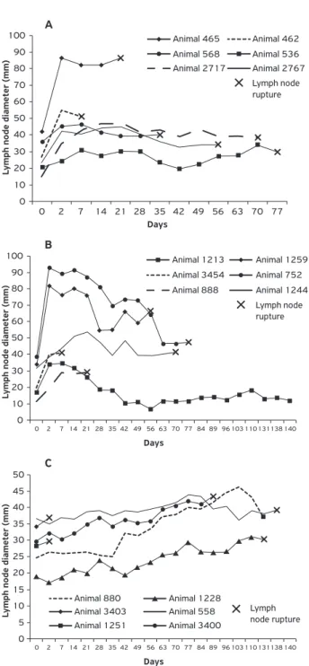
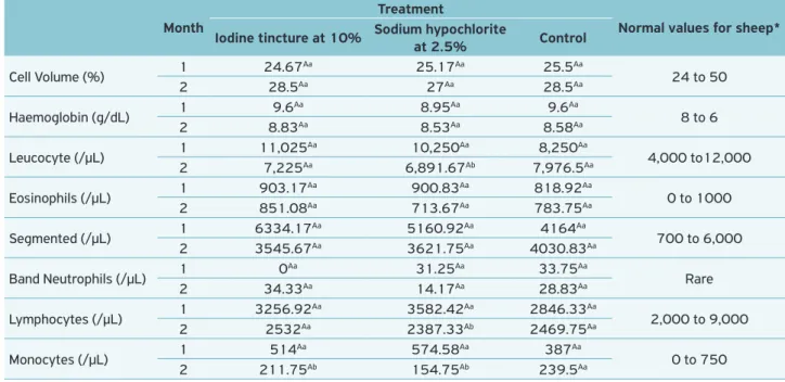
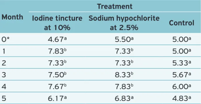
The evolution of clinical case of each animal belonging to the three groups is shown in Figure 1, considering the di-ameter of lymph nodes and presence or absence of rupture in the abscesses. Day 0 corresponds to the size of lymph nodes, measured prior to treatments.
For IT and SH groups, it was considered as satisfactory the treatment which promoted involution of abscess or, after spontaneous rupture of the nodule, did not contain viable
C. pseudotuberculosis in the lesion. As for the conventional treatment, the efficacy was determined according to the non-occurrence of unexpected spontaneous rupture of abscess, considering a follow-up of infected animals every three days.
There was a natural rupture of six abscesses treated with iodine tincture at 10% (Fig. 1A), and in five of them the
after rupture of lymph node. An interesting datum is related to the aspect of the material collected after rupture. All abscesses treated with iodine tincture at 10% contained a fibrinous mate-rial inside it, encapsulated, firm in consistency and far adhered to the adjacent musculature, formed after the completion of the
treatment. In this material, C. pseudotuberculosis was not
con-firmed in any animal. However, in five animals of this group, the caseous material typically observed in abscesses of animals affected by the CL was also present in the lesion. From this
material, there was always confirmation of the agent. It is as-sumed that the quantity of product applied into the lymph nodes of these animals was not enough to cause the elimination of the microorganism over the full extension of the abscess.
As for the group treated with sodium hypochlorite at 2.5%, spontaneous rupture was observed in 5 out of the
6 abscesses treated. In this case, C. pseudotuberculosis was
isolated and identified in the injury of the five animals after rupture of abscess. The confirmation of the etiological agent of CL in the material inside the abscess, after its rupture, makes the use of this treatment impracticable, since this ma-terial represents the main source of infection of the disease over the herd. In the sixth animal from SH group, it was found the involution of abscess after treatment, whose di-ameter reduced to the same value as the contralateral normal lymph node. This result can be explained from analysis of diameter of abscesses of animals, obtained before treatments. The only animal whose lymph node showed regression in size after treatment owned an abscess in a very early stage of development, if compared to other animals. This should be taken into account in assessing the effectiveness of the sug-gested treatment, once the approach recommended for the treatment of any abscess in advanced stage is the complete
draining of all the material formed (Knight et al., 1980).
It is suggested that, if the treatment had been done in an earlier period than the one described, i.e., in an earlier stage of development of abscesses, the results might prove satis-factory. However, if this hypothesis is actually confirmed in future studies, it is assumed that the practicability of the new treatment is compromised, once abscesses in extremely ini-tial stage of development are unlikely to be detected only by periodic inspection of herd, but by monitoring through external palpation of lymph nodes of animals.
Another important factor which should be mentioned concerns the duration of the clinical presentation of the dis-ease. It is observed that, 90 days after treatment, all animals in groups IT and SH did not act anymore as clinical carriers of the disease (Fig. 1A and B). At this time, all lesions in animals belonging to these two groups were already healed, unlike what was observed for animals in group CT, where the resolution of the inflammatory process is concentrated
in the period between the 84th and 140th day of the
experi-ment (Fig. 1C). The information above is confirmed after the evaluation of the average duration, in days, of the clini-cal phase of the disease. IT group had a duration average of 51.33 days, SH group took 52.5 days and CT group lasted 87.67 days, that is, one month more than the other treat-ments. Therefore, if it is conducted some type of adaptation causing it to become satisfactory, shortening the course of the disease would be one more advantage gained with the new technique. A shorter duration of clinical stage of the disease would lower spending on manpower, once after detection of lesions a more frequent monitoring of animals is necessary
Animal 465 100
90 80 70 60 50 40 30 20 10 0
Lymph node diameter (mm)
Days
0 2 7 14 21 28 35 42 49 56 63 70 77 Animal 568
Animal 2717
Animal 462 Animal 536 Animal 2767 Lymph node rupture
Animal 1259 100
90 80 70 60 50 40 30 20 10 0
Lymph node diameter (mm)
Days
0 2 7 14 21 28 35 42 49 56 63 70 77
Animal 752 Animal 888
Animal 3454 Animal 1213
Animal 1244 Lymph node rupture
84 89 96 103 110 131138 140
Animal 3403 50
45 40 35 30 25 20 15 10 5 0
Lymph node diameter (mm)
Days
0 2 7 14 21 28 35 42 49 56 63 70 77
Animal 3400 Animal 1228 Animal 880
Animal 1251
Animal 558 Lymph node rupture
84 89 96 103 110 131138 140
Figure 1. Evolution of the diameter of lymph nodes of animals treated with iodine tincture at 10% (A), animals treated with sodium hypochlorite at 2.5% (B) and animals belonging to the control group (C), over time.
A
B
in order to prevent spontaneous rupture of abscesses and en-vironmental contamination.
Figure 2 shows the percentage of efficacy of both treat-ments compared to the conventional, currently recommended for CL. Regarding the control of environmental
contamina-tion by C. pseudotuberculosis, note that the conventional
treat-ment was more effective than the other two groups. It is im-portant to say that it was considered as efficient, for IT and SH groups, as the treatment which promoted involution of abscess or, after spontaneous rupture of the nodule, did not contain
vi-able C. pseudotuberculosis in the lesion. As for the conventional
treatment, the efficacy was determined according to the non-occurrence of unexpected spontaneous rupture of abscess.
It is important to emphasise that sodium hypochlorite at 2.5% applied into lymph nodes of animals was respon-sible for pain reaction, differently from that observed for io-dine tincture at 10%. In addition, an edema was observed in the region of lymph nodes of animals belonging to both groups, formed shortly after the completion of the treat-ment. This fact can be explained by strong irritation caused by these products when used at high concentrations and in contact with living surfaces such as skin and mucous mem-branes (UFRGS, 2003). It is suggested, therefore, prior use of local anaesthetics, in case future studies will be done con-sidering adaptations of this treatment. Except for one animal in group SH, all abscesses treated progressed to rupture. In all cases, a lesion with exposure of the muscles in the re-gion was formed, requiring a daily monitoring of animals over approximately seven days, until complete wound heal-ing. Treatment of injuries after rupture was done with the application of iodine tincture at 10%, after removal of all purulent material and insect repellent application. Only in one specific case, it was necessary to use antibiotic, healing ointment for treatment of wound, after a local secondary infection was detected through microbiological examination of the material collected.
The hematology average values obtained at the be-ginning and at the end of the experiment are described in Table 1. From data analysis, it was observed that all indexes were within normal limits for sheep, according to values pro-vided by Jain (1993). The comparative analysis between the
100 90 80 70 60 50 40 30 20 10 0
Efficacy (%)
Treatments
Conventional treatment Iodine tincture at 10% Sodium hypochlorite at 2.5%
16.67 16.67
83.33
Figure 2. Efficacy (%) of treatments with iodine tincture at 10%, sodium hypochlorite at 2.5% and conventional treatment.
Table 1. Average values of haematocrit, haemoglobin, leucocytes and absolute averages of segmented neutrophils, band cells, lymphocytes and monocytes of animals in the groups treated with iodine tincture at 10%, sodium hypochlorite at 2.5% and control
group, in the initial (1) and final (2) months of the experiment.
Month
Treatment
Normal values for sheep*
Iodine tincture at 10% Sodium hypochlorite
at 2.5% Control
Cell Volume (%) 1 24.67
Aa 25.17Aa 25.5Aa
24 to 50
2 28.5Aa 27Aa 28.5Aa
Haemoglobin (g/dL) 1 9.6
Aa 8.95Aa 9.6Aa
8 to 6
2 8.83Aa 8.53Aa 8.58Aa
Leucocyte (/µL) 1 11,025
Aa 10,250Aa 8,250Aa
4,000 to12,000
2 7,225Aa 6,891.67Ab 7,976.5Aa
Eosinophils (/µL) 1 903.17
Aa 900.83Aa 818.92Aa
0 to 1000
2 851.08Aa 713.67Aa 783.75Aa
Segmented (/µL) 1 6334.17
Aa 5160.92Aa 4164Aa
700 to 6,000
2 3545.67Aa 3621.75Aa 4030.83Aa
Band Neutrophils (/µL) 1 0
Aa 31.25Aa 33.75Aa
Rare
2 34.33Aa 14.17Aa 28.83Aa
Lymphocytes (/µL) 1 3256.92
Aa 3582.42Aa 2846.33Aa
2,000 to 9,000
2 2532Aa 2387.33Ab 2469.75Aa
Monocytes (/µL) 1 514
Aa 574.58Aa 387Aa
0 to 750
2 211.75Ab 154.75Ab 239.5Aa
Equal uppercase letters in the same row indicate there is no statistically significant difference, and equal lowercase letters in the same column indicate there is no statistically significant difference between values, according to Dunnett’s test at 5% level of significance
Table 2. Averages of serum titers (Log 2) by Serological monitoring in groups treated with iodine tincture at 10%, sodium hypochlorite at 2.5% and the group under conventional
treatment, over five months.
Month
Treatment Iodine tincture
at 10%
Sodium hypochlorite
at 2.5% Control
0* 4.67ª 5.50ª 5.00ª
1 7.83b 7.33b 5.00ª
2 7.33b 7.33b 5.33ª
3 7.50b 8.33b 5.67ª
4 7.67b 7.83b 6.00ª
5 6.17ª 6.83ª 4.83ª
Equal lowercase letters in the same column indicate there is no
statistically significant difference between values, according to Dunnett’s test at 5% level of significance.
*Serology done before treatments of animals.
9 8 7 6 5 4 3 2 1 0
A
v
er
ages of serum titers by SH (Log 2)
Days aſter treatment
0 1 2 3 4 5
Conventional treatment Iodine tincture at 10% Sodium hypochlorite at 2.5%
Figure 3. Averages of serum titers (Log 2) by Serological monitoring of groups treated with iodine tincture at 10%, sodium hypochlorite at 2.5% and the group under conventional
treatment, over five months.
two groups tested and the control group reveals that there is no statistically significant difference (p > 0.05) regarding the evaluated variables. Thus, it is assumed that the toxicity of the products used, pain reaction and stress from all treat-ments did not trigger changes in red blood cell or leucocyte profile of animals. The statistical comparison between the initial and final months of study showed a significant differ-ence (p < 0.05) in SH group for leucocytes, lymphocytes and monocytes, and for monocytes in IT group. The decrease of such values by the end of the experiment can be explained by the resolution of inflammation. It is noteworthy saying that, even though treatments have not provided 100% of efficacy, after obtaining data and information needed for this study, all injuries were properly treated until their complete heal-ing. By the third month of observation, all animals in groups IT and SH already had their lesion completely healed, and the increased values of lymphocytes and monocytes observed in earlier time were probably due to local lymphadenopa-thy and to the proliferation of macrophages for removal of cellular debris during the recovery process (Bush, 1994;
Willard et al., 1994).
It can be seen that changes in blood count described in this study are due, most likely, to the inflammatory process
caused by C. pseudotuberculosis (Gameel; Tatour, 1974).
Therefore, it is important to say that the absence of signifi-cant differences between the groups tested and the control group, besides the permanence of all hematologic values within the normal range for sheep, suppress the possibility of harm to animals in this respect, due to the treatments.
The serological monitoring of animals performed by SHI over the five following months after the completion of treatments is shown in Figure 3. It is possible to see a peak in the serological values of animals belonging to IT and SH groups, in the first month after treatment, different from that observed for the control group. After increase in serology of the two groups mentioned, such values remained
relatively high until the fourth month. Thereafter, there was a slight fall in values. Moreover, it is observed that the sero-logical titration of CT group remained constant throughout the experimental period.
The comparative statistical analysis between months, within each group, showed that months 1, 2, 3 and 4 are different (p < 0.05) from month 0 for IT and SH groups (Table 2). In the CT group, there was no statistically signifi-cant difference between 1–5 months and month 0, confirm-ing what was concluded by the analysis of Figure 3. It is important to remember that SHI is a sensitive but not spe-cific test, since it may indicate positive results from animals
infected by other etiological agents as Staphylococcus aureus,
Arcanobacterium pyogenes or others Gram positive bacillus
(Brown et al., 1987). The absence of significant difference
found for CT group could be justified by the low specificity of the test used in this experiment.
Bulgin (1998) describes the use of “autovaccine” by applying 10–25 mL of formalin (solution at 10%) into the lymph node of animals affected by CL, with the caveat not to use it in animals intended for human consumption.
High values of serology against C. pseudotuberculosis
found in this experiment for IT and SH groups, after the treatment, could be explained by the presence of viable bacteria cells in the lesion, inducing humoral immune re-sponse. The inflammatory reaction caused by the irritant action of iodine tincture at 10% and of sodium hypo-chlorite at 2.5% might also have stimulated cell migra-tion, with greater exposure to antigens and, consequently, increased humoral immunity. It is worth mentioning that
immune activity triggered against C. pseudotuberculosis is
complex and involves both humoral response as well as
cell response (Ellis et al., 1990). Hence the increase in
suggested hypothesis and evaluate the possible protective effect of such products should be conducted.
CONCLUSIONS
It is concluded that the application of iodine tincture at 10% and sodium hypochlorite at 2.5% in the abscess of animals affected by the CL, in a development stage in which lesions are detected by inspection, is not 100% effective for controlling this disease.
Future studies should be carried out in order to evalu-ate the effect of the same treatment in animals which have
not yet marked increase in lymph node, but already show a noticeable lymphadenopathy under palpation.
ACKNOWLEDGEMENTS
To Embrapa Goats and Sheep, for financial support and for having kindly provided the facilities and animals for this experiment. To FUNCAP and CNPq for financial support and provision of scholarships to students enrolled in the study.
REFERENCES
ABNT – BRAZILIAN TECHNICAL STANDARDS ASSOCIATION.
Solução de hipoclorito de sódio comercial – Determinação do
teor de cloro ativo pelo método volumétrico. NBR 9425. Rio de Janeiro, 2005.
ALVES, F.S.F.; PINHEIRO, R.R. Controle da Linfadenite Caseosa
pela aplicação de solução de formol no abscesso. Revista Brasileira de Medicina Veterinária, Rio de Janeiro, v.25, n.3, p.130-132, 2003.
ALVES, F.S.F.; SANTIAGO, L.B.; PINHEIRO, R.R. Linfadenite
Caseosa: o estado da arte. Sobral: EMBRAPA Caprinos e
Ovinos, 2007. 60p. (EMBRAPA-CNPC. Documentos, 74).
Available from: <http://www.cnpc.embrapa.br/admin/ pdf/033204001201512.doc74.pdf.> Acessed on November, 7 2011.
BROWN, C.C.; OLANDER, H.J.; ALVES, F.S.F. Synergistic
Hemolysis-Inhibition Titers Associated with Caseous Lymphadenitis in a Slaughterhouse Survey of Goats and Sheep
in Northeastern Brazil, Canadian Journal of Veterinary Research,
v.51, n.1, p.46-49, 1987.
BROWN, C.C.; OLANDER, H.J. Caseous lymphadenitis of goats
and sheep: a review. Veterinary Bulletin, v.57, p.1-12, 1987.
BULGIN, M.S. Central nervous disorders of sheep. In:
SYMPOSIUM ON SMALL RUMINANTS FOR THE MIXED ANIMAL
PRACTITIONER WESTERN VETERINARY CONFERENCE , I ,
1998, Las Vegas. Proceedings… Las Vegas: 1998.
BUSH, B.M. Interpretation of laboratory results for small animal
clinics. London: Blackwell Scientific Publications, 1994. 515p.
CARTER, G.R.; CLAUS, W.; RIKIHISA, Y. Essentials of veterinary
bacteriology and mycology. 3.ed. Philadelphia: Lea & Febiger, 1986. 261 p.
COLES, E.H. Patologia Clínica Veterinária. 4.ed. Rio de Janeiro:
Saunders, 1993.
DIFFAY, B.C.; MCKENZIE, D.; WOLF, C.; PUGH, D.G. Abordagem e exame de ovinos e caprinos. In: PUGH, D.G. (Ed.). Clínica de ovinos e caprinos. São Paulo: Roca, 2005. cap. 1, p.1-19. ELLIS, J.A.; HAWK, D.A.; HOLLER, L.D.; MILLS, K.W.; PRATT, D.L. Differential antibody responses to Corynebacterium
pseudotuberculosis in sheep with naturally acquired caseous
lymphadenitis. Journal of the American Veterinary Medical
Association, v.196, n.10, p.1609-1613, 1990.
FIGUEIREDO, E.A.P.; SHELTON, M.; PANT, K.P. Goats skins. In: INTERNATIONAL CONFERENCE ON GOAT PRODUCTION AND DISEASE, 3., 1982, Tucson. Proceedings… Scottsdale: Dairy
Goat Journal, 1982. p. 488-490.
GAMEEL, A.A.; TATOUR, G. Haematological and plasma protein
changes in sheep experimentally infected with Corynebacterium pseudotuberculosis. Journal of Comparative Pathology, v.84, p.477-483, 1974.
JAIN, N.C. Essentials of Veterinary Hematology. Philadelphia:
Lea & Febinger, 1993, 417p.
JOIN-LAMBERT, O.E.; OUACHE, M.; CANIONI, D.; BERETTI,
J.L.; BLANCHE, S.; BERCHE, P.; KAYAL, S. Corynebacterium
pseudotuberculosis necrotizing lymphadenitis in a
twelve-year-old patient. Pediatric Infectious Disease Journal, v.25,
p.848-851, 2006.
KNIGHT, H.D. A serological method for the detection of
Corynebacterium pseudotuberculosis infections in horses.
Cornell Veterinary, v.68, n.2, p.220-237, 1978.
KNIGHT, H.D.; HIETALA, S.K.; JANG, S. Antibacterial treatment
of abscesses. Journal of the American Veterinary Medical
Association, v.176, n.10, p.1095-1098, 1980.
KRISHNA, L.; KULSHRESTHA, S.B.; PALIWAL, O.P.
Epididimo-Orchitis in ram due to Corynebacterium ovis. Indian Veterinary
MILLS, A.E.; MITCHELL, R.D.; LIM, E.K. Corynebacterium pseudotuberculosis is a cause of human necrotising
granulomatous lymphadenitis. Pathology, v.29, n.2,
p.231-233, 1997.
MORAES NETO, O.T.; RODRIGUES, A.; ALBUQUERQUE, A.C.A.;
MAYER, S. Manual de capacitação de agentes de desenvolvimento
rural (ADRs) para a Caprinovinocultura. João Pessoa: SEBRAE/
PB, 2003. 114 p. Available from: < http://www.biblioteca.
sebrae.com.br /bds/BDS.nsf/5D120F3826B6458A832
56F58006333A6/$File/NT000A1CF6.pdf>. Acessed on November, 7 2011.
NAIRN, M.E.; ROBERTSON, J.P. Corynebacterium
pseudotuberculosis infection in sheep: role of skin lesions and
dipping fluid. Australian Veterinary Journal, v.50, n.12, p.537-342, 1974.
PATON, M.W.; ROSE, I.R.; HART, R.A.; SUTHERLAND, S.S.; MERCY, A.R.; ELLIS, T.M.; DHALIWAL, J.A. New infection with
Corynebacterium pseudotuberculosis reduces wool production.
Australian Veterinary Journal, v.71, n.2, p.47-49, 1994.
PATON, M.W.; WALKER, S.T.; ROSE, I.R.; WATT, G.T.; MERCY, A.R.; Prevalence of caseous lymphadenitis and usage of caseous
lymphadenitis vaccines in sheep flocks. Australian Veterinary Journal, v.81, n.½, p.91-95, 2003.
PEEL, M.M.; PALMER, G.G.; STACPOOLE, A.M.; KERR, T.G. Human
lymphadenitis due to Corynebacterium pseudotuberculosis:
report of ten cases from Australia and review. Clinical Infectious
Diseases, v.24, n.2, p.185-191, 1997.
SAEG – Sistema para Análises Estatísticas, Versão 9.1: Fundação Arthur Bernardes – UFV – Viçosa, 2007.
SANTIAGO, L.B.; ALVES, F.S.F.; PINHEIRO, R.R.; DOS SANTOS, V.W.S.; RODRIGUES, A.S.; CHAPAVAL, L.; DE BRITO. I.F.; DE SOUSA, F.G.C. Avaliação in vitro da sensibilidade da
Corynebacterium pseudotuberculosis frente a diferentes tipos
de antissépticos e desinfetantes e determinação de sua curva
de crescimento. Arquivos do Instituto Biológico, São Paulo,
v.77, n.4, p.593-600, 2010.
UNIVERSIDADE FEDERAL DO RIO GRANDE DO SUL. Comissão de Segurança-Cbiot. Produtos químicos de uso frequente
no Cbiot. Available from: <http:www.ufrgs.br/cbiot/CS/CS_ Cbiot08.htm>. Accessed on October, 22 2003.
WILLARD, M.D., TVEDTEN, H., TURNVALD, G.H. Small animal
diagnosis by laboratory methods. Philadelphia: W.B. Saunders Company, 1994. 377p.
WILLIAMS, C.S.F. Differential diagnosis of caseous lymphadenitis
in the goat. Veterinary Medicine and Small Animal Clinician, v.75,
n.7, p.1165-1169, 1980.
WILLIAMSON, L.H. Caseous lymphadenitis in small ruminants.