DOI: 10.2298/AVB1206611L UDK 636.:616.14-002:631.466:547.458.5
ENDOMETRITIS THERAPY IN SOWS BY INTRA UTERINE INSTILLATION OF YEAST CELL WALL SOLUTION
LAZAREVI] M*, MILOVANOVI] A**, BARNA T**, MILJAS N*** and MILANOV DUBRAVKA** *University of Belgrade, Faculty of Veterinary Medicine, Serbia
**Veterinary Scientific Institute, Novi Sad, Serbia ***Veterinary Station "Srna", Trebinje, Republic of Srpska
(Received 7thSeptember 2012)
On the basis of our investigations it was possible to conclude that intrauterine treatment of sows with puerperal uterine infections with sterile YCW (Yeast Cell Wall) resulted in significant clinical improvement. The percent of recidivism was the lowest (10%) in groups of sows treated with 10 and 20 g of YCW.
The degree of bacterial CFU (Colony Forming Units) reduction in samples of sows uterine flushings following instillation of YCW (5, 10 and 20 g) was wery high and ranged from 1361 to 1444 times, while in sows treated with Lotagen 2% solution (100 mL) this parametar was only 32.
At the moment of weaning, piglets from sows treated with 10 and 20 g of YCW were heavier when compared to the control and Lotagen group and their DBWG (Daily Body Weight Gain) was higher when compared to the Lotagen and control group.
Treatment of sows by IU instillation of YCW did not influence the number of piglets in the next breeding cycle.
Key words: agalctia, metritis, sows, yeast cell wall
INTRODUCTION
age was significantly smaller and total piglet mortality up to weaning was higher. Noblet et al. (1997) reviewed the importance of sufficient colostrum and milk intake needed for the maintenance of homeothermic balance. As piglets are born with very limited energy reserves they must consume adequate amounts of colostrum and milk shortly after birth. PPDS is a primary cause of neonatal problems (eg, diarrhea, crushing, runting, inanition, poor growth) and affects usually 15-20% of the sows (Merck, 2011).
Puerperal hypogalactia may also be of endocrine and nutritional origin and it was postulated that blood glucose level plays an important role ([amancet al., 1989). In this study authors were able to demonstrate significantly lower levels of blood glucose in primiparous sows that developed agalactia following partitution. However, this phenomenon was not observed in multiparous sows. In another study, [amanc et al. (1992), showed that concentrations of cortisol, tri-iodothyronine, thyroxine and glucose were lower in first litter sows with hypo or agalactia. Nitovski (1993) determined concentrations of the lactogenic hormone prolactin, progesterone, cortisol, insulin, tri-iodothyronine, thyroxine, as well as serum concentrations of glucose, Ca and P in 47 healthy gilts and 17 gilts suffering from hypo or agalactia after partitution. In this study blood samples were collected before mating, in the first third of pregnancy, 10 day before partitution, 1 and 7 days after partitution. The serum concentrations of estimated hormone varied similarly in both experimental groups except for cortisol. In agalactic gilts serum cortisol was significantly lower before and after partitution. This was accompanied with lower serum glucose concentration.
It is reported that uterine resistance to infection is highest in estrus when plasma levels of estrogen are high and progesterone levels are low, and in contrast, susceptibility to infections is high during the luteal phase, i.e. when progesterone levels are high and estradiol is low (Dalin et al., 2004). Wulster-Radclifeet al. (2002) also showed that endogenous and exogenous progesterone reduces in gilts the ability of the uterus to resist infections.
The genetic background of PPDS has been investigated and the estimated heretabilty averaged 0.0879 with a 95% confidence interval. This emphasizes the importance of considering the genetic predisposition and additional factors including hygiene and management conditions (Preissler, 2011). [amanc (2011) points out that nutritional factors may contribute to sows predisposition for PPDS and that special attention must be given to balanced feed mixtures containing a sufficient quantity of crude fibers.
(2010) quoted that 37 of them reported occurrences of PPDS whereas 73 reported no cases of PPDS. The same authors enlisted four main risk factors: (1) moving pregnant sows to the farrowing unit 4 days or less before expected farrowing (2) farrowing induction (3) feeding sowsad libidumduring lactation and (4) frequent farrowing supervision. They concluded that control measures should include optimizing management and feeding practices in order to lower the number of modern pig herds suffering from problems associated with PPDS.
Interesting findings were reported by Van Gelder and Bilkei (2005) who found that mean serum alpha 1-acid glycoprotein (AGP) and serum haptoglobin (HPT) concentrations were significantly higher in sows suffering from PPDS. AGP and serum cortisol concentrations were negatively correlated with litter weight indicating that activation of the cellular immune response in sows negatively affects the growth rate of suckling piglets.
Among other causative agents of hypo and agalactia in sows it is worth mentioning that products of sorghum ergot (Claviceps Africana) and rye ergot (Claviceps purpurea), if present in feed mixtures, may reduce milk production and feed intake resulting in death of piglets. Symptoms may significantly vary depending on the degree of intoxication and it is documented that ergot derivatives suppress prolactine release (Blaneyet al., 2000; Kopinskiet al., 2007). Mannan-oligosaharides are gut active carbohydrates (GAC) derived from the cell wall of yeasts (YCW) and they can adsorb pathogens expressing type-1-fimbrae, reducing their ability to colonize the gastrointestinal tract (Spring et al., 2000). Many trials documented that GAC may bound to and eliminate pathogenic bacteria like Salmonella spp. and E. coli in broilers (Spring et al., 2000) and Clostridia perfringens in turkeys and broilers (Sims et al., 2004). This mode of action resulted in consistent improvements in piglet, sow, broiler and turkey performance (Miguelet al., 2002; Hoogeet al., 2003; Simset al., 2004; Hooge 2004a; 2004b; Rozeboomet al., 2005). One of the important GAC effects, if they are orally administered during the first 24 hours of life, is a positive influence on Ig G absorption in neonatal piglets and calves (Lazarevic, 2005; Lazarevic et al., 2010) and piglets (Hengartneret al., 2005).
In this study we focused our attention to sows with clinically manifested puerperal endometritis and purulent vaginal discharge. Affected animals were treated by intrauterine installation of different doses of sterile Yeast Cell Wall (YCW) Alltech® suspensions, in sterile saline, or 2 % Lotagen solution in order to reduce bacterial count and improve health status of sows without the use of antibiotics.
MATERIAL AND METHODS
Animals
after delivery. Sows with clinical signs of postpartum uterine infections, were treated as described below.
Finally, 50 sows were taken into consideration. Forty of them had clinically expressed signs of postpartum endometritis 2-3 days following partitution and ten of them were healthy. These animals were divided in 4 groups (10 each) and treated as follows only once:
Group YCW I – IU instillation of 5 g YCW in 100 ml of sterile saline Group YCW II – IU instillation of 10 g YCW in 100 ml of sterile saline Group YCW III – IU instillation of 20 g YCW in 100 ml of sterile saline Group L – IU instillation of 100 ml 2 % Lotagen solution Group C – Untreated animals
Group C consisted of 10 healthy animals and no therapy or uterine content sampling for microbial analyses was performed. Only piglets BW was estimated at the same time intervals as for other groups.
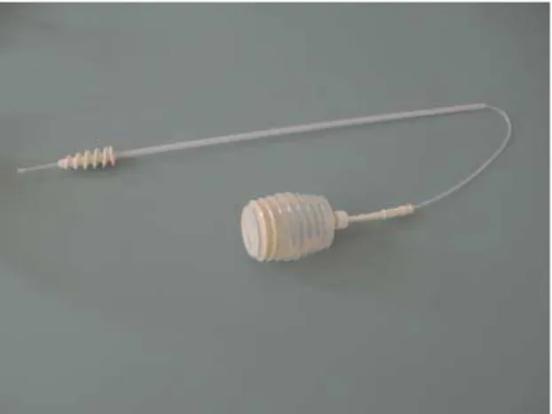
Before treatment, samples of uterine flushing were collected by means of an especially designed catheter for sows (Figure 1) which enables sampling and minimizes the risk of uterine wall perforation (General Medic, Beograd, Srbija). Bacterial count, types of bacteria, bacterial, neutrophyle and eosinophyle presence on the stained smears (May Grünwald-Giemsa method) were determined. Samples were collected again from the same animals two to five days after therapy and the same laboratory tests were repeated.
Body temperature (BT) of the sows was followed daily with laser thermometer (ThermoFlash® PRO-IR ZH-36, France) and when BT was higher than 39.5oC antibiotics were introduced by parenteral route (PenStrep, Avilamycine) in a dose prescribed by the manufacturer. This was performed only in six sows.
A clinical estimation was performed using the originally developed scale as follows:
a. mucopurulent voluminous discharge, reduced appetite, high BT 4 b. mucopurulent voluminous discharge, reduced appetite, normal BT 3 c. discharge in small amount, slightly reduced appetite, normal BT 2
d. no discharge, normalized appetite, normal BT 1
On the first day of therapy and 2-5 days later, body weights of piglets were recorded in order to calculate average BW and daily weight gains. Their BW was also estimated at the moment of weaning. From this data, we calculated daily body weight gains from therapy to second sampling (2-5 days later), and from therapy to weaning.
Since this therapeutic approach has not been described earlier, we have also recorded the number of piglets in treated sows in the next breeding cycle, in order to investigate a possible negative consequence of the applied method.
Housing and feeding
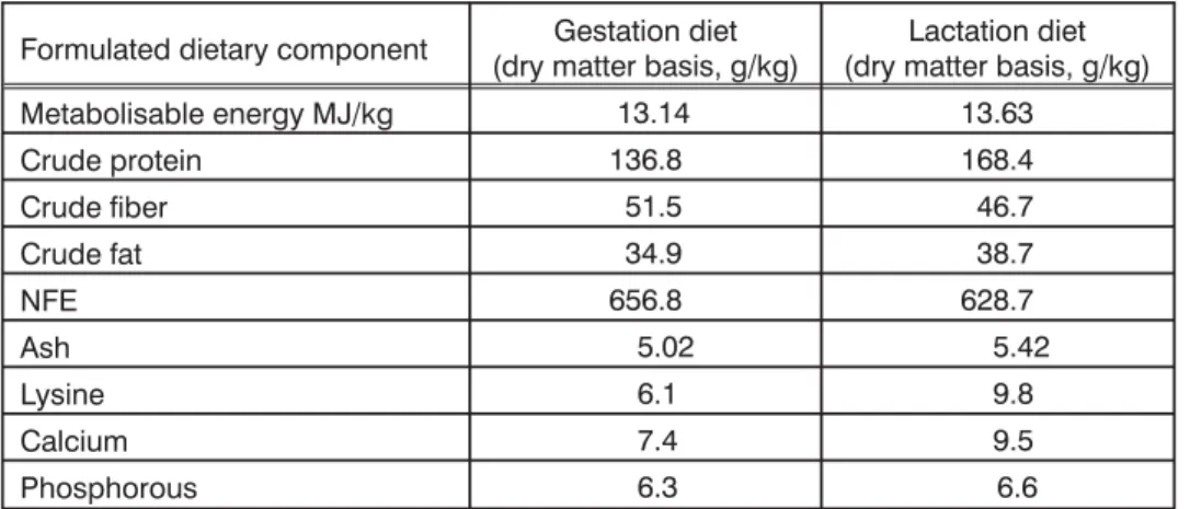
Sows were housed in commercial crates measuring 2.2 m x 1.5 m, with standard creep access for piglets, allowingad libitumsuckling. They were kept under air conditioned environment, 23-24oC and day - light regime 14+10 hrs. Sows were fed corn wheat based diets meeting commercial nutrient specifications (Table 1) according to AEC tables (1993).
Table 1. Composition of sow gestation and lactation diets
Formulated dietary component (dry matter basis, g/kg)Gestation diet (dry matter basis, g/kg)Lactation diet
Metabolisable energy MJ/kg 13.14 13.63
Crude protein 136.8 168.4
Crude fiber 51.5 46.7
Crude fat 34.9 38.7
NFE 656.8 628.7
Ash 5.02 5.42
Lysine 6.1 9.8
Calcium 7.4 9.5
Phosphorous 6.3 6.6
YCW and Lotagen preparation and application
sterile saline was added to each flask and well shaken to form a suspension. Application was performed with sterile catheter, as described above, suitable both for intra uterine sampling and medicament application. In the fourth group of sows, a 100 mL of 2% Lotagen (Byk Gulden, Germany) solution was applied in the same manner. Instillation was performed only once.
Uterine content sampling and bacteriological examination
Samples of the uterine content were collected by syringe aspiration after delivery on days 1 - 3 when clinical signs of infection were evident. This was performed with a sterile catheter that enables safe sampling. Second sampling was performed in the same manner 2 – 5 days following therapy. Samples were placed in labeled sterile plastic tubes and transported to the laboratory. Microbiological analyses of uterine flashings were performed in order to estimate the presence of both aerobic and anaerobic bacteria and their total count (colony forming units – CFU).
All samples were inoculated on Columbia agar (CM331, Oxoid, Basingstoke, UK) containing 5% sheep blood and Mac Conkey agar (CM115, Oxoid, UK). Following inoculation, the plates were incubated aerobically at 37oC for 24 - 48h. The cultures were purified by sub culturing on the nutrient agar (CM3, Oxoid, UK). Each isolate was identified on the basis of colonial morphology, microscopic appearance and biochemical test (Quinet al., 1998).
Total bacterial count (CFU/mL) was determined using the standard plate count method from a series of ten-fold dilutions. Dilutions (101-105) were prepared in buffered peptone water (CM 1049, Oxoid, Basingstoke, UK) and inoculated in the amount of 0.5 mL in Petri dishes containing 5% sheep blood in Tryptone soya agar (CM131, Oxoid). Plates were incubated during 48h at 37oC under aerobic conditions.
Cytology
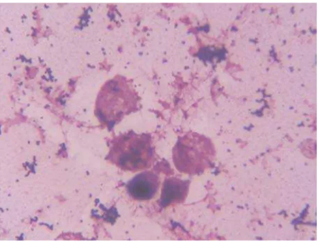
This was performed after drying of smears on microscope slides and standard May Grunwald Giemsa staining. We used Olympus BH-2 direct light microscope with total magnification of 1000 x and an immersion objective. Photomicrographs were taken by digital Olympus BX-40 (Japan) camera. The presence of neutrophiles and eosinophyles, epithelial cells and bacteria in uterine flushing was estimated.
RESULTS
Clinical findings
Using the above described scale, we were able to judge the improvement in the sow's health status following IU instillation of the YCW or Lotagen solution. The data are presented in Table 2.
10 and 20 g of YCW and what is even more important, the percent of recidivism (sows that still had signs of infection 5 days following therapy) was only 10% in these two groups.
Table 2. Clinical improvement in the sow’s health status following IU instillation of YCW (5, 10 and 20 g) and Lotagen 2% solution
Average clinical
score Average clinical score2-5 d after therapy improvement, %Clinical Recidivism%
YCW I 5 g 2.8 ± 0.42 1.3 ± 0.48 53.4 30
YCW II 10 g 3.2 ± 0.63 1.1 ± 0.32 65.7 10
YCW III 20 g 3.4 ± 0.48 1.2 ± 0.32 64.7 10
LOTAGEN 3.4 ± 0.52 1.5 ± 0.53 55.9 50
Bacterial count
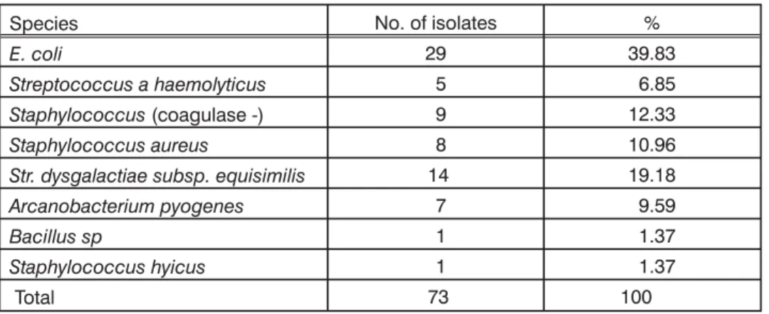
In 40 uterine flushing samples, at the beginning of investigation, we were able to isolate 8 different bacterial species. In 8 sows, we isolated only one dominant bacteria (20%) in three of them (7.5%) three different bacterial species and in remaining 72.5% of animals infection was caused by two bacteria. In eight samples (20%) one bacterial species dominating on the agar plate together with sparse non-specific mixed culture (Staphylococcus coagulase – and Bacillus sp.) was isolated. In two samples we isolated only nonspecific bacteria (Staphylococcus coagulase – and Bacillus sp.). Bacterial species and their abundance are presented in Table 3.
Table 3. Bacterial species isolated from sows uterus and their relative abundance
Species No. of isolates %
E. coli 29 39.83
Streptococcus a haemolyticus 5 6.85
Staphylococcus(coagulase -) 9 12.33
Staphylococcus aureus 8 10.96
Str. dysgalactiae subsp. equisimilis 14 19.18
Arcanobacterium pyogenes 7 9.59
Bacillus sp 1 1.37
Staphylococcus hyicus 1 1.37
Total 73 100
It is evident that the most frequently isolated bacterial species wereE. coli, Streptococcus dysgalactiae subspecies equisimilis, Arcanobacterium pyogenes, Staphylococcus aureus, Staphylococcus (coagulase -) and Streptococcus a
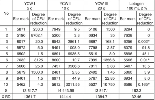
Following IU instillation of YCW we were able to demonstrate a significant reduction in the bacterial count (CFU – colony forming unite) and these data are presented in Table 4. It may be concluded that YCW when applied in the uterus of infected sows significantly reduces the number of CFU. The same effect was observed for Lotagen solution, but to a lesser extent.
Table 4. The degree of bacterial number reduction (CFU) in samples of sows' uterine flushings following instillation of YCW (5, 10 and 20 g) and
Lotagen 2% sol. (100 mL)
No
YCW I
5 g YCW II10 g YCW III20 g 100 mL 2 %Lotagen
Ear mark Degreeof CFU
reductionEar mark
Degree of CFU
reduction Ear mark
Degree of CFU
reductionEar mark
Degree of CFU reduction
1 5871 233.3 7949 9.5 5106 1500 8294 0
2 5190 8702.1 5206 3.3 6634 35 7628 0
3 8017 20.0 8542 2861.1 6897 168.1 6256 0.002*
4 5572 5.0 5491 1008.0 7798 2.87 6079 91.8
5 6502 1.5 6891 6935.5 5519 8.0 5896 45.1
6 7032 3125 8600 12.7 7999 1356.6 5566 0.01*
7 5606 25.0 7457 3566.6 7811 2.83 5407 13.5
8 5679 1500.0 2481 2.35 2492 1.45 5860 3.9
9 8401 1.5 6971 44.9 5767 22.85 6934 8.0
10 5462 4.3 5612 3011.55 5527 10 750 6596 0.165*
S 13 617.7 14 443.95 13 847.1 162.3
X RD 1361.7 1444.4 1384.7 32.46
* values lower than 1 represent elevations of the CFU number, RD – reduction degree
The degree of CFU number reduction was nearly the same in all groups of sows treated with intra uterine instillation of YCW (5, 10 and 20 g). No dose dependent effect was noted and it was evident that the degree of CFU number reduction varied greatly within all groups of sows. Treatment with Lotagen solution, also resulted in clinical improvement, but was not that efficient (degree of CFU reduction was only 32.6) and 50% of sows still had a purulent discharge after 2-5 days (Table 2).
Smears
nuclei do degeneration like karyopycnosis, karyiolysis or lipid degeneration. Such a finding indicates acute inflammation. Lymhocytes, monocytes and uterine epithelial cells were rare. In 30 % of samples we were able to demonstrate chains ofStreptococcaeandCocobacilli.
On the stained smears obtained 2-5 days following therapy, in general, cells were rare and we noticed 1 – 5 epithelial cells and 1 – 10 PMN per visual field accompanied with few lymphocytes. Bacteria were present in one of 15 samples. This phenomenon was observed for all treatments but sows that still had vaginal discharge (Lotagen group) had much more bacteria in the second samples.
Figure 2a. A typical stained smear of vaginal content from sows with clinical signs of puerperal infection. Note the presence of numerous bacteria and neutrophyles
Piglets number and body weights
On the day of therapy, the average number of piglets was lower in the YCW II group when compared to groups YCW I, L and C (p<0.05). After treatment the number of piglets was higher in the control group when compared to groups YCW I and YCW II (p<0.05). The average number of piglets weaned, was highest in the control group (non infected sows) and lowest in the Lotagen treated group. These differences were only numeric. Total piglets loss was highest in the Lotagen treated group (Table 5).
Table 5. Average number of piglets during the trial
Number of pig-lets at the day of
therapy
Number of pig-lets 2 – 5 days after
therapy
Number of
pig-lets weaned Total loss %
YCW I 5 g 10. 8 ± 0.63 10.5 ± 0.85 9.9 ± 1.14 9.16
YCW II 10 g 9.9 ± 0.99 9.8 ± 1.03 9.5 ± 1.18 4.20
YCW III 20 g 10.9 ± 1.37 10.3 ± 0.67 9.5 ± 1.35 14.70
Lotagen 10.9 ± 0.99 10.6 ± 1.07 9.2 ± 1.40 18.40
Control 10.9 ± 0.57 10.9 ± 0.57 10.2 ± 1.40 6.80
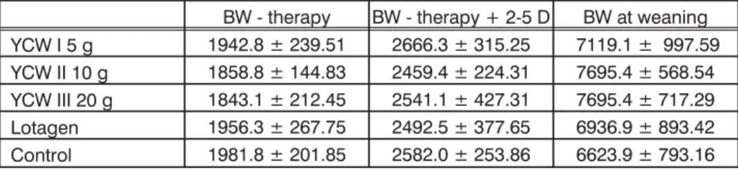
In Table 6, the average values and standard deviations for piglets body weights during the trial are presented, while results of statistical analyses are shown in Tables 6.1, 6.2. and 6.3.
Table 6. Body weights (g) of piglets (X ± SD) at the moment of therapy of sows, 2-5 days after and at weaning
BW - therapy BW - therapy + 2-5 D BW at weaning
YCW I 5 g 1942.8 ± 239.51 2666.3 ± 315.25 7119.1 ± 997.59
YCW II 10 g 1858.8 ± 144.83 2459.4 ± 224.31 7695.4 ± 568.54 YCW III 20 g 1843.1 ± 212.45 2541.1 ± 427.31 7695.4 ± 717.29
Lotagen 1956.3 ± 267.75 2492.5 ± 377.65 6936.9 ± 893.42
Control 1981.8 ± 201.85 2582.0 ± 253.86 6623.9 ± 793.16
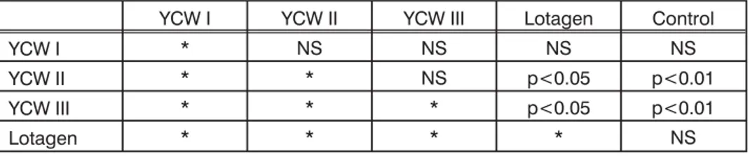
Table 6.1. Statistical analyses of differences in piglets BW at the moment of therapy of sows
YCW I YCW II YCW III Lotagen Control
YCW I * NS NS NS NS
YCW II * * NS NS NS
YCW III * * * NS NS
Lotagen * * * * NS
Table 6.2. Statistical analyses of differences in piglets BW 2 – 5 days after therapy of sows
YCW I YCW II YCW III Lotagen Control
YCW I * NS NS NS NS
YCW II * * NS NS NS
YCW III * * * NS NS
Lotagen * * * * NS
NS – non significant
Table 6.3. Statistical analyses of differences in piglets BW at weaning
YCW I YCW II YCW III Lotagen Control
YCW I * NS NS NS NS
YCW II * * NS p<0.05 p<0.01
YCW III * * * p<0.05 p<0.01
Lotagen * * * * NS
NS – non significant
Statistical differences were not evident for body weights of piglets at the moment of therapy or 2-5 days later (moment of second sampling). However, when we analyzed body weights at the moment of weaning, we were able to conclude that in groups YCW II and YCW III piglets were significantly heavier when compared to piglets in the Lotagen and Control group. Other differences were only numeric. We must stress out that these results are not completely reliable because not all piglets were weaned at the same day of life. Therefore, a more precise parameter was daily body weight gain and these data are presented in Table 5 while results of statistical analyses are shown in Tables 7.1. and 7.2.
Table 7. Daily body weight gain (g) of piglets from the moment of therapy of sows 2-5 days after and during the whole period of observation (x ± SD)
DBWG - therapy – 2-5 days DBWG up to weaning
YCW I 206.4 ± 42.55 221.1 ± 27.85
YCW II 192.1 ± 42.22 230.2 ± 31.93
YCW III 204.5 ± 52.70 241.2 ± 30.91
Lotagen 162.7 ± 31.17 205.7 ± 39.61
Control 199.0 ± 82.26 210.1 ± 38.72
Table 7.1. Statistical analyses of differences in piglets DBWG from the moment of sows therapy to 2-5 days after
YCW I YCW II YCW III Lotagen Control
YCW I * NS NS p<0.02 NS
YCW II * * NS NS NS
YCW III * * * p<0.05 NS
Lotagen * * * * NS
NS – non significant
Table 7.2. Statistical analyses of differences in piglets DBWG from the moment of sows therapy to weaning
YCW I YCW II YCW III Lotagen Control
YCW I * NS NS NS NS
YCW II * * NS NS NS
YCW III * * * p 0.05 p 0.05
Lotagen * * * * NS
NS – non significant
At the end, statistical analyses of piglets DBWG from the moment of sows therapy to weaning revealed significant differences when YCW III group was compared to the Lotagen and control groups (p<0.05).
In order to estimate if the applied treatment had influence on the sow's reproductive performances we have followed the number of piglets born in the next breeding cycle of the same sows. These results are presented in Table 8.
Table 8. Reproductive success of sows, included in the study, in the next reproductive cycle (average values, X ± SD)
Total born Born alive Total loss%
YCW I 5 g, n = 8* 11.90 ± 2.47 10.75 ± 1.67 9.49
YCW II 10 g, n = 9* 12.30 ± 2.69 10.89 ± 1.96 11.78
YCW III 20 g, n = 8* 12.10 ± 2.47 10.40 ± 1.60 14.46
Lotagen, n = 9* 11.78 ± 2.91 10.30 ± 2.18 12.22
Control*, n = 9 13.44 ± 1.59 11.44 ± 0.88 21.50
*From the total of 50 sows 2 died, in the following six months and 5 of them were sold
DISCUSSION
Sufficient colostrum and milk intake is extremely important for piglets and numerous attempts were performed in order to improve milk production in sows. Addition of iodinated casein to sows' diet resulted in higher body weight of piglets at the moment of weaning and also in higher thyroid gland hormone concentrations in the sera of sows (Djurdjevicet al., 1980).
Our results regarding the number and type of bacterial isolates from uterine flushings are in accordance to those reported by Radovic (1997). Treatment of endometritis with antimicrobial agents and antibiotics has various degrees of success and the cost of treatment is high. In cows, milk has to be disposed and there is a possibility for developing microbial resistance along with reduced phagocytic activity of leukocytes. For that reason alternative ways of therapy were investigated by using natural substances as means of activation of natural defense mechanisms in the uterus. One of them was garlic extract (Alium sativum). In this trial administration of PGF 2aserved as a positive control (Sarkar et al., 2006). Authors were able to demonstrate lowering of the cervical mucus pH value accompanied with a significant reduction of bacterial load similar to our findings The total number of isolates was 65, comprising mostly of facultative anaerobic bacteria. Following treatment an elevation of thyroid hormone levels in the plasma from both groups of treated animals was noted, as well as a decline in cortisol levels. It is well known that circulatory levels of thyroxine and tri-iodothyrinine play an important role in correlating the persistent infection, as well as subclinical condition of infection (David et al., 1998). A decrease in their concentration occurred due to constant caloric deprivation or to enhanced endogenous cortisol production (Peterson and Ferguson, 1989).
Another attempt to investigate alternative way of therapy was by using three lactic bacteria strains: Lactobacillus acidophilus, Enterococcus faecalis and Enterococcus cecorum(Xuefenget al., 2011). All tested bacteria exerted a high level of adhesion to immortalized endometrial epithelial cells in vitro and L. acidophiluswas the most efficient. In coculture assays these bacteria significantly reduced adhesion of pathogensE. coliandS. aureus. Authors concluded that the ability of LAB tested for inhibition of adhesion of endometritis-associated pathogens is highly specific and depends on both probiotic and pathogen strains. As prebiotics also prevent bacterial adhesion to epithelial cells in the digestive tract (Springet al., 2000) we may postulate that mannan oligosaccharides were also able to exert the same effect in the lumen of infected sows uterus. Our results clearly show a significant degree of reduction in bacterial count following intrauterine instillation of YCW. Clinical improvement in sows was also evident in our study, as well as piglets higher body weight gains.
Various relatively new medicaments were tested in the therapy of PDA which is usually based on administration of antibiotics and oxytocin. Among them, meloxicam and flunixine were efficient in improving clinical score (Hirschet al., 2003). However meloxicame lowered the mortality rate in piglets by 50% in comparison to reference control group reaching statistical significance (p<0.05) Walleret al. (2002) reported results of the study conducted in 19 Hungarian pig herds over a 4 year period. Authors evaluated the relationship between the duration of vulval discharge and subsequent fertility and litter size. They concluded that both parity 1 and parity 2-8 sows having vulvar discharge in excess of 6 days had significantly lower fertility compared with sows of similar parity where the duration of vulval discharge was less than 6 days. A duration of vulval discharge in 1 parity sows in excess of 6 days significantly reduced litter size (total born and live born) in subsequent farrowings, but not in parity 2-8 sows. Our results confirmed that in treated sows subsequent fertility was not affected and the percent of loss of piglets was highest in the control group.
Address for correspondence: Dr Miodrag Lazarevi}, professor Faculty of Veterinary Medicine
Department of Physiology and Biochemistry Bul. oslobodjenja 18
11 000 Belgrade
E mail: lazarevicmªvet.bg.ac.rs
REFERENCES
1. Alimentation Equilibree de Commentry (AEC) tables 1993, AEC tables – recommendations for animal nutrition, Rhone Poulenc animal nutrition, Commentry, France.
2.Backstrom L, Morkoc CA, Connor J, Larson R, Price W,1984, Clinical study of mastitis-metritis-agalactia in sows in Illinois,JAVMA, 185, 1, 70-3.
3.Blaney BJ, McKenzie RS, Walters JR, Taylor LF, Bewg WS, Ruley MJ et al,2000, Sorghum ergot (Claviceps africana) associated with agalactia and feed refusal in pigs and dairy cattle,Aust Vet J, 78, 2, 102-7.
4. Bostedt H, Maier G, Herfen K, Hospes R, 1998, Clinical examination of gilts with puerperal septicemia and toxemia,Tierarztl Prax Ausg Grosstiere Nutziere,26, 6, 332-8.
5. Dalin MA, Kaeoket K, Persson E,2004, Immune cell infiltration of normal and impaired sow endometrium,Anim Reprod Sci, 82-83, 401-13.
6. David BP, Kumar RV, Paliniswami KS, Kadwad VB, Sivaprasad N, 1989, Estimation of thyroid hormons in animals samples,Ind Vet J,75, 565-6.
7.De Winter P JJ, Verdonck M, de Kruif A, Devries LA, Haesebrouck F,1992, Endometritis and vaginal discharge in the sow, Anim Reprod Sci, 28, 51-8.
8.\ur|evi} \, Maksimovi} A, Stoji} V, Laslo [,1980, Uticaj dodavanja tireoproteina u hrani krma~a na nivo tireodnih hormona u krvnom serumu I porast telesne te`ine njihove prasadi,Vet glasnik, 34, 1, 27-31.
9.Hengartner B, Henggeler M, Kohler S, Spring P,2005, Effects of dietary mannan-oligosaccharides on plasma Ig G concentration in piglets, Proceedings of Antimicrobial Growth promoters, Worldwide Ban on the Horizon, Nordwijk aan Zee, The Netherlands, 101.
11. Hooge DM, Sims MMd, Sefton Ae, Connolly A, Spring P, 2003, Effects of dietary mannan-oligosaccharide with or without bacitracin or virginiamycin on live performance of broiler chickens at relatively high stocking density on new litter,J Appl Poult Res, 12, 461-7. 12. Hooge DM, 2004a, Meta-anlyses of broiler chicken pen trials evaluating dietary mannan
oligosaccharide, 1993-2003,Int J poult Sci, 3, 164-74.
13.Hooge DM,2004b, Turkey pen trials with dietary mannan oligosaccharide meta-anlyses, 1993-2003,Int J Poult Sci, 3, 179-88.
14.Kemper N, Gerjets I,2009, Bacteria in milk from anterior and posterior mammary glands in sows dysgalactia syndrome (PPDS),Acta Vet Scand, 51, 1, 26-34.
15.Klopfenstein C, Farmer C, Martineau GP,2006, Diseases of the Mammary Glands, In Diseases of Swine, IX edition (Eds. Straw BE, Zimmerman JJ, D'Allaire S, Taylor JD), Blackwell Publishing, 57-85.
16. Kopinski JS, Blaney BJ, Downing JA, McVeigh JF, Murray SA, 2007, Feeding sorghum ergot (Claviceps africana) to sows before farrowing inhibits milk production,Aust Vet J, 85, 5, 169-76. 17.Lazarevic M,2005, Mannan oligosaccharide enhances adsorption of colostral Ig G in newborn
calves and piglets, In Proceedings of the 56thMeeting of the European Association for Animal Production, Uppsala Sweden (ed Y van der Honig), Wageningen Academic Press, Wageningen, The Netherlands, 15, 3.
18.Lazarevic M, Spring P, Shabanovic M, Tokic V, Tucker LA,2010, Effect of gut active carbohydrates on plasma Ig G concentrations in piglets and calves,Animal, 4, 6, 938-43.
19. Merck Veterinary Manual, 2011, Postpartum Dysgalactia Syndrome, http//www.merckvetmanual. com/mvm/htm/bc/111105.htm
20. Nitovski A, 1993, Prilog poznavanju etiopatogeneze hipo- i – agalakcije krma~a, Doktorska diseratacija, Fakultet veterinarske medicine, Univerzitet u Beogradu, 171.
21.Noblet J, Dourmad JY, Etienne M and Le Dividich J,1997, Energy metabolisim In pregnant sows and newborn pigs,J Anim Sci, 75, 2708-14.
22.Miguel JC, Rodriguez-Zas SL, Pettigrew JE,2002, Practical responses to Bio-GAC in nursery pigs: A meta analyses, In Nutritional biotechnology in the feed and food industries (ed. TP Lyons and KA Jacques) Nottingham University Press, UK, 425-33.
23. Papadopoulos GA, Venderhaeghe C,Janssens GP, Dewuls J, Maes DG, 2010, Risk factors associated with postpartum dysgalactia syndrome in sows,Vet J, 184, 2, 167-71.
24.Peterson D, Ferguson DC, 1989, Text book of small animal practice, Internal medicine, 3rd(Ed Ettinger SJ), WB Sounders Company.
25.Preissler R, Hinrics D, Reiners K, Loof H, Kemper N,2011, Estimation of variance components for postpartum dysgalactia syndrome in sows,J Anim Breed Genet,129, 2, 98-102.
26. Quinn PJ, Carter ME, Markey BK, Carter GR, 1998: Clinical Veterinary Microbiology, Mosby International, Lynton House, England, 118-224.
27. Radovi} Bisa, 1997, Prilog poznavanju bakterijske etiologije sindroma mastitisa, metritisa i agalakcije kod krma~a uzgajanih na farmama, Doktorska diseratacija, Fakultet veterinarske medicine, Univerzitet u Beogradu, str. 94.
28.Rozeboom DW, Shaw DT, Templeman RJ, Miguel JC, Pettigrew JE, Connoly A,2005, Effects of mannan oligosaccharide and an antimicrobial product in nursery diets on performance of pigs reared on three different farms,J Anim Sci, 83, 2637-44.
29. Sarkar P, Kumar H, Rawat M, Varshney P, Goswami TK, Yadav MC et al, 2006, Effect od administration of garlic extract and PGF 2a on hormonal changes and recovery in endometritic cows,Asian-Aust J Anim Sci, 19, 7, 964-9.
30. Sims MD, Dawson KA, Newman KKE, Spring P, Hooge DM, 2004, Effects of dietary mannan-oligosaccharide, bacitracine metylene disalicylate or both on the live performance and intestinal morphology of turkeys,Poult Sci,83, 1148-54.
32. Spring P, 2004, Impact of mannan oligosaccharide on gut health and pig performance. In: Interfacing immunity, gut health and performance (Ed LA Tucker and JA Taylor-Pickard) Nottingham University Press, Nottingham, UK, 93-105.
33. [amanc H,2011, Poreme}aji funkcije mle~ne `lezde, U: Bolesti svinja, IDP "Nau~na KMD", Beograd, 332-59.
34.[amanc H, Damnjanovic Z, Radoji~i} B, Stoji} V,1992, Cortisol, triiodothyronine, thyroxine and glucose concentrations in the blood of the first litter sows during advanced pregnancy and post partum in relation to hypogalactia and and agalactia, Acta Vet, Beograd, 42, 2-3, 109-14. 35.[amanc H, Stamatovi} S, Damnjanovi} Z, Nitovski A, Mateji} D, 1989, Hipo- i agalakcija krma~a – 1.
Glikemija u visokom graviditetu i postpartalno,Vet Glasnik,43, 3-4, 277- 80.
36.van Gelder KN, Bilkei G,2005, The course of acute-phase proteins and serum cortisol in mastitis metritis agalactia (MMA) of the sow and sow performance,Tijdschr diergeneeskd,Jan 15, 130, 2, 38-41
37.Xuefeng Q, Donging B, Xianjun Z, Sihua W, Yi L, Yapincourse g J, 2011, Probiotic activities of Lab in endometritis: probiotic activities of three lactic acid bacteria strains isolates from healthy bovine cervix: In vitro adherence to immortalized endometrial epithelial cells and antimicrobial properties, J Anim Vet Adv, 10, 4, 484-8.
38. Waller CM, Bilkei GG, Cameron RG,2002, Effect of periparturient diseases accompanied by excessive vulval discharge and weaning to mating interval,Aust Vet J, 80, 9, 545-9.
39.Wulster-Radcliffe, MC, Seals RC, Lewis GS,2002, Progesterone increases susceptibility of gilts to uterine infections after intrauterine inoculation with infectious bacteria,J Anim Sci,81, 1242-52.
TERAPIJA ENDOMETRITISA KRMA^A INTRAUTERINOM INSTILACIJOM PREPARATA NA BAZI ZIDA KVASCA
LAZAREVI] M, MILOVANOVI] A, BARNA T, MILJAS N, MILANOV DUBRAVKA
SADR@AJ
Na osnovu rezultata na{ih istra`ivanja bilo je mogu}e zaklju~iti da tretman krma~a obolelih od puerperalnih infekcija materice, sterilnim preparatom do-bijenim iz zida kvasca (YCW), ima za posledicu zna~ajan klini~ki napredak. Pro-cenat krma~a sa recidivom je bio najni`i (10%) u grupi tretiranoj sa 10 i 20g YCW.
Stepen redukcije broja bakterijskih kolonija (CFU) u uzorcima dobijenih iz materice krma~a posle tretmana YCW preparatom je bio veoma visok i kretao se u opsegu od 1361 do 1444 puta. Kod obolelih krma~a, tretiranih rastvorom Lota-gena (2%), vrednost ovog parametra je bila svega 32.