(Annals of the Brazilian Academy of Sciences)
Printed version ISSN 0001-3765 / Online version ISSN 1678-2690 www.scielo.br/aabc
http://dx.doi.org/10.1590/0001-3765201420140173
In vivo and in vitro effects of fructose on rat brain
acetylcholinesterase activity: an ontogenetic study
CARINE A. GUIMARÃES1, MAIRIS S. BIELLA1, ABIGAIL LOPES1, PEDRO F. DEROZA2, MARIANA B. OLIVEIRA2, TAMIRES P. MACAN1, EMILIO L. STRECK3, GUSTAVO C. FERREIRA4,
ALEXANDRA I. ZUGNO2 and PATRÍCIA F. SCHUCK1 1
Programa de Pós-Graduação em Ciências da Saúde, Universidade do Extremo Sul Catarinense, Unidade Acadêmica de Ciências da Saúde, Laborátorio de Erros Inatos do Metabolismo, Av. Universitária, 1105, Bloco S, Sala 6, Bairro Universitário, 88806-000 Criciúma, SC, Brasil 2Programa de Pós-Graduação em Ciências da Saúde, Universidade do Extremo Sul Catarinense,
Unidade Acadêmica de Ciências da Saúde, Laboratório de Neurociências, Av. Universitária, 1105, Bloco S, Sala 5/ subsolo, Bairro Universitário 88806-000 Criciúma, SC, Brasil 3
Leia-se: Programa de Pós-Graduação em Ciências da Saúde, Universidade do Extremo Sul Catarinense, Unidade Acadêmica de Ciências da Saúde, Laboratório de Bioenergética, Av. Universitária, 1105,
Bloco S, Sala 6, Bairro Universitário, 88806-000 Criciúma, SC, Brasil 4
Instituto de Biofísica Carlos Chagas Filho, Universidade Federal do Rio de Janeiro, Rio de Janeiro, Av. Carlos Chagas Filho, 373, Prédio do CCS, Bloco G, Sala G1-019, 21941-902 Rio de Janeiro, RJ, Brasil
Manuscript received on April 13, 2014; accepted for publication on July 29, 2014
ABSTRACT
Increased fructose concentrations are the biochemical hallmark of fructosemia, a group of inherited disorders on the metabolic pathway of this sugar. The main clinical findings observed in patients affected by fructosemia include neurological abnormalities with developmental delay, whose pathophysiology is still undefined. In the present work we investigated the in vitro and in vivo effects of fructose on acetylcholinesterase (AchE) activity in brain structures of developing rats. For the in vitro experiments, fructose was added at increasing concentrations to the incubation medium. It was observed that fructose provoked an inhibition of acetylcholinesterase activity in cerebral cortex of 30-day-old-rats, even at low concentrations (0.1 mM). For the in vivo experiments, rats were killed 1 h after a single fructose administration (5 μmol/g). Control
group received the same volume of saline solution. We found that AchE activity was increased in cerebral cortex of 30- and 60-day-old rats receiving fructose administration. Finally, we observed that AchE activity was unaffected by acute fructose administration in cerebral cortex, striatum or hippocampus of 15- and 90-day-old rats. The present data suggest that a disruption in cholinergic homeostasis may be involved in the pathophysiology of brain damage observed in young patients affected by fructosemia.
Key words: acetylcholinesterase, brain, fructose, hereditary fructose intolerance.
Correspondence to: Patrícia Fernanda Schuck E-mail: [email protected]
INTRODUCTION
Hereditary fructose intolerance (HFI) or fructose-mia (OMIM ID: 229600) is an autosomal recessive
characterized by increased concentrations of fructose and fructose-1-phosphate in the blood of patients (Scriver et al. 2001). This deficiency may arise from various different mutations in the aldolase B gene situated on chromosome 9q22.3 (Lench et al. 1996, Coffee and Tolan 2010), most of which are point mutations resulting in single amino acid changes that decrease stability or catalytic activity (Bouteldja and Timson 2010). Symptoms appear in the newborn following weaning when fructose-containing foods are first introduced (Bouteldja and Timson 2010) and generally include abdominal pain, vomiting, and diarrhea. Children affected by these disorders may present with a general failure to thrive. Heavy and/or persistent intake of the sugar can lead to hypoglycemia, jaundice, progressive cirrhosis of the liver, renal tubular failure, metabolic acidosis, seizures, coma, and eventually death (Baerlocher et al. 1978, Cox 1993, Laméire et al. 1978, Morris 1968, Odiévre et al. 1978, Steinmann et al. 2001). Recently, a case has been reported with the association of hereditary fructose intolerance and poorly symptomatic Duchenne type muscular dystrophy (Paolella et al. 2012).
It has been previously speculated that the build-up of fructose-1-phosphate is the central element from which the spectrum of pathological pathways irradiates, as this compound fails to be cleaved into glycolysis or gluconeogenesis intermediates (Oberhaensli et al. 1987). The decrease on inorganic phosphate pool also results in impaired glycogenolysis, which, allied to the decreased ATP synthesis, disturbs all cellular processes that rely on phosphorylation or ATP (Oberhaensli et al. 1987, Van Den Berghe et al. 1973). However, the specific fructose toxicity is still poorly known. In the hope to clarify the contribution of fructose on the pathomechanisms of brain damage observed in HFI patients, the aim of the present work was to investigate the in vivo and in vitro effect of fructose on acetylcholinesterase (AchE) activity, an important enzyme involved in the homeostasis of cholinergic system (Igisu et al. 1994, Jha and Rizvi 2009), in different brain structures of young rats.
MATERIALS AND METHODS
REAGENTS
All chemicals were purchased from Sigma (St. Louis, MO, USA). Fructose was dissolved on the day of the experiments in the incubation medium used for each technique with pH adjusted to 7.4.
ANIMALS
Fifteen-, thirty-, sixty- and ninety-day-old male Wistar rats obtained from the Central Animal House of Universidade do Extremo Sul Catarinense, Criciúma, SC – Brazil, were used. The animals were maintained on a 12:12 h light / dark cycle (lights on 07.00 - 19.00 h) in air conditioned constant temperature (22 °C ± 1°C) colony room, with free access to water and 20 % (w/w) protein commercial chow. The experimental protocol was approved by the Ethics Committee on Animal Research of the Universidade do Extremo Sul Catarinense (Protocol 076/2013-2) and followed the Guide for the Care and Use of Laboratory Animals (National Research Council, 2011). All efforts were made to minimize the number of animals used and their suffering.
IN VITROEXPERIMENTS
On the day of the experiments six animals were killed by decapitation without anesthesia and the cerebral cortex was rapidly excised on a Petri dish placed on ice. The brain structures were homogenized in a 100 mM phosphate buffer containing 0.1% Triton X-100, pH 7.5. The homogenates were centrifuged at 800 x g for 10 min at 4 °C. The supernatants were isolated and were preincubated for 1 h at 37 °C in the absence (control group) or presence of different concentrations of fructose (0.1 – 5 mM).
IN VIVOEXPERIMENTS
A total of thirty-six animals were divided into two groups for each age (12 animals of 15-, 30-, 60-, and 90-day-old; 6 animals per group): control group, which received a single injection of saline solution (0.9 g%), and fructose group, which received a single injection of fructose (5 μmol/g body weight). One hour after the administration, the animals were killed by decapitation without anaesthesia, and the brains were rapidly excised on a Petri dish placed on ice. The cerebral cortex, striatum and hippocampus were peeled away from the white matter. Immediately after, the brain structures were kept at -70 °C until being used for enzyme activity determination.
DETERMINATION OF AchEACTIVITY
AchE activity was determined by the method of Ellman et al. (1961). Acetylcholine hydrolysis rate was measured in an incubation medium containing acetylcholine 0.8 mM, 100 mM phosphate buffer (pH 7.5) and 1.0 mM DTNB. Fifty microliters of supernatants were added to the reaction mixture and preincubated for 3 min at 25 °C. The hydrolysis was monitored by the formation of the thiolate dianion of DTNB at 412 nm for 2–3 min (30 s intervals) at 25 °C. The results were expressed as μmol of hydrolysed acetylcholine . h-1. mg of protein-1 and all samples were run in duplicate (Zugno et al. 2008).
PROTEIN DETERMINATION
Protein was measured by the method of Lowry et al. (1951) using bovine serum albumin as standard.
STATISTICAL ANALYSES
Results are presented as mean ± standard error of mean. Assays were performed in duplicate and the mean was used for statistical analyses. Data were analyzed using Student t test for independent samples when comparing two experimental groups (in vivo experiments) or one-way analysis of variance (ANOVA) followed by the post-hoc
Duncan multiple range test when F was significant when comparing three or more experimental groups (in vitro experiments). Differences between groups were rated significant at p < 0.05. All analyses were carried out in an IBM-compatible PC computer using the Statistical Package for the Social Sciences (SPSS) software 16.0.
RESULTS
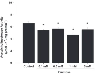
The present work investigated the in vitro effect of increasing fructose concentrations on AchE activity in cerebral cortex of young rats (30-day-old) (Figure 1). We observed that fructose induced a significant decrease in AchE activity even at low concentrations (0.1 mM and higher).
Figure 1 - In vitro effect of fructose on acetylcholinesterase activity in cerebral cortex from 30-day-old rats. Values are means ± standard error of mean for six independent experiments performed in duplicate and are expressed as µmol acetylcholine . h-1. mg protein-1. *p < 0.05 compared to control group (Duncan multiple range test).
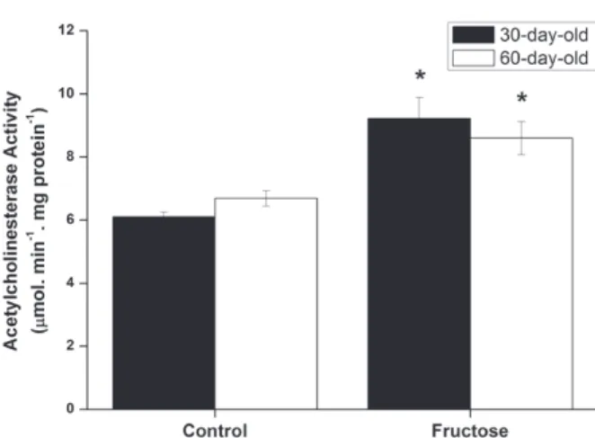
The influence of acute fructose administration on AchE activity in cerebral cortex of rats with 30 and 60 days of life (Figure 2) were also assessed. It was observed that AchE activity was significantly increased in the brain of rats of both ages receiving fructose, as compared to control group.
90-day-old rats (Table I and II, respectively) were finally assessed. AchE activity was not altered in cerebral cortex, striatum or hippocampus by this treatment in 15- and 90-day-old rats.
DISCUSSION
In all types of fructosemia, affected patients may present with CNS abnormalities (Scriver et al. 2001, Steinmann 2007, Wong, 2009, Bouteldja and Timson 2010). Currently, the pathophysiology of brain disturbances in this disease is postulated to be secondary to a fructose-1-phosphate accumulation. Considering that fructose is present at high concen-trations in plasma and tissues of patients affected by fructosemia, and that very little is known regarding the direct toxicity of fructose, we investigated the effect of fructose on AchE activity in cerebral cortex of 30-day-old rats. It was initially observed that fructose provoked an inhibition of AchE activity in vitro at concentrations as low as 0.1 mM.
We then decided to investigate whether the effects observed in vitro for fructose also occur in vivo. Interestingly, AchE activity was increased in cerebral cortex of rats with 30 and 60 days of life receiving acute fructose administration.
Since the clinical onset of the neurological features in fructosemia occurs during the first infancy, we extended our investigation to evaluate the influence of acute fructose administration on AchE activity in various brain structures of suckling rats (15-day-old) and elder rats (90-day-old), without any significant alterations observed. These data point to an age-dependent effect elicited by acute fructose administration enhancing AchE activity. However, it cannot be ruled out that fructose may be metabolized faster in younger animals, as shown for other sugars, resulting in lower fructose in the different brain structures (Cuatrecasas and Segal 1965).
It has been demonstrated that alteration of the cholinergic system occurs during brain development (Herlenius and Lagercrantz 2004). In this context, it is feasible that signaling molecules may act by interfering on transcription and translation through a mechanism of feedback loop (Salgado et al. 2001, Keseler et al. 2005) modulating the levels of proteins, enzyme products, or other molecules related to the Figure 2 - Effect of acute fructose administration (5 µmol/g)
on acetylcholinesterase activity in cerebral cortex from 30-day-old rats (black bars) and 60-day-old rats (white bars). Values are means ± standard error of mean for six independent experiments performed in duplicate and are expressed as µmol acetylcholine . h-1. mg protein-1. *p < 0.05 compared to control group (Student’s t test).
Group Cerebral
Cortex Striatum Hippocampus Control 7.69 ± 1.03 6.94 ± 0.97 7.45 ± 0.87 Fructose 7.02 ± 1.05 7.10 ± 0.99 7.30 ± 0.61
Group Cerebral
Cortex Striatum Hippocampus Control 3.61 ± 1.76 10.50 ± 3.00 6.12 ± 1.92 Fructose 2.44 ± 0.94 16.32 ± 10.73 5.15 ± 1.31
TABLE I
Effect of acute fructose administration (5 µmol/g) on acetylcholinesterase activity in
brain structures from 15-day-old rats.
Values are mean ± standard error of mean for nine independent animals per group. Data were expressed as µmol acetylcholine . h-1 . mg protein-1. No significant differences were detected
between groups (Student t test for independent samples). TABLE II
Effect of acute fructose administration (5 µmol/g) on acetylcholinesterase activity in brain structures from
90-day-old rats.
Values are mean ± standard error of mean for six independent animals per group. Data were expressed as µmol acetylcholine . h-1 . mg protein-1. No significant differences were detected
action of the protein encoded by the gene considered (Krishna et al. 2006). In the present work, we obser-ved that fructose inhibits AchE activity in vitro and enhances this enzyme activity when administered in vivo, indicating that a direct effect elicited by fructose enhancing AchE activity is unlikely. Therefore, it may be speculated that the in vivo increase of AchE activity occurs as a compensatory mechanism, resulting from interactions between genetic and metabolic networks. Alternatively, taking into account that oxidative stress was shown to increase AchE activity (Melo et al. 2003) and that fructose promotes protein oxidative damage and reactive oxygen species generation in vivo (Lee et al. 2009, Semchyshyn et al. 2011, Taleb-Dida et al. 2011), a putative mechanism could involve oxidative stress playing a role in this effect.
At present, we cannot ascertain the exact pathophysiological relevance of our findings. However, it should be mentioned that the findings observed in the present work occurred at concentrations even lower than those found in serum and tissues of patients affected by fructosemia (Levin et al. 1968). Moreover, it has been pre viously demonstrated that some meta-bolites, which accumulate in other inborn errors of the metabolism with neurological involvement, are also able to interfere on AchE activity (Ratnakumari et al. 1995, Schulpis et al. 2006, Zugno et al. 2008).
The present study provides evidence that fructose elicits a dual effect on AchE activity in cerebral cortex of rats. Considering that alterations on AchE activity has been related to progressive neurological decline (Beeri et al. 1995, García-Ayllón et al. 2008), and that increased fructose intake was recently appointed as a risk factor for dementia (Stephan et al. 2010), our data suggest that a disruption in the cholinergic system may be involved in the pathophysiology of the neurological symptoms observed in patients affected by fructosemia. However, more studies are necessary in order to investigate the influence of fructose on the cholinergic system, including those involved
in the maintenance of acetylcholine levels in the synaptic cleft (synthesis, release, degradation and reuptake), as well as on the quantity, distribution and functionality of cholinergic receptors.
ACKNOWLEDGMENTS
This work was funded with grants from Conselho Nacional de Desenvolvimento Científico e Tecnológico (CNPq), Universidade do Extremo Sul Catarinense (UNESC) and Núcleo de Excelência em Neurociências de Santa Catarina (NENASC Project/PRONEX).
RESUMO
Concentrações elevadas de frutose são a principal carac-terística bioquímica da frutosemia, um grupo de doenças hereditárias na via metabólica deste carboidrato. Os prin-cipais achados clínicos observados nos pacientes afetados pela frutosemia incluem anormalidades neurológicas com retardo do desenvolvimento, cuja fisiopatologia ainda não está definida. No presente trabalho investigou-se os efeitos in vitro e in vivo da frutose sobre a atividade da enzima acetilcolinesterase (AChE) em diferentes estruturas cerebrais de ratos em desenvolvimento. Para os experimentos in vitro, a frutose foi adicionada em concentrações crescentes ao meio de incubação. Observou-se que a frutose inibiu a atividade da AChE em córtex cerebral de ratos de 30 de vida, mesmo em baixas concentrações (0,1 mM). Para os experimentos in vivo, os ratos sofreram eutanásia 1 hora após uma administração única de frutose (5 µmol/g; subcutânea). O grupo controle recebeu o mesmo volume de solução salina. A atividade da AChE encontrou-se aumentada em córtex cerebral de ratos com 30 e 60 dias de vida que receberam administração de frutose. Finalmente, não se observou diferença significativa entre os grupos controle e frutose em cérebro de animais de 15 e 90 dias de vida. Os resultados do presente trabalho sugerem que um desequilíbrio na homeostase colinérgica pode estar envolvido na fisiopatologia do dano cerebral observado em pacientes jovens afetados pela frutosemia.
REFERENCES
BAERLOCHER K, GITZELMANN R, STEINMANN B AND GITZELMANN-CUMARASAMY N. 1978. Hereditary fructose intolerance in early childhood: a major diagnostic challenge. Helv Paediat Acta 33: 465-487.
BEERI R, ANDRES C, LEV-LEHMAN E, TIMBERG R, HUBERMAN T, SHANI M AND SOREG H. 1995. Transgenic expression of human acetylcholinesterase induces progressive cognitive deterioration in mice. Curr Biol 5: 1063-1071.
BOUTELDJA N AND TIMSON DJ. 2010. The biochemical basis of hereditary fructose intolerance. J Inherit Metab Dis 33: 105-111.
COFFEE EM AND TOLAN DR. 2010. Mutations in the promoter region of the aldolase B gene that cause hereditary fructose intolerance. J Inherit Metab Dis 33:715-725.
COX TM. 1993. Iatrogenic deaths in hereditary fructose intolerance. Arch Dis Childhood 69: 423-415.
CUATRECASAS P AND SEGAL S. 1965. Mammalian Galactokinase: developmental and adaptive characteristics in the rat liver. J Biol Chem 240: 2382-2388.
ELLMAN GL, COURTNEY KD, ANDRES VJ AND FEATHER-STONE RM. 1961. A new and rapid colorimetric determination of acetylcholinesterase activity. Biochem Pharmacol 7: 88-95. FROESCH ER, WOLF HP, BAITSCH H, PRADER A AND LABHART
A. 1963. Hereditary fructose intolerance. An inborn defect of hepatic fructose-1-phosphate splitting aldolase. Am J Med 34: 151-167.
GARCÍA-AYLLÓN MS, CAULI O, SILVEYRA MX, RODRIGO R, CANDELA A, COMPAN A, JOVER R, PÉREZ-MATEO M, MARTÍNEZ S AND FELIPO V. 2008. Brain cholinergic impairment in liver failure. Brain 131: 2946-2956. GRUCHOTA J, PRONICKA E, KORNISZEWSKI L, STOLARSKI
B, POLLAK A, ROGASZEWSKA M AND PLOSKI R. 2006. Aldolase B mutations and prevalence of hereditary fructose intolerance in a Polish population. Mol Genet Metab 87: 376-378.
HERLENIUS E AND LAGERCRANTZ H. 2004. Development of neurotransmitter systemsduring critical periods. Exp Neurol 190: S8-S21.
HERS HG AND JOASSIN G. 1961. Anomalie de l'aldolase hepatique dans l'intolerance au fructose. Enzymol Biol Clin 1: 4-14.
IGISU H, MATSUMURA H AND MATSUOKA M. 1994. Acetylcho-linesterase in the erythrocyte membrane. J Uoeh 16: 253-262.
JHA R AND RIZVI SI. 2009. Age-dependent decline in erythrocyte acetylcholinesterase activity: correlation with oxidative stress. Biomed Pap Med Fac Univ Palacky Olomouc Czech Repub 153: 195-198.
KESELER IM, COLLADO-VIDES J, GAMA-CASTRO S, INGRAHAM J, PALEY S, PAULSEN IT, PERALTA-GIL M AND KARP PD. 2005. EcoCyc: a comprehensive database resource for Escherichia coli. Nucleic Acids Res 1: 334-337.
KRISHNA S, ANDERSSON AM, SEMSEY S AND SNEPPEN K. 2006. Structure and function of negative feedback loops at the interface of genetic and metabolic networks. Nucleic Acids Res 34: 2455-2462.
LAMÉIRE N, MUSSCHE M, BAELE G, KINT J AND RINGOIR S. 1978. Hereditary fructose intolerance: A difficult diagnosis in the adult. Am J Med 65: 416-423.
LEE O, BRUCE WR, DONG Q, BRUCE J, MEHTA R AND O'BRIEN PJ. 2009. Fructose and carbonyl metabolites as endogenous toxins. Chem Biol Interact 178: 332-339.
LENCH NJ, TELFORD EA, ANDERSEN SE, MOYNIHAN TP, ROBINSON PA AND MARKHAM AF. 1996. An EST and STS-based YAC conting map of human chromosome 9q22.3. Genomics 38: 199-205.
LEVIN B, SNODGRASS GJAI, OBERHOLZER VG, BURGESS EA AND DOBBS RH. 1968. Fructosemia: Observations on seven cases. Am J Med 45: 826-838.
LOWRY OH, ROSEBROUGH NJ, FARR AL AND RANDALL RJ. 1951. Protein measurement with the Folin phenol reagent. J Biol Chem 193: 265-267.
MELO JB, AGOSTINHO P AND OLIVEIRA CR. 2003. Involve-ment of oxidative stress in the enhanceInvolve-ment of acetylcho-linesterase activity induced by amyloid beta-peptide. Neurosci Res 45: 117-127.
MORRIS RCJ. 1968. An experimental renal acidification defect in patients with hereditary fructose intolerance. II. Its distinction from classic renal tubular acidosis: its
resemblance to the renal acidification defect associated
with the Fanconi syndrome of children with cystinosis. J Clin Invest 47: 1648-1663.
OBERHAENSLI RD, RAJAGOPALAN B, TAYLOR DJ, RADDA GK, COLLINS JE, LEONARD JV, SCHWARZ H AND HERSCHKOWITZ N. 1987. Study of hereditary fructose into-lerance by use of 31P magnetic resonance spectroscopy. Lancet 2: 931-934.
ODIÉVRE M, GENTIL C, GAUTIER M AND ALAGILLE D. 1978. Hereditary fructose intolerance in childhood: diagnosis, management and course in 55 patients. Am J Dis Child 132: 605-608.
PAOLELLA G, PISANO P, ALBANO R, CANNAVIELLO L, MAURO C, ESPOSITO G AND VAJRO P. 2012. Fatty liver disease and hypertransaminasemia hiding the association of clinically silent Duchenne muscular dystrophy and hereditary fructose intolerance. Ital J Pediatr 38: 64.
RATNAKUMARI L, QURESHI IA, MAYSINGER D AND BUTTERWORTH RF. 1995. Developmental deficiency of the cholinergic system in congenitally hyperammonemic spf mice: effect of acetyl-L-carnitine. J Pharmacol Exp Ther 274: 437-443.
SALGADO H, SANTOS-ZAVALETA A, GAMA-CASTRO S, M ILLAN-ZARATE D, DIAZ-PEREDO E, SÁNCHEZ-SOLANO F, P ÉREZ-RUEDA E, BONAVIDES-MARTÍNEZ C AND C OLLADO-VIDES J. 2001. RegulonDB (version 3.2): transcriptional regulation and operon organization in Escherichia coli K-12. Nucleic Acids Res 29: 72-74.
SCRIVER C, BEAUDET A, SLY W AND VALLE D. 2001. The metabolic and molecular basis of inherited disease. New York: Mc Graw-Hill, p. 1489-1520.
SEMCHYSHYN HM, LOZINSKA LM, MIEDZOBRODZKI J AND LUSHCHAK V. 2011. Fructose and glucose differentially affect aging and carbonyl/oxidative stress parameters in Saccharomyces cerevisiae cells. Carbohydr Res 346: 933-938.
STEINMANN B. 2007. Disorders of fructose metabolism. In: Goldman L and Ausiello DA (Eds), Cecil medicine, chapter 22. Elsevier Health Sciences, Amsterdam. STEINMANN B, GITZELMANN R AND VAN DEN BERGHE G.
2001. Disorders of fructose metabolism. In: SCRIVER C, BEAUDET A, SLY W and VALLE D (Eds), The metabolic and molecular basis of inherited disease, Mc Graw-Hill, Inc, New York, p. 1489-1520.
STEPHAN BC, SAVVA GM, BRAYNE C, BOND J, MCKEITH IG AND MATTHEWS FE. 2010. Optimizing mild cognitive impairment for discriminating dementia risk in the general older population. Am J Geriatr Psychiatry 18: 662-673.
TALEB-DIDA N, KROUF D AND BOUCHENAK M. 2011. Globularia alypum aqueous extract decreases hypertriglyceridemia and ameliorates oxidative status of the muscle, kidney, and heart in rats fed a high-fructose diet. Nutr Res 31: 488-495.
VAN DEN BERGHE G, HUE L AND HERS HG. 1973. Effect of administration of the fructose on the glycogenolytic action of glucagon. An investigation of the pathogeny of hereditary fructose intolerance. Biochem J 134: 637-645. WONG DA. 2009. Fructose intolerance, hereditary. In: Lang F
(Ed), Encyclopedia of molecular mechanisms of disease,. Springer-Verlag, Berlin.