UNIVERSIDADE DA BEIRA INTERIOR
Ciências da Saúde
Purification of DNA vaccine to prevent or treat
cervical cancer
Ana Rita Santos Simões
Dissertação para obtenção do Grau de Mestre em
Ciências Biomédicas
(2º ciclo de estudos)
Orientadora: Profª. Doutora Ângela Sousa
Co-orientadora: Profª. Doutora Diana Costa
Acknowledgements
I wish to thank all of those who contributed to the fulfilment of this work, directly or indirectly.
First, a special thank you to my supervisor Professor Doctor Ângela Sousa for all the guidance, support and help that you gave me throughout this year. Without your valuable knowledge and belief in me, this work would not have been possible.
To my co-supervisor Professor Doctor Diana Costa, for the shared knowledge and suggestions, for the constant availability. Thank you for always being patient and encouraging me.
To the University of Beira Interior, particularly to the Health Sciences Research Centre, where the entire research project was developed.
To my lab colleagues, especially Margarida Almeida, for your constant help, availability and patience at any time over the entire year, for sharing the knowledge that made me able to accomplish this work and for always answering my questions.
To Patrícia Pereira and Joana Tomás, for helping me on the last stage of my work. Your advice was essential to obtain some of the results.
To Engª Ana Paula for accompanying me during the acquisition of SEM and TEM images, and to Catarina Ferreira for accompanying me during the Confocal Microscopy visits.
To my dear friends who were always there for me, for all the talks, the coffees and the laughs.
I am also extremely grateful to my family, my parents and brother, for giving me the chance to grow professionally and personally and for always believing in me. You make me the person I am today. For all your love, support and advice, a very big thank you.
To my boyfriend André for all the patience and support you give me every day. You give me the strength to never give up and even far away you are always there for me. Thank you for making me smile
.
Resumo alargado
A infeção causada pelo Vírus do Papiloma Humano (HPV) é uma doença sexualmente transmitida, que afeta tanto homens como mulheres a nível mundial. Em último caso, a infeção causada pelo HPV pode levar ao aparecimento de massas tumorais. De facto, o ácido desoxirribonucleico (DNA) do HPV foi encontrado em 99,7% dos casos de cancro do colo do útero, provocando mais de meio milhão de mortes. A progressão do cancro é devida à expressão das oncoproteínas E6 e E7, consideradas tumorogénicas pela sua capacidade de alterar o ciclo celular, sendo estas responsáveis pela replicação viral e transformação e imortalização das células hospedeiras. Atualmente, existem apenas duas vacinas comercializadas contra a infeção pelo HPV: a Gardasil® e a Cervarix®. Estas vacinas profiláticas ativam unicamente a imunidade humoral, pela geração de anticorpos contra o HPV e são somente preventivas, ou seja, apenas são efetivas antes de ocorrer a infeção. Assim, as vacinas terapêuticas têm a promissora vantagem de conseguir eliminar lesões pré-existentes e até tumores.
Surgem então algumas estratégias terapêuticas inovadoras, como a terapia génica e as vacinas de DNA, que ativam tanto a resposta humoral como a celular, permitindo a prevenção e o tratamento de doenças como o cancro do colo do útero. Nas vacinas de DNA, o uso do DNA plasmídico (pDNA) como vetor não viral torna-se bastante apelativo, não só pela sua baixa toxicidade e elevada segurança, mas também pela simples produção e aplicação. A produção destas vacinas requer a purificação à escala preparativa do pDNA superenrolado (sc), considerada a isoforma biologicamente ativa. É, por isso, necessário explorar diversas estratégias de purificação de forma a obter o maior rendimento e pureza do pDNA sc.
A cromatografia de afinidade com aminoácidos tem demonstrado ser uma abordagem promissora, pois permite a interação seletiva entre ligandos específicos e as biomoléculas de interesse, à semelhança de interações biológicas que ocorrem naturalmente entre proteínas a aminoácidos no organismo. Para além disso, o uso de monolitos como suporte cromatográfico tem vindo a demonstrar que estes suportes são uma excelente alternativa aos convencionais, visto terem uma maior capacidade de ligação para moléculas de grandes dimensões e que possibilitam a utilização de fluxos mais elevados, diminuindo o tempo de retenção da biomolécula de interesse, evitando assim a sua degradação.
Assim, o presente trabalho teve como primeiro objetivo explorar diferentes estratégias de eluição cromatográficas, utilizando um monolito de arginina com um braço espaçador, no sentido de purificar o pDNA sc a usar numa vacina de DNA contra o cancro do colo do útero. Inicialmente, foram realizados vários ensaios, quer em condições de eluição iónicas quer hidrofóbicas, para avaliar o comportamento cromatográfico e a influência dos diferentes grupos imobilizados no monolito de epóxi. Depois, o monolito de arginina com um braço
espaçador foi caracterizado em termos de capacidade dinâmica de ligação (2.53 mg/mL obtido a 10% da curva “breakthrough”), confirmando que este suporte apresenta maior capacidade de ligação do que um suporte convencional (0.133 mg/mL), modificados com o mesmo ligando (arginina). Por outro lado, este valor é menor que o valor de capacidade de ligação obtido com o monolito de arginina (3.55 mg/mL), provavelmente devido à eletronegatividade do braço espaçador que promove repulsão pelo pDNA. Para avaliar a seletividade do suporte, vários ensaios foram realizados utilizando amostras de plasmídeo pré-purificado com o kit comercial (isoformas circular aberta, linear e sc), manipulando a concentração de cloreto de sódio (NaCl) e o pH do tampão de eluição. Os resultados comprovaram que é possível obter a isoforma sc purificada, apesar da sua recuperação ser ligeiramente sacrificada. Posteriormente, prosseguiu-se para a purificação do pDNA sc a partir de uma amostra mais complexa de lisado de Escherichia coli (E. coli). Diferentes estratégias de eluição foram abordadas, incluindo a manipulação de NaCl e pH, assim como a adição de arginina no tampão de eluição como agente de competição. Após várias otimizações, a estratégia que melhor resultou na purificação da isoforma de interesse foi a de um gradiente por passos com o tampão de equilíbrio a 680 mM de NaCl em tampão 10 mM tris e 10 mM EDTA (Tris-EDTA), pH 7 e o tampão de eluição a 649 mM e 1 M de NaCl em Tris-EDTA, pH 7,5. Esta estratégia cromatográfica permitiu obter o plasmídeo sc com 93,3% de pureza e 72% de recuperação. A aplicabilidade do monolito de arginina com um braço espaçador na purificação do plasmídeo à escala preparativa também foi avaliada, tendo-se recuperado o plasmídeo com 98,5% de pureza. As impurezas (DNA genómico, proteínas e endotoxinas) das frações recolhidas de pDNA sc, tanto na escala laboratorial como na preparativa, foram quantificadas, estando os resultados dentro dos valores recomendados pelas agências reguladoras. Assim sendo, o monolito de arginina com um braço espaçador permitiu uma rápida e eficaz separação do pDNA sc, recorrendo a baixas concentrações de sal, tanto numa escala laboratorial como preparativa.
Por outro lado, sabe-se que apenas um em mil plasmídeos apresentados às células eucarióticas conseguem alcançar o núcleo e levar à expressão do gene de interesse. Desta forma, torna-se crucial desenvolver estratégias que permitam a proteção do pDNA e que facilitem a sua entrada no núcleo. O uso de nanopartículas tem revelado ser uma valiosa solução, pois além de protegerem o pDNA da degradação enzimática, permitem uma entrega específica e, consequentemente, um aumento na transfeção celular. Assim sendo, este trabalho teve como segundo objetivo a formulação de nanopartículas de carbonato de magnésio (MgCO3) e gelatina, funcionalizadas com os ligandos de manose e galactose para
direcionar as nanopartículas para as células alvo (células dendríticas). Em termos da morfologia, as imagens obtidas na microscopia eletrónica de varrimento (SEM) e na microscopia eletrónica de transmissão (TEM) permitiram concluir que todos os sistemas adquirem uma forma arredondada. Foi também calculada a eficiência de encapsulação (EE) dos diferentes sistemas com diferentes quantidades de pDNA, constatando-se que o sistema
com 5 μg de pDNA possibilitou uma melhor encapsulação (cerca de 87%). Para além disso, a gelatina permitiu diminuir o tamanho médio das nanopartículas e a funcionalização com os ligandos de manose e galactose não aumentou significativamente o tamanho das nanopartículas de gelatina, estando os valores entre 99,7 nm e 237,4 nm. Por fim, os valores do potencial zeta foram positivos, o que sugere uma interação facilitada das nanopartículas com a membrana celular que é carregada negativamente, possibilitando uma transfeção mais eficiente. Todos os sistemas estudados apresentam características promissoras para um
uptake celular adequado, o que foi comprovado pela transfecção de células HeLa.
Em conclusão, o presente trabalho mostrou que o monolito de arginina com braço espaçador permitiu a purificação do pDNA sc com um bom grau de pureza e recuperação e as nanopartículas de MgCO3 provaram ser um sistema de entrega eficiente, sendo uma estratégia
promissora para o desenvolvimento de uma vacina de DNA eficaz contra infeções provocadas pelo HPV.
Palavras-chave
Cromatografia de afinidade; HPV; MgCO3; Monolito de arginina com braço espaçador;
Abstract
Human Papillomavirus (HPV) is worldwide sexually transmitted and associated with 99.7% of cervical cancer. The cancer progression is due to the expression of the oncoproteins E6 and E7, which can alter the cell cycle and are responsible for the viral replication and transformation of host cells. The vaccines available are only preventive ones, it being necessary to develop therapeutic ones, to prevent and treat a pre-existent infection.
Deoxyribonucleic acid (DNA) vaccination along with the use of plasmid DNA (pDNA) as a non-viral vector arises as a good strategy that can activate both humoral and cellular immune responses, allowing the prevention and treatment of HPV infections. The combination of the amino-acid affinity chromatography (AC) with the innovative monolithic supports appears as a promising approach to obtain highly purified supercoiled (sc) pDNA – the active biological conformation – with high purity and recovery. This allows the selective interaction of specific ligands to the target biomolecule adding to the higher capacity of monoliths when compared to conventional chromatographic supports. Monoliths also allow the use of high flow rates, which allows a fast purification procedure and decreases the retention time of the target biomolecule, avoiding its degradation. In the present work, different elution strategies (manipulation of sodium chloride (NaCl) concentrations and/or pH and competition) were explored, in order to purify the supercoiled HPV-16 E6/E7MUT pDNA, by using the arginine
monolith with spacer arm. The best elution strategy applied on both laboratorial and preparative scales allowed the removal of impurities within the regulatory agency recommendations, with 93.3% and 98.5% of purity degree, respectively. This reinforces the applicability of this monolith for the sc pDNA purification.
Moreover, only one in thousands naked plasmids presented to the cells reach the nucleus and are expressed. The use of nanoparticles is a valuable strategy that permits the protection of the pDNA by avoiding the enzymatic degradation and facilitates the specific delivery, enhancing the cellular transfection. Thus, different magnesium carbonate (MgCO3) systems
were characterized regarding its encapsulation efficiency (around 87%), morphology (round shape), size (99.7-237.4 nm) and zeta potential (positive). These data suggest that the developed nanoparticles are suitable for cellular uptake and thus appropriate for therapeutic applications. Additionally, in vitro studies accompanied with confocal microscopy were performed, which revealed that all the formulated systems are able to transfect eukaryotic cells.
Keywords
Affinity chromatography; Arginine monolith with spacer arm; DNA vaccine; HPV infection; MgCO3 nanoparticles; Supercoiled plasmid DNA.
Table of contents
Chapter I - Introduction ... 1
1.1 Human Papillomavirus ... 1
1.1.1 HPV structure and genome organization ... 1
1.1.1.1 E6 oncoprotein... 4 1.1.1.2 E7 oncoprotein... 5 1.1.2 Preventive Vaccines ... 6 1.1.3 Therapeutic vaccines ... 7 1.2 DNA technology ... 7 1.2.1 Gene therapy ... 8 1.2.2 DNA vaccines ... 9
1.2.3 DNA delivery systems ... 10
1.2.3.1 Viral vectors ... 11
1.2.3.2 Nonviral vectors ... 12
1.3 Plasmid DNA ... 13
1.3.1 pDNA manufacture ... 14
1.3.2 pDNA purification ... 15
1.3.2.1 Size exclusion chromatography ... 15
1.3.2.2 Anion exchange chromatography ... 16
1.3.2.3 Hydrophobic interaction chromatography ... 16
1.3.2.4 Affinity chromatography ... 16
1.3.2.4.1 Amino acid-DNA AC ... 17
1.3.3 Monoliths: innovation on chromatographic supports ... 18
1.4 Nanotechnology ... 18
1.4.1 Cellular trafficking of pDNA – barriers to cross ... 19
1.4.2 Nanoparticles ... 20
1.4.2.1 Mg2CO3 Nanoparticles ... 21
Chapter II - Global aims ... 23
Chapter III – Materials and methods ... 25
3.1 Materials ... 25
3.2 Methods ... 26
3.2.1 Bacterial growth conditions ... 26
3.2.2 Alkaline lysis with Qiagen Kit ... 26
3.2.3 Modified alkaline lysis ... 26
3.2.4 Affinity Chromatography ... 27
3.2.5 Agarose gel electrophoresis ... 27
3.2.6 Supercoiled plasmid DNA quantification ... 27
3.2.7 Protein quantification ... 28
3.2.8 Genomic DNA quantification ... 29
3.2.9 Endotoxin quantification ... 29
3.2.10 Nanoparticle synthesis ... 30
3.2.11 Nanoparticles morphology ... 30
3.2.12 Encapsulation Efficiency ... 30
3.2.13 Nanoparticles Size and Zeta (ζ) Potential ... 31
3.2.14 Cell Culture ... 31
3.2.15 Cell Cytotoxicity ... 31
3.2.16 FITC-pDNA staining ... 32
3.2.17 Transfection ... 32
Chapter IV – Results and Discussion ... 33
4.1 HPV E6/E7MUT plasmid DNA purification ... 33
4.1.1 Epoxy monolith modification ... 33
4.1.2 Dynamic binding capacity ... 34
4.1.3 Separation of HPV E6/E7MUT plasmid DNA isoforms ... 36
4.1.4 Purification of HPV E6/E7MUT plasmid DNA from a complex E. coli lysate sample .. 38
4.1.5 Recovery and purity quantification of the recovered peaks ... 43
4.1.6 Preparative chromatography ... 43
4.1.7 Host impurities assessment in the purified sc pDNA ... 45
4.2 Nanotechnology ... 46
4.2.1 MgCO3 Nanoparticles synthesis ... 46
4.2.3 Encapsulation efficiency ... 47
4.2.4 Galactose Encapsulation Efficiency ... 48
4.2.5 Nanoparticles morphology ... 48
4.2.6 Nanoparticles size ... 51
4.2.7 Zeta (ζ ) potential ... 52
4.2.8 Transfection studies ... 52
Chapter V - Conclusions and future perspectives ... 55
List of Figures
Chapter I - Introduction
Figure 1 - Genome and structural organization of the HPV-16.. ... 2
Figure 2 - Representation of the E6 protein-p53 tumor suppressor protein interaction.. ... 5
Figure 3 – Representation of the E7 protein-pRb protein.. ... 6
Figure 4 - Representation of the mechanisms of both humoral and cellular immune responses.. ... 10
Figure 5 - Representation of the three essential stages to obtain pure sc pDNA... 14
Figure 6 - Representation of the construction of the plasmid DNA. ... 15
Figure 7 - Representation of the cellular trafficking of pDNA within the nanoparticles and the barriers that it has to overcome. ... 20
Figure 8 – Several examples of nanoparticles with diferent materials, sizes and structures. . 20
Chapter II -Materials and methods
Figure 9 - Representation of HPV-16 E6/E7 pDNA. ... 25Figure 10- Calibration curve with pDNA standards. ... 28
Figure 11 - Calibration curve with Bovine Serum Albumin standards. ... 29
Figure 12 - Calibration curve of E. coli DH5α genomic DNA standards. ... 29
Figure 13– Calibration curve of endotoxins standards. ... 30
Chapter IV - Results and discussion
Figure 14 - Breakthrough curve and void volume of arginine monolith with spacer arm.. .... 35Figure 15 – Chromatographic profile of the pre-purified pDNA sample in the arginine monolith with spacer arm, at a laboratorial scale, (stepwise gradient of 584 (A), 596 (B) and 620 (C) mM and 1 M NaCl in Tris- EDTA, pH 8.0). ... 37
Figure 16 –Chromatographic profile of the pre-purified pDNA sample in the arginine monolith with spacer arm, at a laboratorial scale (stepwise gradient of 680 mM and 1 M NaCl in Tris- EDTA, pH 7.0). ... 38
Figure 17 –Chromatographic profile of the E. coli lysate sample in arginine monolith with spacer arm, at a laboratorial scale (stepwise gradient of 680 mM and 1 M NaCl in Tris-EDTA, pH 7.0). ... 39
Figure 18 - Chromatographic profile of the E. coli lysate sample in arginine monolith with spacer arm, at a laboratorial scale (stepwise gradient of 680 mM, 710 mM (A), 800 (B) and 1 mM NaCl in Tris-EDTA, pH 7.0). ... 40
Figure 19 - Chromatographic profile of the E. coli lysate sample in arginine monolith with spacer arm, at a laboratorial scale (stepwise gradient of 680 mM, 680 mM + 0.01 M arginine and 1 mM NaCl in Tris-EDTA, pH 7.0).. ... 41 Figure 20 - Chromatographic profile of the E. coli lysate sample in arginine monolith with spacer arm, at a laboratorial scale (stepwise gradient of 680 mM in 10 mM Tris-EDTA buffer, pH 7.0, 649 mM and 1 M NaCl in Tris-EDTA, pH 7.5) ... 42 Figure 21 - Chromatographic profile of the E. coli lysate sample in arginine monolith with spacer arm, at a preparative scale under overloading conditions (stepwise gradient of 670 mM in Tris-EDTA buffer, pH 7.0, 649 mM and 1 M NaCl in Tris-EDTA, pH 7.5). ... 44 Figure 22 - Morphology of the different system studied. ... 50 Figure 23 – Transfection ability for the different studied systems. ... 53
List of Tables
Chapter I - Introduction
Table 1 - A summary of the Human Papillomavirus Open Reading Frames. ... 4
Table 2 - Advantages of DNA vaccination. ... 9
Table 3 - Advantages and disadvantages of the main viral vectors. ... 12
Chapter IV - Results and discussion
Table 4 - Evaluation of the retention time of four epoxy monoliths. ... 33Table 5 - Regulatory agency specifications. ... 45
Table 6- Protein, gDNA and endotoxins measurement in the sc pDNA recovered fraction from the laboratorial and preparative chromatography approaches. ... 46
Table 7 – Average %EE of the different pDNA based nanoparticles. ... 48
Table 8 - Average %EE of galactose of the pDNA based nanoparticles. ... 48
Table 9 - Average size of the different pDNA based nanoparticles. ... 51
Table 10 - Average zeta potential of the different pDNA based nanoparticles. ... 52
List of Acronyms
AC AIDS
Affinity chromatography
Acquired immunodeficiency syndrome
AP 1 Activator protein 1
APC Antigen-presenting cell
ATPase Adenosine triphosphatase
bp Base pairs
BCA Bicinchoninic acid
BSA Bovine Serum Albumin
ºC Celsius
CaCO3 calcium carbonate
cccDNA CO2
Covalently closed circular DNA Carbon dioxide Cys Cysteines CR CT DBC Conserved Regions Control
Dynamic binding capacity
DC Dendritic cell
DNA Desoxirribonucleic acid
E6AP E6-associated protein
E. coli
EDTA
Escherichia coli
Ethylene-diamine tetraacetic acid EMEA
EU FBS
European Agency for the Evaluation of Medical Products
Endotoxin units Fetal bovine serum FDA
FITC
Food and Drug Administration Fluorescein isothiocyanate isomer I
g Gram
gDNA Genomic DNA
Glu Gluteine h HCl Hour Chloride acid HPV Human Papillomavirus Kbp KDa
kilo base pairs Kilo Dalton
KH2PO4 Monopotassium phosphate
K2HPO4 Dipotassium phosphate
KRF 1 Keratinocyte-specific transcriptional
Kv Kilovolt
LAL Limulus amebocyte lysate
LCR Long control region
Leu Leucine M mAU Molar Miliabsorbance units MgCl2 Magnesium chloride
MgCO3 Magnesium carbonate
MHC Major histocompatibility complex
min Minute mL Milliliter mM MTT Millimolar 3-[4,5-dimethyl-thiazol-2-yl]-2,5-diphenyltetrazolium bromide
NaCl Sodium chloride
NaCO3 Sodium carbonate
NaOH Sodium hydroxide
NF-I/CTF Nuclear factor
nm Nanometer
oc Open circular
OD600 Optical density at 600 nm
ORF PBS
Open reading frame Phosphate buffered saline
PCR Polymerase chain reaction
pDNA pRb
Plasmid DNA
Retinoblastoma protein
RNA Ribonucleic acid
rpm Revolutions per minute
sc Supercoiled
SD Standard deviation
SDS SEM
Sodium dodecylsulfate Scanning electron microscopy TAE
TEM
Tris, acetic acid, EDTA
Transmission electron microscopy Tris
Tris-EDTA
Tris(hydroxymethyl) aminomethane 10 mM Tris-HCl and 10 mM EDTA UV v:v Ultraviolet Volume:volume w/w Mass/mass μg Microgram μL Microliter μm Micrometer
Chapter I – Introduction
1.1 Human Papillomavirus
HPV infection is a widespread and most common sexually transmitted disease, that affects both women and men [1]. Even though the risk of this infection remains during a woman’s lifetime, it is often acquired by adolescents and young adults, especially in ages between 15 and 19 years old [2]. It can infect the anogenital region and other mucosal sites of the body and it can lead to vulvar/vaginal precancerous lesions, genital warts and respiratory papillomatosis and also different kinds of cancer, such as vulvar, vaginal, anal, penile or cervical cancer [2, 3]. This last one represents the second largest cause of cancer death of women worldwide. In fact, the HPV DNA has been found in 99.7% of cervical cancer, being the most common cause of mortality by this type of cancer – more than half a million cases, 270.000 ending with death [4, 5]. Furthermore, recent studies suggest that this type of infection might also affect fertility and change the efficacy of assisted reproductive technologies [1]. As listed before, HPV infections cause a large spectrum of epithelial lesions. This happens because there are more than 150 different HPV types that have been already identified based on DNA sequence analysis and each is associated with an infection at a specific epithelial site [1, 3]. Of these, more than 30 cause cervical epithelium lesions and some of these lesions can ultimately lead to cancer [6].
HPVs belong to Papovaviridae family [7]. They are divided into several groups or genera. The two main ones are the Alpha and the Beta Papillomaviruses. Beta Papillomaviruses are normally associated to cutaneous infections and Alpha Papillomaviruses are associated to genital/mucosal infections and they represent the largest group of HPVs. Some Alpha HPVs, such as HPV-2, can also include some cutaneous lesions, like common skin warts and are hardly ever related to cancer [6, 7]. According to their tendency to cause cervical cancer, this group is then subdivided into those that have low risk, intermediate risk and high risk [8]. Inside the high risk group, also called oncogenic group, HPV-16 is the most prevalent one and is responsible for nearly 60% of cervical cancer and HPV-18 is the second most common, causing 10-20% of this type of cancer [3, 9]. The low risk type of HPVs usually causes genital warts and it almost never progresses to cancer. The HPVs that belong to the other genera cause mostly cutaneous papillomas and verrucas, but not any type of cancer [3]. Seeing that HPV can induce a diversity of lesions and cancers and it is the responsible for the majority of cervical cancers, it has been studied increasingly and so have been the ways to prevent and treat his consequences, like vaccination.
1.1.1 HPV structure and genome organization
HPV is small, with approximately 55 nm in diameter, does not have any envelope and has an icosahedral capsid composed of 72 capsomers. His DNA is double-stranded and circular, with
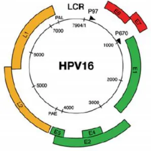
around 8000 base pairs (bp) [10, 11]. HPV genome is divided into three main regions: the long control region (LCR), that covers about 10% of the genome, the region of the early genes, over 50% of the genome, and the region of the late genes, almost 40% of the genome (figure 1) [12] . These two last regions are generally called open reading frames (ORFs) [10].
Figure 1 - Genome and structural organization of the HPV-16. The HPV-16 genome is represented as a
black circle with the early (p97) and late (p670) promoters marked by arrows. The early genes (E1, E2, E4 and E5) are represented in green and the early genes (E6 and E7) in red. The late genes (L1 and L2) are represented in yellow. The LCR is presented between yellow and red regions [6].
The LCR is a segment of about 850 bp next to the origin of viral replication. This part of the genome does not encode any protein, but despite that it is also relevant since it has several binding sites for a lot of different transcriptional repressors and activators, including the activator protein 1 (AP1), the keratinocytic-specific transcription factor 1 (KRF l), nuclear factor (NF-I/CTF) and some viral transcriptional factor that are encoded by the early region. For this reason, the LCR regulates the transcription of the early and late regions, hence it controls the expression of viral proteins and infectious particles. The host range of specific HPV types is quite determined by LCR, since it has the capacity for binding so many transcription factors [11].
The segments of the genome that actually encode proteins are called ORFs. The late gene region has two of this ORF and encodes for two proteins: the L1 protein and the L2 protein. These two proteins are the structural components that form the viral capsid and are only expressed in productive infected cells [10, 13]. The L1 protein is the major viral capsid protein and is highly conserved through the different Papillomavirus species. In turn, the L2 protein is the minor viral capsid protein and has much more sequence variation amongst HPV types than the L1 protein [11]. Their expression is tightly regulated and linked to the differentiation of infected epithelial cells [4].
The early gene region encodes for viral replication and cellular transformation and it consists of six ORFs: the E1, E2, E4, E5, E6 and E7 (table 1) [10, 12]. Depending on the HPV type, the E4, E5 and E7 genes usually encode for a single polypeptide, while the E1, E2 and E6 genes can suffer different splicing and so be expressed as several related polyproteins [14].
The E1 gene is expressed in 68 and 27 kDa polypeptides. The 68 kDa protein has adenosine triphosphatase (ATPase) and helicase activities and it can bind to specific sequences within the LCR so the DNA replication starts [14]. The HPV E2 gene encodes for two proteins that, along with E1, are necessary for extrachromosomal DNA replication [11]. The E2 proteins have from 370 to 430 amino acids in length and DNA binding domains, that can function as transcriptional activators or repressors – they are the major regulator in virus transcription and genome replication [14].
The HPV E4 protein seems to have an important role on the maturation, replication of the virus and the release on the HPV particles and, like the L1 and L2 capsid proteins, it is only expressed in later stages of the infection, at the assembly of the complete virions [11, 14]. Apparently, this protein does not transform the cells, but it can associate with cellular membranes and accumulate in the cytoplasm, inducing the collapse of the cytoplasmic cytokeratin network, in human keratinocytes, promoting the necessary conditions to the release of the virions [15].
The HPV E5 is a small polypeptide with highly conserved 44-80 amino acids [14]. Usually, the E5 gene is not expressed in cervical carcinoma cells, suggesting that is not essential in the malignant transformation of the host cell and thus its exact role in human cancers is yet to be known. Despite this, it is already established that E5 interacts with cell membrane receptors, like epidermal growth factor, platelet-derived growth factor β and colony stimulating factor, stimulating the cell proliferation of HPV infected cells [11, 15].
Lastly, the E6 and E7 genes express two oncoproteins indispensable for the viral replication and the host cell immortalization and transformation [15]. These are pleiotropic proteins, since they can make transmembrane signaling, regulate cell cycle, immortalize primary cell line and regulate chromosomal stability [16].
These two proteins are considered tumorigenic, because they have the capacity to bind to some tumor suppressor proteins, like p53 and retinoblastoma protein (pRb), consequently preventing the HPV infected cell’s apoptosis and enhancing their malignant conversion [13]. The function of E6 and E7 will be detailed in next chapters.
Table 1 - A summary of the HPV ORFs (adapted from [14]).
Viral protein Function
L1 Major capsid protein L2 Minor capsid protein E1 Viral DNA synthesis E2 Transcription regulation
E4 Disrupts cytokeratins, late protein E5 Interacts with growth factor receptors
E6 Transforming protein; binds and initiates p53 degradation E7 Major transforming protein; binds pRb, p103 and p107
1.1.1.1 E6 oncoprotein
The E6 protein has approximately 150 amino acid residues and 18 kDa and it can be found in the nuclear matrix and other nonnuclear membranes [17, 18]. E6 and E7 proteins bind to zinc ion through the coordination of cysteine residues [19]. The E6 contains four Cys-X-X-Cys motifs that form two zinc-binding domains, joined by an interdomain linker of 36 amino acids [19, 20]. E3 ubiquitin ligase, also known as E6 associated protein (E6AP), forms a complex with both E6 and some target proteins. The motif through which there is a binding between E6, E6AP and the target proteins is referred to as LXXLL motif and is conserved throughout the E6 proteins of numerous Papillomaviruses. Another motif that all high risk E6 proteins have is referred to as XT/SXV and it is responsible for the binding to specific domains on cellular proteins that are known as PDZ proteins. Some other proteins, namely p53, Bak and procaspase 8, do not have the LXXLL nor the PDZ domain, nevertheless they bind to E6 protein, possibly through other yet undefined motifs or indirectly through binding to E6AP or other E6 associated proteins. As mentioned above, the E6 protein can interact with a wide number of target proteins, but the most well studied E6-protein interaction is with the p53 tumor suppressor protein [19].
p53 is a nuclear protein that functions as a transcriptional factor and regulates the transcription of various downstream target genes, which controls cell cycle arrest, apoptosis, DNA repair, senescence and metabolism [21]. The increase of p53 is triggered by cellular damage and it activates pathways for DNA repairs, cell arrest and/or apoptosis [19]. When there is damage in the DNA, p53 induces cell growth arrest in the G1 phase of the cell cycle, so it can be repaired. This is important for the genomic integrity maintenance [11].
Therefore, by targeting p53 for degradation and so interfering with its biological function, E6 promotes cell transformation and proliferation. This degradation occurs via the formation of the complex that includes p53, E6 and E6AP, causing the transfer of ubiquitin peptides to p53, which marks it for degradation by a proteasome. Low risk HPV E6 cannot target p53 for degradation by the proteasome as it happens with the high risk HPV E6. In this way, the E6 disturbs the control of the cell cycle progression, leading to an increase of tumor cell growth at the end (figure 2) [16].
Figure 2 - Representation of the E6 protein-p53 tumor suppressor protein interaction. The E6 binds
to E6-AP and to p53, which gets marked for ubiquitination mediated by the E6-AP and then suffers proteasomal degradation [16].
1.1.1.2 E7 oncoprotein
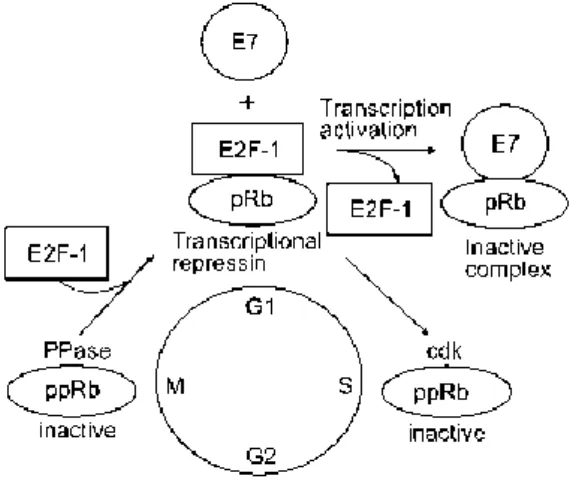
The E7 protein has approximately 100 amino acids and 25 kDa and its found mostly in the nucleus, but also in the soluble cytoplasmic fraction and nucleolus [22, 23]. There are three domains in the E7 protein called conserved regions (CR) – (CR1, 2 and 3) [24]. Two of these regions, the CR1 and CR2, share homology with SV40 T antigen and adenovirus E1A [25]. This homology is also conserved amongst different HPV E7 proteins and are separated by a non-conserved sequence of variable size and amino acid composition [22]. Both CR1 and CR2 are necessary for cellular transformation [26]. CR1 can stimulate the cellular transformation in a pRb-binding independent way, whereas CR2 associates with pRb through the conserved LXCXE (Leu-X-Cys-X-Glu) motif [25]. The HPV E7 protein is capable of associating with a group of proteins known as pocket protein family, such as the pRb and its related proteins p107 and p130, which act as negative regulators of cell growth, including in the G0/G1, G1/S and G2/M transitions [16, 18].
In a normal biological situation, when the pRb is hypophosphorylated, it binds to transcription factors of the E2F family. As these transcriptional factors are responsible for regulating the
expression of cellular genes that are involved in the DNA synthesis, like the DNA polymerase, by blocking its functions, the hypophosphorylated pRb negatively controls the progression of the cell cycle. As mentioned earlier, the E7 is able to form complexes with the pRb and its related proteins, preventing them to bind to the E2F that is now free to activate and stimulate the expression of several host genes necessary for DNA replication, allowing the cell to proliferate [11, 18]. It has been shown that the ability to disrupt the E2F-pRb complex is bigger for the high risk HPV E7 than it is for the low risk HPV E7 (figure 3) [25].
Figure 3 – Representation of the E7 protein-pRb protein. In physiologic conditions, the
hypophosphorylated pRb form complexes with E2F, negatively regulating the progression of the cell cycle. When the infection occurs, the E7 binds to the hypophosphorylated pRb, which inhibits its binding to the E2F that stays free to stimulate transcription of DNA synthesis genes, allowing the cell cycle to progress [16].
1.1.2 Preventive Vaccines
It has been reported that HPVs are associated with approximately 99% of cervical cancers, particularly HPV type 16. Thus, HPV is a potential target for development of vaccines, being necessary a basic understanding of HPV biology [4]. There are two types of vaccines that can be used in cervical cancer or other HPV-associated malignancies. The first strategy is to prevent infection with preventive vaccines that are based on HPV virus-like particles containing HPV structural proteins and can generate neutralizing antibodies to block HPV infection. The second one is to eliminate HPV infection by inducing a virus-specific T cell-mediated response by the use of therapeutic vaccines [4, 10].
At present, there are two preventive vaccines (bivalent and quadrivalent) against HPV infection, also called prophylactic vaccines, both approved by the Food and Drug Administration (FDA) [3]. The quadrivalent one is called Gardasil®, developed by Merck (NJ, USA), and is expressed in yeast Saccharomyces cerevisiae [27]. Gardasil® acts successfully against infection by four of the most clinically relevant HPV types: the low risk HPV-6 and 11 and the high risk HPV-16 and 18 [10, 28]. The bivalent vaccine is called Cervarix®, developed
by GlaxoSmith-Kline (GlaxoSmith-Kline Biologicals, Rixensart, Belgium), and is expressed in an insect cell system [27]. This vaccine protects against HPV-16 and 18 and also does partial cross-protection against HPV-31 and 45, which are phylogenetically related to the two previous ones [28].
Both Gardasil® and Cervarix® contain a non-infectious recombinant L1 virus-like protein with the aim to generate neutralizing antibodies against major capsid protein, L1 [4]. They are highly immunogenic, have high avidity for the systemic antibody response and are capable of producing memory of B cell response, which enables the cell to make a rapid burst of antibodies upon a secondary exposure [10, 29]. Despite the L1 is not expressed in the basal cells infected with HPV, it still is deeply studied and targeted for preventive vaccines [4]. However, preventive vaccines are limited in their action to few types of HPVs, they do not have therapeutic effects against pre-existing HPV infections nor HPV-associated lesions and the vaccination program has relatively high cost [4, 30].
1.1.3 Therapeutic vaccines
As the name implies, preventive vaccines only have a preventive effect and can only be applied before the infection, unlike the therapeutic ones that can eliminate pre-existing lesions and even malignant tumors. To do that, it is important to select the ideal target antigen. As described above, HPV early proteins are expressed throughout the virus life cycle and help regulate progression of the disease. In particular, the HPV E6 and E7 proteins are of great interest for potential targets as they are essential to induce and maintain the cellular transformation and malignancy [31].
Nevertheless, before using E6 and E7 in DNA vaccines for human application and regarding safety, there is the need to eradicate their oncogenic potential. There are two ways to accomplish this: the first and most used one is to introduce point mutations that have been reported to prevent interaction of E6 with p53 and E7 with pRb. The second method is called ‘gene-shuffling’ and involves the rearrangement of the primary gene sequences, so the ligand binding domains are disrupted. To be sure that no loss of possible T-cell epitopes is caused, the original sequence junctions that are destroyed are added as an appendix [32].
1.2 DNA technology
In the last decades, the knowledge about genes and their function augmented significantly, allowing the discovery of recombinant DNA technology and gene cloning, in the 80s, and the increase in genomics data, in the 90s [33]. The decoding of the entire human genome has provided the knowledge to define some disease-causing genetic factors and the association between DNA (genes) and proteins have generated a fullness of potential therapeutic opportunities based on engineered genes and cells [33, 34].
Despite the evolution on the Biotechnology field, a great number of diseases are yet to be conquered, with millions of people dying each year due to the inefficiency of the current therapeutic methods [35]. To overcome the demands of present and emerging public health problems, some innovative therapeutic strategies are being developed, like gene therapy and DNA vaccines, that seem to be really promising [36].
1.2.1 Gene therapy
Gene therapy is the transfer of genetic material (DNA) to cells that have defective or mutant genes, creating a therapeutic effect, by either assisting or replacing the genetic defects or by overexpressing proteins that are therapeutically useful [33, 36]. By the definition of United States FDA, gene therapy is the product “that mediate their effects by transcription and/or translation of transferred genetic material and/or by integrating into the host genome and that are administered as nucleic acids, viruses, or genetically engineered microorganisms” [37]. Therefore, gene therapy uses genes as a medicine to cure, or at least to improve the clinical status of a patient, a broad spectrum of serious acquired and inherited diseases, namely cancer, acquired immunodeficiency syndrome (AIDS), cardiovascular diseases, infectious diseases and other [38, 39]. However, this is a complex process, since there is the need to ensure the arrival of the transgene into the nucleus without suffering any degradation. To overcome this obstacles, namely the degradation and the passing through the plasma membrane to the nucleus, it is necessary to use a gene delivery system [39]. This topic will be discussed later.
Gene therapy is generally classified into two categories according to the nature of the targeted cell: germ line gene therapy and somatic gene therapy. In the first one, the functional gene is inserted in the reproductive cells, like sperm or zygote, and thus it will be integrated into the individual genome and the modification might pass along to the next generation. In the second one, the transgene is inserted in the somatic cells (non-reproductive cells), narrowing the effects and modifications to the specific individual, not passing to the next generation. So far, the legislation only allows the use of somatic gene therapy, due to ethical reasons [37, 39]. The somatic gene therapy may also be divided into two different approaches: ex vivo, where the cells are removed from the patient’s body, genetically manipulated and then returned to the patient’s body, and in vivo, where the cells manipulation occurs in the patient’s body. Both are under investigation and their great goal is to successfully deliver therapeutic genetic material to the target cells [40].
It is important to refer that up until now cancer composes over 60% of all ongoing clinical trials on gene therapy, followed by monogenetic and cardiovascular disease, being by far the most common disease treated by gene therapy [37].
1.2.2 DNA vaccines
Looking back to the past century, it can be said that the development and widespread use of vaccines against a large number of infectious agents have been a great triumph of medical science. It all started over 200 years ago when Jenner succeeded to show that prior exposure to cowpox could prevent infection by smallpox, emerging the concept of vaccination [41]. Despite this progress, as mentioned above, some diseases still cause death to millions of people. Hence, the world urgently requires new technologies able to respond quicker and that are able to be developed faster for new vaccines. An opportunity to answer this matter relies on DNA vaccines [42]. The DNA vaccination is a recent therapeutic strategy that is based on the use of a vector that encodes one or more antigens corresponding to the protein(s) of interest under a promoter, capable of function in the transferred cells [43]. Compared to the conventional vaccines, DNA vaccines have some advantages that are worth to considered, as briefly presented in table 2. A big difference between them is that the gene-based vaccines can generate both humoral and cellular immune responses [44].
Table 2 - Advantages of DNA vaccination (adapted from [45]).
Advantages of DNA vaccines comparing with conventional vaccines
Design DNA vector optimization through codon and ribonucleic acid (RNA) structure changes
Can generate effective cytotoxic T lymphocyte and antibody responses Can be engineered to express tumor antigenic peptides or proteins
Enables prolonged expression of antigens and enhancement of immunologic memory Safety Unable to revert into virulent forms, unlike live vector–based vaccines
Capacity for repeated administration safely and effectively No significant adverse events in any clinical trial
Stability Temperature-stable Long shelf life
Manufacture Suitable for large scale production at high purity Rapid production and formulation
Easy to store and transport
The DNA vaccination is capable of inducing the adaptive immunity, while producing antibodies and activating helper T cells and cytotoxic T cells, and even the innate immunity [46]. Upon DNA vaccine transfection, the host cell transcribes, translates and expresses the viral antigen [47]. When professional antigen presenting cells (APCs) encounter an exogenous
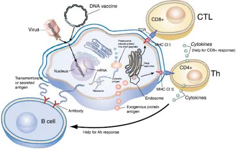
and foreign antigen, they take it up into their endolysosomal pathway. The protein is then processed and degraded to peptide fragments that are loaded and presented on the cell surface by the major histocompatibility complex (MHC) class II [35, 48]. This antigen peptide-MHC class II complex is recognized by specific helper T cells, the CD4+ T cells that can produce cytokines that will help in other cell activities. For example, they can help B cells generate effective antibody responses and/or help cytolytic T lymphocyte responses, depending on the cytokine [35, 41]. This via is known as humoral response (figure 4). Considering the antibody responses, B cells recognize and respond to extracellular antigens or exposed extracellularly antigens that belong to transmembrane proteins [35].
On the other hand, there is the cellular response, where the foreign protein can be intracytoplasmic and so it is processed by the proteosome into peptide fragments. The peptide fragments associate to MHC class I molecules, transported to the cell membrane and then are presented on the APC surface. This peptide-MHC class I complex is recognized by cytolytic T lymphocytes, the CD8+ T cells, that become activated also by the action of co-stimulatory molecules (figure 4) [35, 48].
Figure 4 - Representation of the mechanisms of both humoral and cellular immune responses. DNA
vaccination aims these two responses. If the foreign antigen is exogenous, it will be taken up by professional APCs into its endolysosomal pathway. The protein is degraded to peptide fragments, which are presented by the MHC-II and recognized by CD4+ T cells. These are activated to produce cytokines that help B cells to become activated and produce antibodies (humoral response) and also help cytolytic T cells response. The antigen can likewise be intracytoplasmic, being degraded by the proteosome into fragment peptides that are presented by the MHC-I to the CD8+ T cell, leading to its activation (cellular response) [35].
1.2.3 DNA delivery systems
The delivery of naked DNA to target cells for therapeutic purposes has its drawbacks, as it is susceptible of degradation by endonucleases, its crossing through the cell membranes is
limited by its net negative surface charge and large hydrodynamic diameter and if it is endocytosed it may be degraded by the endolysosome. To ensure that none of these occurs, it is imperative to use a DNA delivery system [49]. The ideal DNA delivery system, also known as vector, must not trigger a strong immune response, it must be capable of transporting the DNA independently of its size and deliver the transgene to target cells, it must be episomal or integrate into a specific genome region without randomly integration and it must be easily prepared, not expensive and available at high concentrations commercially [39]. Currently, there are two types of available vehicles for gene delivery: the viral and nonviral vectors [50].
1.2.3.1 Viral vectors
Viruses have a number of biological properties that made them one of the first choices for gene delivery vehicles: they can recognize and enter cells, specifically penetrate into the host cell nucleus and then take advantage of the cellular machinery and express its own genetic material and replicate it in the host cell and spread to other cells [39, 51]. Before using a virus as a gene transfer vector, it must be modified by genetic engineering, in order to reduce patho/immunogenicity. To accomplish this, the dispensable and pathogenic genes are removed and replaced by the gene(s) of interest. On the other hand, the viral genes that are necessary for the assembly of viral particles, the packing of the viral genome into particles and the therapeutic gene delivered to the target cells still remain in the vector construction [39, 50]. The main viruses used on gene delivery are adenovirus, adeno-associated virus, lentivirus, retrovirus and also herpes simplex virus. These viral vectors are the most used system to transfer genes, because they have high transfection efficiency, but they also have some downsides, as briefly presented in table 3 [39].
Table 3 - Advantages and disadvantages of the main viral vectors [34].
Viral vector Advantages Disadvantages
Adenovirus High transfection efficiency Transfects proliferating and non-proliferating cells
Substantial clinical experience
Strong immune response Insert size limit of 7.5 kbp
Difficult to manufacture and quality control Poor storage characteristics
Short duration of expression Retrovirus High transfection efficiency
Fairly prolonged expression Low immunogenicity
Substantial clinical experience
Low transfection efficiency in-vivo Insert size limit of 8 kbp ex-vivo Transfects only proliferating cells Difficult manufacture and quality control Safety concerns (mutagenesis)
Lentivirus Transfects proliferating and non-cells
Transfects haematopoietic stem cells
Very difficult manufacture and quality control
Poor storage characteristics Insert size limit of 8 kbp No clinical experience
Safety concerns (origins in HIV)
Adeno-associated virus Efficient transfection of wide variety of cell types in-vivo Prolonged expression
Low immunogenicity
Difficult manufacture and quality control Insert size limit of 4.5 kbp
Safety concerns (mutagenesis) Limited clinical experience
1.2.3.2 Nonviral vectors
As presented in table 3, the viral vectors have disadvantages that have to be seriously considered, like the capacity to cause several immune responses. This has led to the need of finding safer alternatives and nonviral vector delivery systems are emerging as a favorable solution to overcome some of the viral vector drawbacks [52]. Comparing to viral vectors, the nonviral vectors are relatively safe, have low immunogenicity and less toxicity, have easy formulation and assembly and can be prepared in large quantities at low cost. Furthermore, they are capable of transferring different and larger therapeutic genes, with no limit on size,
and because of their stability they can be stored for long periods [34, 39]. Unfortunately, their use in large amounts is limited by their low transfection efficiency [39].
There are two categories when considering nonviral DNA delivery systems: the physical and the chemical one. The physical approach is applied when the DNA delivery into the target cells is made by the use of physical forces that weakens the cell membrane, causing it to be temporarily permeable, which facilitates the diffusion of the transgene. This process is not mediated by a carrier. The physical methods include needle injection, electroporation, gene gun, ultrasound and hydrodynamic injection. The chemical approach occurs when the DNA is delivered into the target cell nucleus by a carrier that can be prepared by several types of chemical reactions [39, 53]. Within this group, the most studied strategy so far has been the formulation of DNA into condensed particles by using cationic lipids or cationic polymers. Hence, these particles suffer cell endocytosis, macropinocytosis or phagocytosis as intracellular vehicles, from which a small part of the DNA is released into the cytoplasm and migrates into the nucleus, where the therapeutic gene is expressed [53]. Our research group has been conducting several studies with nanoparticles, including chitosan nanoparticles for the delivery of p53 sc pDNA [54]. The subject-matter of nanoparticles will be discussed later.
1.3 Plasmid DNA
Within the huge variety of vectors for gene delivery, there is the pDNA. Because of its safety, its easiness of production on a large-scale, its simple application and also the fact that it does not cause toxicity, the pDNA has received an increased attention and has gained a huge interest for therapeutic applications [49]. It is used to deliver the desired genetic information into the target cells and to induce the production of the relevant proteins. Consequently, in the past few years, the use of pDNA as a delivery system on approved gene-therapy protocols has increased exponentially, representing 64.4% of the gene therapy clinical trials in 2016 [55].
Plasmids are double-stranded DNA molecules that are covalently closed. Each strand is a linear polymer of deoxyribonucleotides that are linked by phosphodiester bonds, negatively charged at pH>4 [42]. The two strands wind in an anti-parallel sense around each other and around a common axis that forms the double helix structure, stabilized by hydrogen bonds and stacking forces [56]. This structure has a hydrophilic backbone, composed by sugars and phosphate groups, and a hydrophobic interior, composed by planar aromatic bases stacked on each other. pDNA can have different sizes and normally they are small (2 to 20 kbp and a molecular weight of 106 to 107 Daltons), although they are very large when compared with proteins [56, 57]. Despite this, in the future it will be needed multigene vectors, including extensive control regions, that may require the production of larger plasmids [58]. The pDNA molecule can be coiled in space, causing the formation of a higher order molecule known as sc pDNA [56]. The active sc pDNA form, also called covalently closed circular DNA (cccDNA), is
the main one, but under stress or unfavorable environment conditions, such as extreme pH or high temperature, it can generate other forms [57]. The other topological pDNA conformations can be the open circular (oc) or the linear form, caused by single-stranded and double-stranded nicks, respectively [42]. Linear and oc pDNA isoforms are formed by random nick(s) that might damage at different gene locations, such as the promoter or gene coding regions that become destroyed, and so they are inefficient to induce the expression of the therapeutic gene [57]. Therefore, the sc pDNA is the most appropriate and desired form for therapeutic applications [59].
By estimates, it is known that only one per thousand plasmid molecules presented to the cells reaches the nucleus and is expressed [60]. Thus, there is the need to improve the current strategies of sc pDNA production, so it may be of high copy number, highly pure and successfully delivered to the targets cells. Moreover, it is also important to develop adequate delivery systems that protect pDNA vector from degradation and also that facilitates the entrance and delivery to the nucleus of the higher number of pDNA.
1.3.1 pDNA manufacture
The manufacture of pDNA is divided into three different stages: upstream processing, fermentation and downstream processing (figure 5). Firstly, there is the construction and selection of an appropriate plasmid vector and production of microorganisms, followed by selection and optimization of the fermentation conditions and cell growth and then the isolation and purification steps, with the aim of producing large quantities of stable and highly purified sc pDNA [61].
Figure 5 - Representation of the three essential stages to obtain pure sc pDNA [61].
When the purpose is pDNA vaccination, the design of the pDNA vector must include some typical elements, such as an origin of replication for efficient propagation in the adequate
host cell, a selectable marker like an antibiotic resistance gene for growth selection, a strong eukaryotic promoter to drive expression, a polyadenylation signal to terminate the transcription and the transgene that encodes the antigen of interest (figure 6) [62].
Figure 6 - Representation of the construction of the plasmid DNA [62].
The typical host used in fermentation is E. coli. To obtain the pDNA, the cells are usually disrupted by alkaline lysis [56]. Unfortunately, the pDNA only represents around 3% mass/mass (w/w) of the E. coli extract [63]. To eliminate some of the impurities, it follows a primary isolation process that involves clarification and concentration steps [57]. This allows the removal of some impurities like RNA, genomic DNA (gDNA), endotoxins and proteins [63]. Lastly, a chromatographic purification step is used to separate sc pDNA from structurally related impurities, like relaxed and denatured pDNA, gDNA, low molecular weight RNA and endotoxins [61].
1.3.2 pDNA purification
With the progress in therapeutic approaches, emerges the need to develop a good pDNA purification process, in order to obtain sc pDNA homogeneity near to 100% and to follow the quality specifications recommended by the regulatory agencies, such as FDA and the European Medicines Evaluation Agency (EMEA) [56, 64]. At present, liquid chromatography is the central technology used in pDNA purification, as it is simple, robust, versatile and it has high resolution and high reproducibility [57, 65]. Despite its plusses, chromatography has a challenge when it comes to the separation of pDNA from the contaminants, because they share similar characteristics, like the negative charge (RNA, gDNA, endotoxins), similarity in size (gDNA, endotoxins) and hydrophobicity (endotoxins) [63]. Numerous chromatography processes have been developed by exploring different properties including charge, molecular size, hydrophobicity and affinity [56].
1.3.2.1 Size exclusion chromatography
Size exclusion chromatography can fractionate and purify plasmids from a clarified lysate based on the wide variety of molecular mass. The larger molecules, like pDNA and gDNA, are
incapable of penetrate the pores, eluting first, so they can be separated from the smaller ones, like RNA, endotoxins and proteins [42, 57]. But because the lysate is a complex mixture of different molecules, the resolution here is limited, as well as the isolation of sc pDNA in one single step [57].
1.3.2.2 Anion exchange chromatography
Anion exchange chromatography is based on the interaction between the negatively charge phosphate groups in the DNA backbone and the positively charged ligands on the stationary phase [56]. After the binding occurs, it is applied an increasing salt concentration to displace and elute the different nucleic acids by order of an increasing overall net charge, which is function of chain length and conformation [65]. Given that sc pDNA is more compact and has higher charge than the oc pDNA, it is possible to separate these two isoforms [42]. Nevertheless, this type of chromatography presents poor selectivity towards pDNA and impurities, like RNA, gDNA and endotoxins, due to their similar binding affinities, making the purification of pDNA insufficient [66].
1.3.2.3 Hydrophobic interaction chromatography
Hydrophobic interaction chromatography relies on the differences in the hydrophobic interactions of pDNA, single-stranded nucleic acid and endotoxins, using high salt concentration for the biomolecules retention [57]. To elute the bound species, the salt concentration of the mobile phase is decreased, weakening the hydrophobic interactions and the elution occurs by increasing the hydrophobicity order. This property is mainly defined by base composition, size and structure [56, 57]. This technique is inefficient on separating different pDNA isoforms and in addition the use of high salt concentration, which is associated with higher costs and environmental impact, is also a downside [57, 67].
1.3.2.4 Affinity chromatography
Affinity chromatography (AC) is a separation technique that exploits natural biological processes like molecular recognition for the selective purification of target biomolecules based on their biological function or chemical structure [42]. The high specificity and efficiency of affinity interactions allow this method to eliminate additional steps, to increase yields and to improve process economics [57, 66]. Nonetheless, it has some limitations, as the fragility and low binding capacity of the biological ligands. To overcome this, synthetic ligands were designed, combining the selectivity of natural ligands with the high capacity and durability of synthetic systems [57].
This purification method separates biomolecules based on reversible interactions between the target one and its specific ligand that is immobilized on the chromatographic matrix [57]. Under appropriate pH and ionic strength, the sample is injected onto the column and the target biomolecule binds to the specific ligand [68]. After, elution steps are performed, being
specific with a competitive ligand or non-specific with a change in pH, ionic strength or polarity, depending on the matrix and the characteristics of the biomolecule [57].
The interactions between target biomolecule and its ligand result of the combination of electrostatic interactions, hydrophobic interactions, van der Waals forces and/or hydrogen bonding. As these interactions are so specific, they represent a crucial advantage of AC, because they allow to obtain high selectivity and high resolution [57].
Within AC, there are several types like immobilized metal-ion, triple-helix, polymyxin B, protein-DNA and amino acid-DNA [57]. For the purpose of this project, we will only focus on the last method.
1.3.2.4.1 Amino acid-DNA AC
The use of amino acids in AC has already demonstrated to be efficient on the successful biorecognition of the sc isoform, by using a single purification step.
Amino acids have been of great use in biotechnology applications, since they are natural compounds that can be safely used in pharmaceutical applications [42, 69]. Besides, based on atomic studies, amino acids preferentially promote specific interactions with nucleic acid bases, especially the positively charged ones like histidine, lysine and arginine [69]. The use of these amino acids as ligands has allowed an efficient purification of sc pDNA and the recognition of this isoform proved the presence of specific interactions between pDNA molecule and the amino acid-based matrices [70].
In fact, our research group has already showed the successful application of some amino acids for the purification of pDNA. For instance, the use of lysine and histidine for the separation of sc pDNA from a clarified lysate sample resulted on a high purity degree of this biomolecule of interest, being in accordance with the regulatory agencies specifications [64, 66, 71]. Nonetheless, the overall recovery yield of these two strategies was low: 45% and 40%, respectively [66, 71]. On the contrary, the use of arginine on the affinity chromatography resulted on a recovery of 79% of sc pDNA and also a high purity degree, under mild elution conditions, thus representing a smaller environmental impact [72].
Taking this into account, the arginine amino acid reveals itself to be a good affinity ligand to purify the sc pDNA, due to its high selectivity for the sc pDNA recognition and thus high recovery and purity. Despite this, the conventional stationary phases have some drawbacks, like the low capacity, working at low flow rates that results in longer retention time and possible degradation of the target biomolecule [73]. Consequently, raises the need to explore alternative chromatographic supports.
1.3.3 Monoliths: innovation on chromatographic supports
As discussed above, the studies with affinity matrices have revealed positive results, however there are still some limitations to be surpassed. Monoliths have been gaining attention owing to their appealing properties. Considered the fourth generation of the chromatographic stationary phases, a monolith is a continuous and highly porous bed; the pore size is adjustable considering the desired application, depending on polymerization process [68, 70, 74]. They are polymerized into a column as a single unit and thus the scale-up and scale-down variations in packing quality and the need to repack the column because of the appearance of air bubbles are eliminated [74]. Monoliths exhibit increased permeability and interconnectivity, allowing high mass transfer and more access to binding sites for the target biomolecule, having a very high binding capacity for pDNA [68]. Additionally, this innovative support has flow independent resolution, allowing the same separation and resolution even working at high flow rates. This enables a very fast separation and reduced retention time, which ensures less biomolecule degradation [70, 73, 74].
Bearing all this positive characteristics in mind along with the amino acids’, combining a monolithic support with amino acids ligands or derivatives for the purification of sc pDNA using AC has been a promising and favorable strategy.
For instance, our research group has already demonstrated that arginine monolith allows the separation of a sc pDNA, at a laboratorial scale, with 86% of purity. This support was also characterized in terms of dynamic binding capacity (DBC), which has presented a higher value that the equivalent conventional support [75]. On the other hand, agmatine and histamine monoliths also revealed themselves to be efficient at separating sc pDNA, with 99.6% and 96.66% of purity, respectively [76, 77]. Thus, the monolithic approach allows higher selectivity for the sc pDNA and higher binding capacity.
1.4 Nanotechnology
Nanotechnology is a recent and promising field that involves multiple disciplines and within it there is the Cancer Nanotechnology, which includes the use of nanoparticles to detect and treat cancerous cells [78, 79]. As previously mentioned, estimates indicate that only one per thousand plasmid molecules presented to the cells reaches the nucleus and is expressed [60]. Thus, there is the need to create and develop strategies that help protect vectors, like pDNA, from degradation and that facilitates the entrance into the nucleus. Nanoparticles emerge as a good solution, offering many advantages over the delivery of free pDNA: they protect the therapeutic cargo against enzymatic degradation; they allow more specific targeting and delivery; by targeting to specific cells, there is an improvement of distribution and reduction of side-effects; the therapeutic cargo is more probably delivered to the desired intracellular compartment, with an improvement of cellular penetration [80–82].
![Table 1 - A summary of the HPV ORFs (adapted from [14]).](https://thumb-eu.123doks.com/thumbv2/123dok_br/18901441.935337/24.892.114.589.132.544/table-summary-hpv-orfs-adapted.webp)
![Figure 2 - Representation of the E6 protein-p53 tumor suppressor protein interaction. The E6 binds to E6-AP and to p53, which gets marked for ubiquitination mediated by the E6-AP and then suffers proteasomal degradation [16]](https://thumb-eu.123doks.com/thumbv2/123dok_br/18901441.935337/25.892.322.608.316.609/figure-representation-suppressor-interaction-ubiquitination-mediated-proteasomal-degradation.webp)
![Table 3 - Advantages and disadvantages of the main viral vectors [34].](https://thumb-eu.123doks.com/thumbv2/123dok_br/18901441.935337/32.892.102.753.119.982/table-advantages-disadvantages-main-viral-vectors.webp)
![Figure 5 - Representation of the three essential stages to obtain pure sc pDNA [61].](https://thumb-eu.123doks.com/thumbv2/123dok_br/18901441.935337/34.892.121.719.728.1024/figure-representation-essential-stages-obtain-pure-sc-pdna.webp)
![Figure 6 - Representation of the construction of the plasmid DNA [62].](https://thumb-eu.123doks.com/thumbv2/123dok_br/18901441.935337/35.892.260.661.203.411/figure-representation-construction-plasmid-dna.webp)
![Figure 8 – Several examples of nanoparticles with diferent materials, sizes and structures [92]](https://thumb-eu.123doks.com/thumbv2/123dok_br/18901441.935337/40.892.246.598.793.1097/figure-examples-nanoparticles-diferent-materials-sizes-structures.webp)

