Ciências da Saúde
Pilot-Model for oxidative post-competition
recovery in swimmers
Luís Daniel Machado Crisóstomo
Dissertação para obtenção do Grau de Mestre em
Ciências Biomédicas
(2º ciclo de estudos)
Orientador: Prof. Doutor Luiza Breitenfeld Granadeiro
Co-orientador: Prof. Doutor Aldo Costa
Pilot-model for oxidative post-competition recovery in swimmers | Luís Crisóstomo
III
“Para ser grande, sê inteiro: nada Teu exagera ou exclui. Sê todo em cada coisa. Põe quanto és No mínimo que fazes. Assim em cada lago a lua toda Brilha, porque alta vive.”
Pilot-model for oxidative post-competition recovery in swimmers | Luís Crisóstomo
V
Aknowledgments
After a year totally dedicated to the research and the writing of this thesis, there are plenty people I would like to thank, and my most honest fear is that I can be unfair and forgot someone who helped me in this great accomplishment.
First I want to thank my project coordinators, Prof. Dr.ª Luiza Granadeiro and Prof. Dr. Aldo Costa, for all their work, support, patience and knowledge, which without it this project would never be possible. I also thank them for everything they taught to me along this year, from such different areas as Sport Science and Genetics, or training methods and laboratory practices. I hope our work encourage future MSc Students to link different areas of knowledge on their thesis, since the whole Human knowledge isn’t locked in a specific Science, it is the sum of the knowledge of us all.
In troubled times like this, in a troubled country, I have to thank all my funders, everyone that contributed to this huge economic investment that is attending the University, especially when we are through such a serious economic crisis. In this point my main gratefulness goes to my family, who underwent great sacrifices to provide me the opportunity to nourish my potential. Also, they were the ones who taught me the most, the ones who seed my curiosity and my thirsty for knowledge, along with a huge subject spectrum, even from their humble beginnings.
I have also to thank the Portuguese Government and SASUBI (Social Action Services of University of Beira Interior) for their social funding which they granted to me since my first year at University. It obliged me to a bureaucratic race almost every one of the 5 years spent to complete my Licenciature and Master Degrees on Biomedical Sciences, but without it, I would never be able to ingress University, even with the biggest efforts from me and my family.
I also want to thank Dr.ª Ana Cristina Ramalhinho for introducing me at the lab procedures when I was still “lost” in the whole-new environment of a Research Centre, and to Dr. Paulo Saldanha who taught me the basics of the RT-PCR technique. A word of appreciation to all the researchers who share the lab with me, for all the support for my never-ending inquisitions about the lab organization, simple tasks and their useful advices.
At last, but not least, a very hearty acknowledgement to all my friends, classmates or colleagues who I have met during this 5-year long journey. We helped each other when we were in need, we lived the most daring and amazing stories, we shared our knowledge and beliefs, and with all of this, we grew so much stronger. They are also a part of this thesis, because anyone you met leaves a part of itself on you. Thus, I’m a puzzle composed of all those uniquely shaped pieces, and that is my real dimension.
Pilot-model for oxidative post-competition recovery in swimmers | Luís Crisóstomo
VII
Resumo alargado
O treino desportivo com o objetivo de performance competitiva coloca os atletas sob um forte risco de desequilíbrio oxidativo, conhecido por stress oxidativo. A produção de radicais livres e espécies electrofílicas, como as Espécies Reativas de Oxigénio (ROS), são uma constante no metabolismo normal do organismo, no entanto, a maior taxa metabólica exigida pela demanda energética do exercício físico intenso, provocam uma produção de tais espécies a um nível superior às defesas antioxidantes disponíveis. Nesta situação de stress oxidativo, os radicais livres e ROS provocam danos a fulcrais estruturas e macromoléculas celulares, reagindo forte e rapidamente com estas, ameaçando a homeostasia celular.
Para controlar a ação nefasta dessas agressões oxidativas, os organismos possuem mecanismos de defesas antioxidantes, podendo estas ser de origem endógena ou exógena. Entre as defesas antioxidantes endógenas encontram-se proteínas expressas pelas células, e cuja expressão pode ser influenciada pelo ambiente oxidativo celular, como é o caso das Glutationa S-Transferases (GST). Desta forma, situações que criem stress oxidativo, como no treino desportivo, ativam a expressão das defesas antioxidantes.
Assim sendo, o treino desportivo regular e bem planeado, de forma a evitar danos constantes ao organismo, deve ativar uma resposta deste de forma a protege-lo dessa agressão, preparando-o previamente para essa agressão. Essa preparação pode ser verificada através da expressão génica de fatores antioxidantes endógenos. Além disso, certos genótipos podem revelar-se vantajosos nesta proteção, nomeadamente os genótipos associados às várias isoformas das GSTs. Nestes, constam vários e frequentes genótipos Null (ausência do gene), o que permite uma grande variabilidade entre indivíduos para a disponibilidade de isoformas de GSTs.
O objetivo deste trabalho foi precisamente verificar a distribuição de genótipos Null/Present para duas isoformas de GSTs, a GSTM1 e a GSTT1, numa amostra de 20 nadadores portugueses de nível nacional. Para comparação de genótipos, foi recolhida semelhante informação a partir de um grupo de controlo constituído por 52 indivíduos aleatórios. Além disso, observou-se a expressão relativa de GSTT1 ao longo de 5 momentos distintos ao longo da época de Inverno (preparação geral, preparação específica, fase taper e dois momentos pós-competição) em 3 desses atletas, e a expressão relativa, também de GSTT1, 48h e 72h após uma competição, para 8 desses atletas.
Para conseguir alcançar isto, foi necessário montar uma técnica totalmente nova para recolher as amostras de forma rápida, fiável e praticável nas condições de treino, e otimizar todos os procedimentos laboratoriais para conseguir processar essas amostras de forma eficiente e rigorosa. As amostras foram recolhidas em papel de filtro de análises clínica, através de uma picada no dedo dos nadadores, antes do início do treino do dia definido previamente para recolha de amostras. As amostras foram ainda conservadas em invólucros
Pilot-model for oxidative post-competition recovery in swimmers | Luís Crisóstomo
VIII
individuais para cada recolha a cada momento e de cada atleta, numa câmara-fria 4°C, no Centro de Investigação em Ciências da Saúde (CICS) da Faculdade de Ciências da Saúde (FCS) da Universidade da Beira Interior (UBI).
Para genotipagem dos nadadores em amostra, DNA foi extraído da amostra de sangue em papel utilizando o método do Chelex 100. Após extração, o DNA foi usado para amplificação enzimática da sequência específica dos genes da GSTM1 e GSTT1, pela técnica de PCR. Por fim, os resultados foram corridos por electroforese em gel de agarose, usando Green-safe como fator de marcação de DNA, e os resultados foram visualizados à luz ultravioleta num transiluminador. A presença de GSTM1 foi identificada pela presença de uma banda com cerca de 215bp, enquanto a presença de GSTT1 foi identificada pela presença de banda aos 473bp.
Para análise da expressão génica, RNA foi isolado a partir das amostras de sangue em papel, pelo método do Trizol. O RNA era correspondente a cada um dos momentos de recolha. De seguida o RNA foi convertido a cDNA através da técnica de transcriptase reversa, utilizando a enzima M-MLV. Por fim, o cDNA foi amplificado pela técnica de RT-PCR, para o gene GSTT1, tendo ainda como controlo a amplificação da β-Actin, também para cada um dos momentos de recolha e fazendo duplicados por uma questão de rigor. A expressão foi calculada através das curvas de amplificação de RT-PCR e utilizando o método ΔΔCT.
Não foram encontradas distribuições de genótipos GSTM1 e GSTT1 Null/Present estatisticamente significativas entre a nossa amostra de teste e o grupo de controlo. No contexto da expressão relativa de GSTT1, verificou-se que variações muito acentuadas ao longo da época desportiva ou após um exercício foram prejudiciais à performance física dos nadadores. Encontramos também algumas diferenças na recuperação das nadadoras, mantendo uma expressão mais alta e por um maior período de tempo após o exercício físico intenso que os homens. Além disso, verificou-se uma tendência para os indivíduos GSTM1 Null manterem os níveis de expressão relativa de GSTT1, ao longo da época e após um exercício intenso, mais estáveis, o que parece favorecer o seu rendimento. Conclui-se ainda que a análise da evolução da expressão relativa de GSTT1 em vários treinos, após uma competição ou outro exercício de elevada intensidade, pode ajudar a perceber qual a forma atual de um nadador.
Palavras-chave
Glutationa S-Transferases, Espécies Reactivas de Oxigénio, treino desportivo, expressão génica.
Pilot-model for oxidative post-competition recovery in swimmers | Luís Crisóstomo
X
Abstract
Physical exercise have several health benefits, but it can also be a source of cellular damage. The energetic demands of physical exercise and training promote an increase on metabolic rate, and its pathways may produce secondary harmful compounds that will cause cellular damage. Some of those compounds are the free radicals and Reactive Oxygen species, which are highly instable molecules that react quickly, oxidizing important functional molecules such as proteins, membrane lipids and DNA, in a condition known as oxidative stress. To dampen the action of these molecules, the cells express antioxidant defence proteins. One of the most ubiquitous and polymorphic of those is the family of Gluthatione S-Transferases (GSTs). The great physical load of competitive training creates serious oxidative stress on athletes so, it is expected that their expression of GSTs will vary throughout the season to overcome such aggression, quickly recovering from one training session and preparing the antioxidant defence for the next one.
Our main objective was to verify if the expression of a GST (GSTT1) varies throughout the season, as expected theoretically, and how it fluctuates after a competition. We also check if the distribution of the GSTM1 and GSTT1 Null/Present genotypes had some influence in the preparation and performance of our sample, consisting in 20 national level swimmers. A control group of 52 random individuals was also used to compare genotype distribution.
We collected blood samples in analytic filter paper, at 5 different moments throughout the winter season. DNA was isolated from a sample of each individual, amplified by PCR for our interest genes, and ran in agarose gel by electrophoresis to genotype our 20 swimmers. RNA was isolated from all the samples of a swimmer and converted in cDNA by reverse transcriptase. The relative expression of GSTT1 was done using β-actin (a housekeeping gene) as a control gene and the first collected sample of the swimmer as control condition, by the RT-PCR technic. Three swimmers were accessed for the whole 5 moments, while eight were only evaluated their expression at 48h and 72h after competition.
The results showed little influence in the distribution of genotype from swimmers to controls. The expression results show influence of the GSTT1 expression profile throughout the season and after an intense exercise with sport performance and as a fitness check tool.
Keywords
Pilot-model for oxidative post-competition recovery in swimmers | Luís Crisóstomo
XII
Index
1. Introduction 1
1.1. GSTs 1
1.1.1. Physiologic function and distribution 1
1.1.2. Acting mechanisms 2
1.1.3. GST classification 9
1.1.4. Structure and dimerization 11
1.1.5. Chromosomic location 12
1.1.6. Nrf2 and GST 14
1.2. Oxidative stress and physical exercise 16
1.2.1. Oxidative stress 16
1.2.2. Oxidative damage linked to physical exercise 18
1.2.3. Oxidative stress and GST 20
2. Objectives 21
2.1. Main objective 21
2.2. Secondary objectives 21
3. Materials and Methods 22
3.1. Subjects 22
3.2. Study design 22
3.3. Data collection and storage 24
3.4. Sample genotyping 24
3.4.1. DNA extraction 25
3.4.2. Polymerase Chain Reaction (PCR) 25
3.4.3. Electrophoresis 27
3.5. Gene expression 28
3.5.1. RNA extraction 28
3.5.2. cDNA synthesis 29
XIII
3.5.4. Relative expression calculus 33
3.6. Statistical analysis 34 3.6.1. Genotyping 34 3.6.2. Relative Expression 35 4. Results 36 4.1. Genotyping 36 4.2. Relative expression 38 5. Discussion 44 5.1. Genotype determination 44 5.2. Gene Expression 44 6. Conclusion 47 7. Bibliography 48 8. Annexes 56 8.1. Authorization Chart 56
Pilot-model for oxidative post-competition recovery in swimmers | Luís Crisóstomo
XV
Figure list
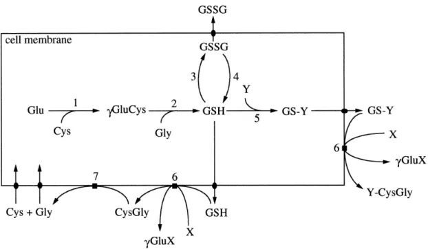
Figure 1: Pathways of GSH metabolism. Y represents a GSH substrate, while X represents a γ-Glutamil moiety acceptor. Enzymes involved: 1) γ-Glutamylcysteine Synthetase; 2) Glutathione Synthetase; 3) Glutathione Peroxidases; 4) Glutathione Reductase; 5) Glutathione S-Transferases; 6) γ-Glutamil Transferase; 7) Ectopeptidases. Adapted from (8). 3
Figure 2: Simplified Mercapturic Acid Pathway. The GSH conjugation whit a xenobiotic is catalyzed by a GST, resulting a Glutathione S-Conjugate. Adapted from (27). 4
Figure 3: Aromatic Amino Acids Catabolism. GSTZ1-1 has Maleylacetoacetate Isomerase activity, catalyst of the penultimate reaction in this catabolic pathway. 6
Figure 4: Synthesis of Steroid Hormones. GSTA3-3 acts as a highly efficient keto-steroid isomerase and only exists at steroid producing tissues. 7
Figure 5: Regulation of Prostaglandins signalling by action of GSTs, and the transcription factors that may be activated or inhibited by them. Adapted from article (4). 9
Figure 6: Ball-and-socket Interactions between GST monomers. Red and blue regions are the ball and the socket, respectively. Adapted from article (2). 12
Figure 7: Schematic representation of the Nrf2-Keap1 gene expression regulation. The Keap1 complex, due to its E3 ubiquitin ligase activity, constantly bind and marks Nrf2 for degradation. When electrophile substance are present, due to oxidative stress, keap1 cysteine residues get alquilated, releasing Nrf2 that can migrate to the nucleous, where it recognizes the ARE element of cytoprotective genes and promotes their expression. Adapted
from article (40). 15
Figure 8: The electron transport chain, in the inner membrane of mitochondrion. Electrons are transferred through proteins by a series of oxireductions, with reduction potential progressively higher. The electron energy differential through the chain is used to pump electrons to the inner mitochondrial membrane, by complexes I, III and IV, creating a chemiosmotic gradient. After complex IV, electrons are captured by oxygen, which reacts with hydrogen to form water. This whole transport, very well balanced can, nevertheless, let 0.4% to 4% of electrons escape and form ROS. Adapted from (47). 17
Figure 9: Catabolism of purines. This metabolic pathway creates ROS by the Xanthine oxidase activity (Bold compounds have recognized electrophilic activity). Damage caused to DNA will
Pilot-model for oxidative post-competition recovery in swimmers | Luís Crisóstomo
XVI
trigger this catabolism, in order to eliminate the damaged purine bases, but the action of xanthine oxidase results in peroxide, contributing to oxidative stress. 19
Figure 10: Well displacement on the RT-PCR plate. The wells represented as green squares were relative to the target gene, GSTT1, while the blue-coloured squares were relative to the housekeeping gene, β-actin. The wells with an inner rotated square were the Negative Controls, in which cDNA had been replaced by water. The numbering represents the sample collection order (1 for the first sample moment, and so on). This plate was repeated for each
individual and for each target gene. 31
Figure 11: Optimization of annealing temperature and amplification cycles for GSTT1. Sample 1 and sample 2 correspond to two different individuals whose RNA was previously extracted and converted in cDNA. Annealing temperature was set to 58.0° C and were done 60 amplification cycles. NTC-1 is the Non-template control. In spite of its amplification, it occurs
later than our samples. 32
Figure 12: Melting curves for GSTT1 and β-Actin genes. Each line correspond to a sample well, and all graphics are expressed as fluorescence units by °C. Correct amplification at a sample was considered when its melt peak was between the 80°C and 90°C. a) Dissociation lines for GSTT1 wells; b) Melting curve peaks for GSTT1; c) Dissociation lines for β-Actin wells; d)
Melting curve peaks for β-Actin. 33
Figure 13: Gel images captured with UV light, for genotype analysis. On left side, an example of a null and a present genotype for GSTM1. On the right side, two examples for present GSTT1 genotype. In this case, bands migrated further than expect, so their length doesn’t match with the DNA ladder. That situation is likely due to partial DNA degradation by
haemoglobin contamination. 36
Figure 14: GSTT1 expression throughout the Winter season, for three different swimmers. Units are expressed in expression fold, comparing to the housekeeping-gene, β-actin. Baseline is the moment 1, Specific is the moment 2, Taper is the moment 3, and 48h and 72h are the fourth and fifth moments, relative to the post-competition recovery. 39
Figure 15: GSTT1 expression tendency after a competition, for eight different swimmers. Units are expressed in expression fold, comparing to the housekeeping-gene, β-actin. Our baseline is the first collected sample, then 48h and 72h are the samples collected at that
time after the competition. 40
Figure 16: GSTT1 expression lines after the division by 2 groups, according to their evolution at the first 48h. Lines in blue correspond to male swimmers, and those in rose to female swimmers. a) swimmers who dropped their GSTT1 expression after the first episode; b) swimmers who had increased expression after the same episode. 40
XVII
Figure 17: GSTT1 expression lines after the division by 3 groups, according to their expression evolution at the 48h-72h period. Lines in blue correspond to male swimmers, and those in rose to female swimmers. a) Swimmers who greatly increased GSTT1 expression after the second episode; b) Swimmers with moderate expression increase; c) Swimmers who dropped or maintained their GSTT1 relative expression at this interval. 41
Figure 18: Expression tendency lines for our sample's swimmers, evidencing the groups divided by number of physical episodes. The orange lines are for swimmers with only one episode (the training), while black lines stand for swimmers who had two episodes (the competition 48h
before the training). 42
Figure 19: Gene expression comparison between the one episode group and the two episodes group, in matter of gender influence. The rose lines mark female swimmers, while the blue lines correspond to male swimmers. a) Swimmers submitted to 2 episodes (competition + training); b) Swimmers submitted to only one episode (training). 42
Figure 20: Comparison of the GSTT1 expression in the two episode group versus the single episode group, for the influence of the GSTM1 genotype. GSTT1 genotype has not included because it not a differentiation factor, as all our swimmers were GSTT1 Present. Blue lines represent GSTM1 Null individuals and magenta lines are GSTM1 Present. a) Swimmers submitted to 2 episodes (competition + training); b) Swimmers submitted to only one episode
Pilot-model for oxidative post-competition recovery in swimmers | Luís Crisóstomo
XIX
Table list
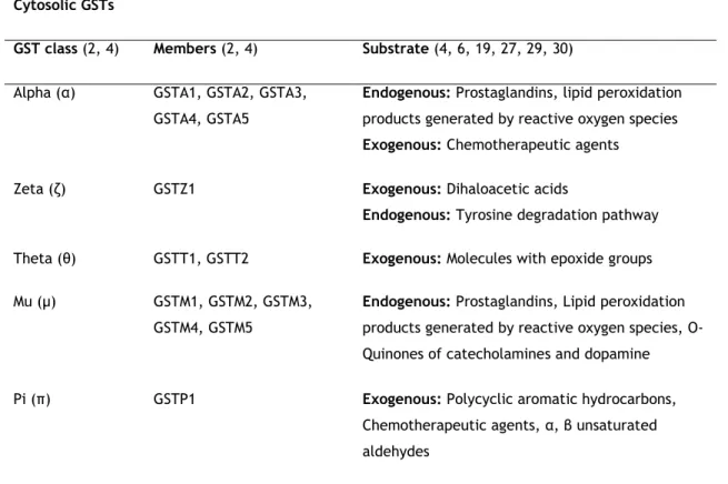
Table 1: Families and classes of GSTs, their members and substrate. 10
Table 2: Cytosolic GSTs’ genes, chromosomic location, alleles, mutations and their effects. 13
Table 3: Training characteristics. WV (weekly volume); A0 (slow/regenerative swimming speed); A1 (aerobic, low intensity swim); A2 (swim speed at lactic anaerobic threshold); A3 (pace above the lactic anaerobic threshold); A4 (maximal aerobic potency pace); LT (Lactic Tolerance); LP (Lactic Potency); PR (Potency Resistance). 23
Table 4: Reagents per PCR reaction tube, for acessing GSTM1 and GSTT1 null/present
genotypes in swimmers. 26
Table 5: GSTM1 null/present genotype distribution comparison between swimmers and
controls. 37
Table 6: GSTT1 null/present genotype distribution comparison between swimmers and controls. † P-value calculated by Fisher’s Exact Test, since one class count less than 5 cases.
37
Table 7: Combination of GSTM1 and GSTT1 null/present genotypes distribution between
swimmers and controls. 37
Table 8: Combination of GSTM1 and GSTT1 null/present genotypes distribution between within swimmers, according to their distance speciality. (1)SDS vs MDS; (2)SDS vs Controls;
(3)MDS vs Controls 37
Table 9: Combination of GSTM1 and GSTT1 null/present genotypes distribution between swimmers and controls according to their gender. (1)Male vs Male; (2)Female vs Female; (3)Male
vs Controls; (4)Female vs Controls. 38
Table 10: Individual characteristics for the 8 swimmers eligible for GSTT1 relative expression analysis. All of them compared on the 5 sample collect moments. The nr of races and best position are related to the winter short course National championships. *Number of events (number of eliminatories); #Classifications achieved in all events, from the best to the worst.
Pilot-model for oxidative post-competition recovery in swimmers | Luís Crisóstomo
XXI
Abbreviation list
15d-PGJ2 15-deoxy-Δ12, 14-prostaglandin J 2 AA Amino Acid Ala AlanineAMV Avian Myeloblastosis Virus ARE Antioxidant Response Element ATP Adenosine Triphosphate
bp Base Pair(s)
cDNA Complementary DNA
CICS Health Sciences Research Centre CnC Cap’n’Collar C T Cycle threshold Cul3 Cullin 3 Cys Cysteine DDCT d-Dopachrome Tautomerase DHA Dehydroascorbic Acid DNA Deoxyribonucleic Acid DTT Dithiothreitol
FCS Faculty of Health Sciences
Glu Glutamate
Gly Glycine
GSH Glutathione
GSSG Oxidized Glutathione GST Glutathione S-Transferase HWE Hardy-Weinberg Equilibrium
Ile Isoleucine
JNK c-Jun NH2-terminal kinase
kb Kilobase
Keap1 Kelch-like ECH Associated Protein 1 LP Lactic Potency
LT Lactic Tolerance LTC4 Leukotriene C4
MAPEG Membrane-Associated Proteins of Eicosanoids and Glutathione metabolism MDS Middle-Distance Swimmer
M-MLV Moloney-Murine Leukaemia Virus MRP Multidrug Resistance Protein
NFE2L2 Nuclear factor erythroid-2-related-factor 2 gene Nrf2 Nuclear factor erythroid-2-related-factor 2 PCR Polymerase Chain Reaction
PGX
y Prostaglandin Xy (Xy represent the different isoforms)
PR Potency Resistance PR Potency Resistance Prx VI 1-cys Peroxiredoxin Rbx1 Ring-box 1
Pilot-model for oxidative post-competition recovery in swimmers | Luís Crisóstomo
XXII
ROS Reactive Oxygen Species RT Reverse Transcriptase
RT-PCR Real Time Polymerase Chain Reaction SDS Short-Distance Swimmer
SNP Single Nucleotide Polymorphism UBI University of Beira Interior
Val Valine
1
1.
Introduction
The physical exercise practice naturally obliges to an oxygen consumption rate increase, comparing to the basal state, so the body can obtain the energy required to perform the necessary motor and sensorial actions. However, energy attainment, by complete lipid and carbohydrate metabolization at mitochondrial cristae, involves the electron chain transporter, a process that may cause Reactive Oxygen Species (ROS), since oxygen is the final electron receptor. ROS are very reactive chemical species that strongly and quickly react, for instance, with lipids and nucleic acids, oxidizing them. Thus, physical exercise potentiates ROS production, arising oxidative stress at cells. In order to protect cells from such menace, there are detoxifying enzymes, such as Glutathione S-Transferases (GSTs), whose expression is mediated by several transcription factors, amongst them Nrf2. Thus, to keep organism’s homeostasis, it is expected that a competitive athlete has changes at gene expression for the transcription factors which regulate antioxidative protection, so he can be prepared against the extraordinary oxidative stress he is subjected to.
In this work, we investigate the Null/Present genotype distribution for two GSTs (GSTM1 and GSTT1) for a swimmer group and a control group, and evaluate the fluctuations of the GSTT1 relative expression along different training phases on a sports season and after a competition.
1.1.
GSTs
Glutathione S-Transferases are a multifunctional and ubiquitous dimeric phase II detoxifying enzyme superfamily (1-3). They are usually divided into 3 main families, according to their intracellular location: Cytosolic, Mitochondrial and Membrane-Associated Proteins of Eicosanoids and Glutathione metabolism (MAPEG family) (2, 4).
1.1.1.
Physiologic function and distribution
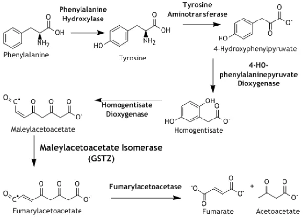
The main function of GSTs is to metabolize and detoxify chemical electrophiles, carcinogens, pollutants and products of oxidative stress (2, 4), that are responsible to damage at membrane lipids and DNA (4). However, besides cellular detoxification and antioxidant protection, GSTs take also an important role at eicosanoids, prostaglandins and steroids metabolism, tyrosine catabolism and apoptosis (2-5). Theta class enzymes have a highly
Pilot-model for oxidative post-competition recovery in swimmers | Luís Crisóstomo
2
specific sulphatase activity (2), while Zeta class has maleyloacetoacetate isomerase activity (the penultimate step at phenylalanine and tyrosine catabolism) (2, 4). The role at prostaglandins metabolism is ensured by Sigma class, which acts as a Prostaglandin D synthase, a mediator of allergic and inflammatory responses (2, 4), and by the MAPEG family, that are involved at the synthesis of leukotrienes, too (2, 4). This role at Eucasanoids and Prostaglandins has a major impact at gene expression, due the activation of divergent regulatory pathways (figure 6) (2, 4). Omega class enzymes are crucial at ascorbic acid maintenance at brain, since they show dehydroascorbate reductase activity (2, 4, 6). All classes have also a peculiar ability, called ligandin function (2). This property is the ability to bind several non-substrates, such as bilirubin, steroids and xenobiotics, often but not always at the dimer interface. Nevertheless, when a non-substrate is bound, even at non-active site, the active site is affected, and rends the enzyme either inhibited or inactive (2). At last, GSTP1 conjugates with chemotherapeutic drugs, being highly expressed at cancer cells that resist medication, but also regulate a c-Jun NH2-terminal kinase (JNK) pathway, that is
involved at apoptosis mechanisms (5). Thus, GSTP1 has both importance at preventing cancer evolution, and on cancer spreading (2, 5).
Although GSTs are ubiquitously expressed by cells, not all subclasses are expressed evenly throughout all tissues. A good example is the hepatic tissue, which expresses GSTM1 but does not GSTP1 (7).
1.1.2.
Acting mechanisms
GSTs are known to typically catalyse the conjugation of Glutathione (GSH) and toxic compounds, such as xenobiotics, electrophile substances and oxidative stress products, but they can also take part in isomerization and reduction reactions of those compounds (2). Glutathione is the most abundant tripeptide in cells, and it is constituted by glutamate (Glu), cysteine (Cys) and glycine (Gly). It is synthetized at the cytosol by action of the γGluCys ligase, resulting the dipeptide γGluCys, after combined with Gly by the glutathione synthase (figure 2) (4, 8); both enzymes require ATP (Adenosine-triphosphate) and are substrate-inhibited by high GSH concentrations (8).
3
Figure 1: Pathways of GSH metabolism. Y represents a GSH substrate, while X represents a γ-Glutamil moiety acceptor. Enzymes involved: 1) γ-Glutamylcysteine Synthetase; 2) Glutathione Synthetase; 3) Glutathione Peroxidases; 4) Glutathione Reductase; 5) Glutathione S-Transferases; 6) γ-Glutamil Transferase; 7) Ectopeptidases. Adapted from (8).
The conjugation reaction attenuates the oxidative effects that free radicals cause to cellular structures, reducing them to more stable and less reactive chemical species, more soluble and easier to excrete (2-4, 9). The typical conjugation reaction (figure 3) involves the activation of the thiol group (-SH) of cysteine to react with a xenobiotic or other compound for excretion (2). This chemical group is also known by mercaptan (from Latin, mercurium captans, meaning capturing mercury), so this reaction may be referred as Mercapturic Acid Pathway too (4). After conjugation, it follows several steps to complete substrate’s excretion (Figure 2). First, the glutamil moiety or glutathione conjugate is removed by the γ-Glutamiltransferase (γGT), remaining CysGly or CysGly conjugate (8). The conjugates are N-acetylated to further elimination by the transmembranar MRP (Multi Resistance-associated Proteins), which are members of the C family of ABC transporters (4).
Pilot-model for oxidative post-competition recovery in swimmers | Luís Crisóstomo
4
Figure 2: Simplified Mercapturic Acid Pathway. The GSH conjugation whit a xenobiotic is catalyzed by a GST, resulting a Glutathione S-Conjugate. Adapted from (27).
However, at some circumstances, the resulting products from this pathway are even more reactive than the substrate (4). Amongst the chemical functional groups that may lead to this effect are the short-chain alkyl halides that contain two functional groups (4, 10) and the 1,2-dihaloethanes (4, 11). Both chemical species result into unstable glutathione conjugates which possess an electrophilic centre capable of modifying DNA (4, 10, 11). Some compounds formed from plant glucosinolates are conjugated with GSH by GST to yield thiocarbamates, which are exported out of the cell by MRP (4). Outside the cell, they may spontaneously degrade into isothiocyanates, releasing GSH, and then be reimported to be once more conjugated with GSH (4). This creates a cycle that leads to a fatal depletion of intracellular GSH, occurring protein thiocarbomylation, a condition that leads to cell death (4). There are also cases of tissue-specific toxicity for some chemicals. For instance, haloalkanes and haloalkenes conjugation, which occurs mainly at liver, can generate reactive thiols at kidney, through the action of renal cysteine conjugate β-lyase (2, 4, 12).
The products of oxidative stress are subproducts of the partially reduced O
2, such as
the superoxide anion (Oଶି), the hydrogen peroxide (H
2O2) and the hydroxyl radical (HO•), and
they arise not only through the oxidative phosphorylation of aerobic respiration, but also through 5-lipoxygenase, cyclooxygenase, cytochrome P450 and xanthene oxidase catalysed
5
reactions (4). Even when combined with antioxidant defence proteins, the degradation of those species can result into products that are also cytotoxic and mutagenic (4, 13). GST isoenzymes are one of the enzymes that protect cells against the subproducts of the oxidative stress (4). GST’s are particularly involved in protection against the electrophiles derived from membrane lipids’ oxidation, because they conjugate GSH to several end-products of lipid peroxidation, such as 4-hydroxy-alkenals of between 6 and 15 carbon atoms in length, 2-alkenals acrolein and crotonaldehyde, phosphatidylcholine hydroperoxide, cholesteryl hydroperoxides (14) and fatty acid hydroperoxides (4). Besides, GST’s can indirectly combat those damages through regeneration of 1-cys Peroxiredoxin (Prx VI), an enzyme that reduce phospholipid hydroperoxides to their respective alcohols (4). Prx VI acts by oxidizing his Cys residue at position 47 to sulfenic acid, and later GST reactivates it through glutathionylation followed by spontaneous reduction of the mixed disulphide by GSH (4, 15). The base propenals that result from nucleotides oxidation are also detoxified by GSTs, as well as the products of catecholamine oxidation, which are harmful due to Oଶି production by redox cycling (4, 16, 17).
GSTs intervene in the degradation of aromatic amino acids (figure 4), namely the Zeta Class, which is a maleylacetoacetate isomerase (2, 4, 18, 19), as previously mentioned. Phenylalanine is degraded in a six-step reaction to fumarate and acetoacetate, that is an intermediate compound of Krebs Cycle, also known as Citric Acid Cycle. The intermediates of this reaction are Tyrosine, 4-Hydroxyphenylpyruvate, Homogentisate, Maleylacetoacetate, and Fumarylacetoacetate, thus GST class Zeta catalyses the penultimate step of this reaction (Maleylacetoacetate to Fumarylacetoacetate) (2, 4). GSTO1 and GSTO2 are also important for cells metabolism, since they help maintain the ascorbate homeostasis, due to its Dehydroascorbate reductase activity (2, 6). This discovery was surprising, because it is the only known GST-dependent reaction that GSH does not bind the G site; instead, Dehydroascorbate (DHA) binds to the G site, and GSH forms a disulfide bond with a cysteine in the active site (2, 6). This cysteine residue donates an electron to DHA that will trigger the capture of hydrogen from GSH, resulting Ascorbate (2, 6). However, class-omega isoenzymes doesn’t have the same substrate affinity, and so GSTO2 shows more affinity to DHA than GSTO1, maybe due to the size of the active-site cavity (2, 6).
Pilot-model for oxidative post-competition recovery in swimmers | Luís Crisóstomo
6
Figure 3: Aromatic Amino Acids Catabolism. GSTZ1-1 has Maleylacetoacetate Isomerase activity, catalyst of the penultimate reaction in this catabolic pathway.
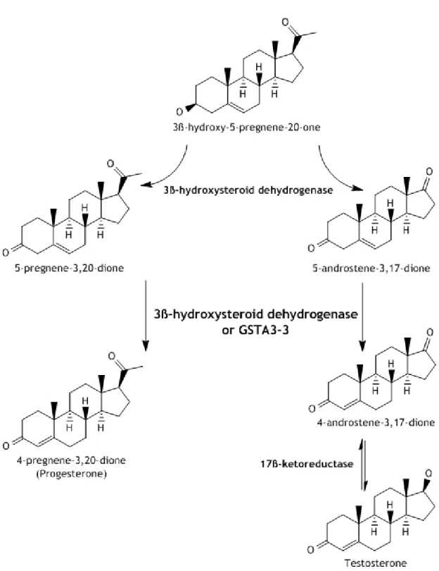
Alpha GSTs isoenzymes participate in Synthesis of Steroid Hormones. Both testosterone and progesterone are synthetized from 3β-hydroxi-5-pregnene-20-one, a cholesterol metabolite (2, 4). This compound can be converted to Δ5-androstene-3,17-dione, an
intermediate of the testosterone pathway, or Δ5-pregnene-3,20-dione, an intermediate of the
progesterone pathway, by action of 3β-hydroxysteroid dehydrogenase (2, 4, 20). Then, both these 3-keto-Δ5-steroids are converted to their 3-keto-Δ4-steroid isomers, also by action of
3β-hydroxysteroid dehydrogenase, which has found to exhibit keto-steroid isomerase activity (2, 4). However, in steroidogenic tissues, GSTA3-3 has found to have a 230-fold higher catalytic efficiency for isomerization of 3-keto- Δ5-steroids than 3β-hydroxysteroid
7
Figure 4:Synthesis of Steroid Hormones. GSTA3-3 acts as a highly efficient keto-steroid isomerase and only exists at steroid producing tissues.
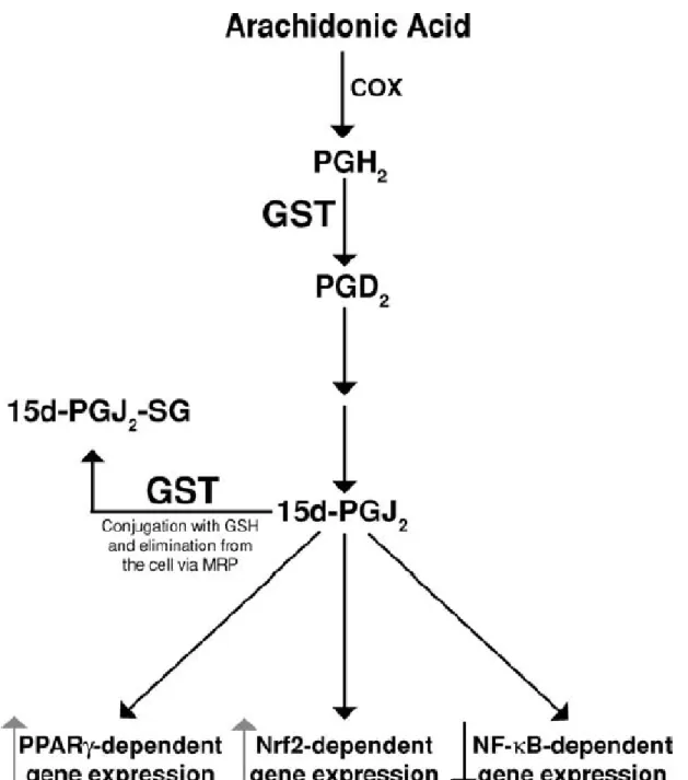
GSTs also play an important role at the synthesis and inactivation of eicosanoids, a group of metabolites derived from arachidonic acid. Many GSTs were suggested to be responsible for the isomerization of the Prostaglandin H
2 (PGH2) to Prostanglandins D2 (PGD2)
and E
2 (PGE2), or responsible for its reduction to PGH2α, but now is clear that certain GSTs are
very specific for some of those reactions (4). For instance, sigma-class GST has found to have GSH-dependent Prostaglandin D
Pilot-model for oxidative post-competition recovery in swimmers | Luís Crisóstomo
8
brain’s cytosolic GSTM homodimers (GSTM2-2 and GSTM3-3) present PGE
2 synthase activity (4,
22) and the MAPEG family enzymes show PGF
2α synthase activity (4, 23). The influence of
GSTS1 is the most interesting of the above-mentioned, since it also participates in the formation of the downstream cyclopentone prostaglandin 15-deoxy-Δ12, 14-prostaglandin J
2
(15d-PGJ2), which acts as a signalling molecule (figure 6), rather than the typical pro-inflammatory role of most prostaglandins (4). Cyclopentone prostaglandins, like 15d-PGJ
2, are
also substrates for GSH conjugation by several GSTs (GSTA1-1, GSTA2-2, GSTM1-1, GSTP1-1) (24), and thus their elimination is facilitated through Multidrug Resistance Protein (MRP) 1 and MRP3 transporters (25). Leukotrienes, another eicosanoid class, are critically influenced by GSTs, namely by the MAPEG family, in which several members intervene in the Leukotriene C4 (LTC4) synthesis (group I and group II enzymes) (4), and in the 5-lipoxygenase activation, uniquely done by FLAP and MGTS2 (4, 26).
9
Figure 5: Regulation of Prostaglandins signalling by action of GSTs, and the transcription factors that may be activated or inhibited by them. Adapted from article (4).
1.1.3.
GST classification
GSTs are classified, as above mentioned, according to their cell location. There are 3 major families: cytosolic or soluble, mitochondrial and peroxisomal, and microsomal or MAPEG (2-4).
Pilot-model for oxidative post-competition recovery in swimmers | Luís Crisóstomo
10
Cytosolic GSTs are especially polymorphic, being further divided into 7 different classes, according to their amino acid sequence, identified by Greek letters: α (alpha, five members), ζ (zeta, one member), θ (theta, two members), µ (mu, five members), π (pi, one member), σ (sigma, one member) and ω (omega, 2 members) (2-4, 27, 28). Some authors include also the class κ (kappa, one member), in spite of being a mitochondrial GST, because it is also soluble, have other similar structure and may appear on cytosol (3, 4, 29). Those classes are the only yet discovered in mammals, however there are plenty more GSTs classes for plants and even for other animal classes, such as insects (30). Classes should be designated by the name of the Greek letter (Alpha, Mu, etc.), abbreviated as Latin capital letters (A, M, and so on) (28). Class members are numbered by Arabic numerals and a GST protein dimer is distinguish by their monomers (e.g. GSTM1-M1 is a dimer of two subunits 1 in the Mu class) (28).
In spite of this huge variability, those enzymes have roughly 30% sequence homology (27); more than 40% sequence homology amongst the same class, but less than 25% between proteins from different classes (2). These similarities point that all cytosolic GSTs share a mutual precursor, from which had evolved divergently (29). Since GSTK1 share an N-terminal amino acid sequence motif solely with GST class Theta, but share the motif II with all other cytosolic GST classes, it suggests that GSTK1 is the precursor of GST class Theta, and class Theta is the precursor of classes Alpha, Mu, Pi and Sigma (29).
Table 1: Families and classes of GSTs, their members and substrate. Cytosolic GSTs
GST class (2, 4) Members (2, 4) Substrate (4, 6, 19, 27, 29, 30)
Alpha (α) GSTA1, GSTA2, GSTA3,
GSTA4, GSTA5
Endogenous: Prostaglandins, lipid peroxidation products generated by reactive oxygen species Exogenous: Chemotherapeutic agents
Zeta (ζ) GSTZ1 Exogenous: Dihaloacetic acids
Endogenous: Tyrosine degradation pathway
Theta (θ) GSTT1, GSTT2 Exogenous: Molecules with epoxide groups
Mu (µ) GSTM1, GSTM2, GSTM3,
GSTM4, GSTM5
Endogenous: Prostaglandins, Lipid peroxidation products generated by reactive oxygen species, O-Quinones of catecholamines and dopamine
Pi (π) GSTP1 Exogenous: Polycyclic aromatic hydrocarbons,
Chemotherapeutic agents, α, β unsaturated aldehydes
11
Sigma (σ) GSTS1 Endogenous: Prostaglandins, Lipid peroxidation
products generated by reactive oxygen species
Omega (ω) GSTO1, GSTO2 Endogenous: Dehydroascorbate
Mitochondrial GSTs
Kappa (κ) GSTK1 Endogenous: Lipid peroxidation products
generated by reactive oxygen species MAPEG GSTs
gp I MGST2, FLAP, LTC
4S Endogenous: Leukotriene, non-enzymatic binding
of arachidonic acid
gp II MGST3 Endogenous: Leukotriene
gp IV MGTS1, PGES1 Endogenous: Prostaglandins
1.1.4.
Structure and dimerization
Cytosolic GSTs have a well-conserved secondary and tertiary structure (2). A single unit is composed by an N-terminal α/β domain (the G site, for binding GSH), and an all-α-helical domain (the H site, for binding hydrophobic substrates) (2, 3, 31). The G site is a thioredoxin-like domain that contains four mixed β-sheet strands, whose strand 3 is antiparallel to the others; the H site consists of five to six alpha helixes (2). However, the G site only achieve its full catalytic activity once GSTs dimerize, because it locates on a cleft between both subunits (31). Some classes differ only by the number of such elements (like the extra α9 helix of classes alpha, theta and omega), others have unique features, like the C-terminal mu-loop, the N-terminal extension of classes mu and omega, and the long loop between helixes α4 and α5 of theta enzymes (2). These variations lead to a wide range of available substrates for GSTs (3).
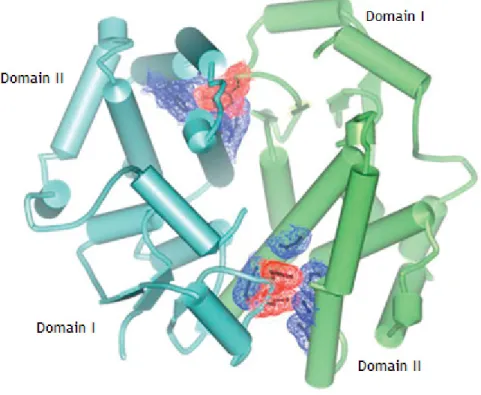
GSTs always form dimers, both homodimers or heterodimers, i.e., GST dimers may be an association between two equal GSTs subunits (GSTM1-M1, for instance), of the same class (GSTM1-M2) or from different classes (GSTM1-P1) (7), which increases complex’s protein stability and increases the range of available substrates (2, 4, 7). Recent studies point that heterodimers always are formed by same class GSTs, though. The dimer is formed by ball-and-socket or lock-and-key interactions, organized around a twofold axis, and through an extensive interface (2). These interactions occur between the domain I and the domain II of each subunit, particularly residues F52 and M51 from a loop between an α-helix and a β-sheet from a subunit interacts with an hydrophobic pocket between two α-helixes of the other
Pilot-model for oxidative post-competition recovery in swimmers | Luís Crisóstomo
12
subunit, as a typical ball-and-socket interaction (Figure 2) (2). There is also a region of hydrophilic interaction between subunits at near base of the twofold axis, involving residues of two α-helixes (2).
Figure 6: Ball-and-socket Interactions between GST monomers. Red and blue regions are the ball and the socket, respectively. Adapted from article (2).
It is not known yet how occurs the formation of GST heterodimers, but it seems to happen in vitro, under non-denaturating conditions, between GSTs classes Mu and Pi (7). Moreover, any GST class may have affinity to form heterodimers with subunits from different classes, since the amino acids residues at dimer interface are well conserved (7). In vivo, heterodimer association within different GST classes may be limited due to biological activity; for example, GSTM1-P1 heterodimers should not be possible at liver because it does express class Mu enzymes but not class Pi, but may be possible at other tissues that express both classes (7). In the breast, the predominant GST is the Pi class (32).
1.1.5.
Chromosomic location
GSTs genes can be divided into two gene superfamilies: the cytosolic or soluble enzymes genes (including the mitochondrial Kappa class) and the microsomal or MAPEG enzymes genes (1). Each class has its own gene family, and each one is located at a different chromosome (1). For soluble GSTs, alpha on chromosome 6, mu on chromosome 1, theta on
13
chromosome 22, pi on chromosome 11, zeta on chromosome 14, sigma on chromosome 4, kappa (chromosomal location not known, probably mitochondrial) and omega on chromosome 10 (1). There are polymorphisms in several of those genes, but the focus is on those which present allelism, namely the mu, theta and pi families.
The mu class gene family are on a 100kb cluster at chromosome 1p13.3, in tandem (5’-GSTM4-GSTM2-GSTM1-GSTM5-GSTM3-3’) (1, 32). There are three known polymorphisms at GSTM1, resulting into three possible alleles: GSTM1*0, GSTM1*A and GSTM1*B (1, 4). GSTM1*0 represents the deletion of the gene, and homozygotes for this allele can’t express the protein; GSTM1*A and GSTM1*B differ only by a base pair (bp) at exon 7, their monomers can form dimers it one another, and show equal catalytic efficiency (1). On the other hand, null genotype is associated with increased breast cancer risk (3, 4). GSTM3 has two different alleles, GSTM3*A and GSTM3*B, the latter having a 3 bp deletion at exon 6. This deletion creates a recognition motif for YY1 transcription factor, so the regulation of those two alleles may be different (1, 32).
GSTT1 and GSTT2 are located on chromosome 22q11.2, 50kb apart each other, and have similar structures (5 exons and same intron/exon boundaries) (1, 32). GSTT1 may be deleted, the GSTT1*0 allele, and 1 at each 5 individual amongst Caucasian population is homozygote for it, so can’t produce the encoded enzyme (1). GSTT2 gene is side-to-side to d-dopachrome tautomerase gene (DDCT), sharing a common bidirectional promoter on the sequence between the two genes and are both duplicated in an inverted repeat (1, 32). Several polymorphisms are described for GSTT2, including an amino acid substitution and a nonsense mutation, but it was not found any functional implications (1).
The single gene for class Pi (GSTP1) is at 11q13, is 2,8kb long and has 7 exons, encoding a protein of 209 amino acids (32). This gene is regulated by a CpG island at the promoter, which appear to be hypermethylated in one third of the primary breast cancer, inactivating the gene (32). There are two SNP’s (Single Nucleotide Polymorphism) at this gene that cause amino acid substitution and shift substrate specificity: one at codon 105, exon 5 (Ile/Val) and another at codon 113, exon 6 (Ala/Val) (32).
However, there are much more information about mutations and SNPs present at the several GST’s genes. Table 2 depicts a brief compilation of most information available in the scientific bibliography about these genes, their mutations, alleles and effects on proteins.
Table 2: Cytosolic GSTs’ genes, chromosomic location, alleles, mutations and their effects. GST class Genes (1, 2, 28) Chromosomic location (1, 27, 28, 33) Alleles (1, 27)
Mutation nature or effect (4)
Alpha (α) GSTA1 GSTA2 GSTA3 GSTA4 GSTA5 6p12.1 GSTA1*A GSTA1*B GSTA2*A GSTA2*B-E “Wild type”
Base substitution, low protein levels
Pilot-model for oxidative post-competition recovery in swimmers | Luís Crisóstomo
14
Base and AA substitution
Zeta (ζ) GSTZ1 14q24.3 GSTZ1*A-D Four different base and AA
substitutions Theta (θ) GSTT1 GSTT2 22q11.2 GSTT1*0 GSTT1*A GSTT2*A-B Gene deletion “Wild type”
Base and AA substitution
Mu (µ) GSTM1 GSTM2 GSTM3 GSTM4 GSTM5 1p13.3 GSTM1*0 GSTM1*1x2 GSTM1*A-B GSTM3*A GSTM3*B GSTM4*A GSTM4*B Gene deletion
Gene duplication, protein overexpression Base/AA substitution “Wild type” 3bp deletion, Protein unchanged “Wild type”
Base substitution on intron, protein unchanged
Pi (π) GSTP1 11q13 GSTP1*A-D Four different base and AA
substitutions
Sigma (σ) GSTS1 4q22.3 GSTS1*A
GSTS1*B
Inversion, “reference” protein levels
Inversion, protein unchanged Omega (ω) GSTO1 GSTO2 10q25.1 GSTO1*A/C GSTO1*B/D GSTO2*A-B Base/AA substitution
Base/AA deletion and Base/AA substitution
Base/AA substitution
1.1.6.
Nrf2 and GST
There is a positive feedback between Nrf2 and GSTs. Nrf2 recognizes genes with the ARE element in its promoter region, increasing their expression (34-36). GSTs do have such element, and so their expression may be influenced by the activation of the transcription factor Nrf2 (36). On the other hand, some cytosolic GSTs intervene in the metabolism of prostaglandins, particularly 15d-PGJ
2, a group of pro-inflammatory biomolecules that can
activate Nrf2, and thus the induction of gene expression through the antioxidant response element (ARE) (2, 4, 34).
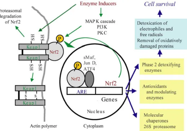
15
Nrf2 is widely expressed by all cells, however, is function is suppressed by the conjugation with Keap1 (Kelch-like ECH Associated Protein 1), with whom forms cytosolic protein complexes (34, 35, 37, 38). Keap1 is also associated to two other proteins, Cullin 3 (Cul3) and Ring-box 1 (Rbx1), that form together a complex E3 ubiquitin ligase which ubiquitinates Nrf2, to further proteasome degradation, keeping it at low concentration at cytosol (34, 38, 39). When cells are exposed to oxidative stress or electrophile substances, cysteine residues at Keap1 are alquilated, acting as sensors (38, 39). This alquilation is thought to promote structural changes on E3 ubiquitin ligase complex, inactivating it, and thus interrupting Nrf2 ubiquitination, which accumulates freely on cytosol (38-40). At last, Nrf2 migrates to the nucleus, where it dimerizes with a small Maf protein, to recognize and bind to the Antioxidant Response Element (ARE), a cis element that consist on a nucleotide sequence present at the promoter of most cytoprotective genes (41).
Figure 7: Schematic representation of the Nrf2-Keap1 gene expression regulation. The Keap1 complex, due to its E3 ubiquitin ligase activity, constantly bind and marks Nrf2 for degradation. When electrophile substance are present, due to oxidative stress, keap1 cysteine residues get alquilated, releasing Nrf2 that can migrate to the nucleous, where it recognizes the ARE element of cytoprotective genes and promotes their expression. Adapted from article (40).
The gene that encodes Nrf2 transcription factor is known as NFE2L2. It is a ubiquitously expressed gene, but its highest expression occurs, in descending order, in the kidney, muscle, lung, heart, liver and brain (42). As previously referred, Nrf2 is a transcription factor whose function is to recognize and bind the ARE element, a regulatory cis-element which exists at the promoter region of most cytotoxicity-protective and phase II detoxification enzymes
Pilot-model for oxidative post-competition recovery in swimmers | Luís Crisóstomo
16
genes. This element core sequence is 5’-TGACNNNGC-3’ (36). As for its active-form structure, Nrf2 forms protein dimers with small Maf proteins, resulting in a Cap’n’colar (CnC) leucine-zipper transcription factor (36, 41, 42).
1.2.
Oxidative stress and physical exercise
1.2.1.
Oxidative stress
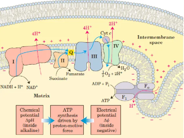
Oxidative stress is a condition that results from an unbalanced production of free radicals when compared to the body’s antioxidative capacity to dampen their harmful action (43-45). The production of free radicals is impossible to avoid, since the main metabolic pathways in humans produce such reactive compounds. The electron transport chain at the mitochondrion’s inner membrane (figure 8), a common step for both aerobic respiration and lipid oxidation, is one of the principal sources for ROS, a family of chemical compounds that cause oxidative stress. In this step, electrons are transferred throughout 4 protein complexes, which use the electromotive energy from them to pump hydrogen ions (H+) to the
mitochondrion’s intermembrane space, creating an acidic mean and a chemiosmotic gradient (46-48). Electrons can’t be left loose into cytoplasm, because they would cause damage to cellular structures, and therefore are received by oxygen atoms, the final electron acceptor, after chain’s protein Complex IV. A fifth protein complex, a transmembrane complex denominated by ATP-synthetase A, uses that chemiosmotic gradient to create ATP, using a proton-motive force to combine ADP and inorganic phosphate (47-49).
The electron transfer in this chain is only possible because each electron acceptor as a higher reduction potential than the one before (47, 48). However, some electrons can escape the chain before reaching the final acceptor, and may be conjugated by oxygen and oxygenated compounds into Reactive Oxygen Species such as hydroxide (HO⦁), peroxide (H2O2) and superoxide (O2
⦁
-) ions (50, 51-). The complex II, ubiquinone, and cytochrome C are the main sources for electron leakage that promotes ROS, and about 0.4% to 4% of all O2 used by the cell forms superoxide ions (47, 51, 52).
17
Figure 8: The electron transport chain, in the inner membrane of mitochondrion. Electrons are transferred through proteins by a series of oxireductions, with reduction potential progressively higher. The electron energy differential through the chain is used to pump electrons to the inner mitochondrial membrane, by complexes I, III and IV, creating a chemiosmotic gradient. After complex IV, electrons are captured by oxygen, which reacts with hydrogen to form water. This whole transport, very well balanced can, nevertheless, let 0.4% to 4% of electrons escape and form ROS. Adapted from (47).
ROS are highly unstable and reactive chemical species, and often are responsible for the induction of other free-radicals, through reactions triggered by their action (51). The reactivity of the free radical is due to the unpaired electron in their electronic orbit, a condition that causes instability due to charge unbalance between positive charges (protons) and negative charges (electrons). To balance this condition, radicals react quickly with other molecules so their electronic orbit become fully filled, “stealing” electrons from other molecules and causing Oxidative Damage. Rather than inducing more free radicals, these compounds several damages to cells, such as membrane lipid peroxidation, damage to nucleic acids bases and proteins, inflammatory process, which can compromise its viability (50, 51).
Detoxification enzymes are able to attenuate those damages by reacting with free radicals before they react with macromolecules, or by catalysing reactions between free radicals and antioxidative compounds (45). Drug metabolism is commonly divided into 3 phases: the phase I, modification of drugs to react with furthers cell defensive agents; phase II, conjugation of the modified drugs with cell protective molecules to dampen its action; and phase III, excretion of the drugs due to more modifications to facilitate its elimination from
Pilot-model for oxidative post-competition recovery in swimmers | Luís Crisóstomo
18
the organism. ROS and free radical are reactive for themselves, so phase II detoxification enzymes are the most important agents to dampen its effects. Therefore, GSH and GST are very important to maintain oxidative balance. GST is an enzymatic catalyst that uses GSH and a reactive compound as substrates (reactive compounds depend on GST isoform, see table 1), and the result of the reaction is oxidized glutathione (GSSG) and the reduced, now less reactive molecule. Once the reserves of GSH are depleted due to an excessive production of ROS, oxidative stress will arise (45). Hopefully, GSH is swiftly regenerated by a flavoprotein GSSG reductase, using NADPH to donate an electron to GSSG, resulting NADP+ and GSH (45).
1.2.2.
Oxidative damage linked to physical exercise
Physical exercise requires an increase in energy production by the body, through ATP synthesis and utilization. For mammals, and particularly for humans, the principal energetic pathway is the aerobic respiration, which is the main endogenous ROS source (44, 53). To face such energetic demand, the amount of respiration cycles and oxygen intake increase, thus augmenting the quantity of ROS produced (44, 50, 54), to a level that our body’s antioxidant defences can’t handle (43, 50).
Furthermore, ROS produced by the electron transport chain cause cellular damages that have cumulative effects (50), inducing further production of ROS, throughout other metabolic pathways such as the respiratory burst of lymphocytes or xanthine oxidase activity (figure 9) (44, 53, 54). Exhaustive exercise may impair the immune system (50, 55), rapidly increasing the number of circulating neutrophils but reducing the number of lymphocytes, which might be reflected in the increased risk for upper respiratory tract infections (44). Erythrocytes also contribute to the increased ROS production during exercise, because superoxide anion is produced as result of the first step of heme oxidation (44, 54). Besides, in spite of every cell is susceptible to severe impairment due to ROS (particularly due to peroxidation), erythrocytes are especially susceptible due to the lack of repair mechanisms, causing haemolysis (44). This oxidative effect may be significantly greater in runners due to the trauma-induced haemolysis at their feet, which releases free haemoglobin, highly oxidative due to the iron-containing heme group, to the blood stream (43). Oxidative stress tend to lead, ultimately, to activation of apoptosis (44), and this phenomenon is associated to the inflammatory acute phase response, which is, in its turn, linked to neutrophil increasing number (55). It is interesting to notice that, this neutrophil exercise-induced activation is coupled with a decrease in the antioxidant activity (55). Thus, we can consider that there is a positive feedback in exercise-induced oxidative stress.
Exercise, increasing metabolic rate and thus body temperature, also increases the rate of ROS produced in the mitochondrion of myocytes, as they undergo increased electron uncoupling (54). Shorter efforts, such as for short distances and anaerobic training, due to lactate production, cause blood and muscle acidosis and this is associated to higher
19
catecholamines’ levels (56, 57). In its turn, catecholamines are easily oxidized, and that reaction produces ROS (54, 56). Besides this effect, anaerobic exercise contributes to oxidative stress by purine degradation, stimulating xanthine oxidase activity (figure 9) (54, 56). The high intensity this training provoke periods of blood shunt due to the intense muscle contraction, causing brief moments of ischemia, followed by reperfusion, along with mechanical muscle damage (57, 58). Proteins are then prone to get carbonylated due to free radicals, causing structural and functional changes (58, 59). Local radical insult will cause death to some myocytes, releasing calcium to the extracellular mean, interfering with the homeostasis of this apoptotic metal (57, 58). Once more, to “clean” cell fragments is activated the inflammatory response and neutrophils will be recruited, contributing with more oxidative species due to their respiratory burst (57, 58).
Figure 9: Catabolism of purines. This metabolic pathway creates ROS by the Xanthine oxidase activity (Bold compounds have recognized electrophilic activity). Damage caused to DNA will trigger this catabolism, in order to eliminate the damaged purine bases, but the action of xanthine oxidase results in peroxide, contributing to oxidative stress.
All those damages have a functional effect. Inflammation and apoptosis are correlated to overtraining in athletes (50). Lipid peroxidation and protein carbonylation cause impairment of muscle cells and is reflected in strength and endurance decrease (58). Hopefully, physical exercise, creating this situation of oxidative stress repetitively, may promote physiological adaptions according to duration and intensity of those electrophilic assaults (43, 50). Despite all damage linked to exercise that ultimately increases oxidative stress, there are evidences that their continued assault upregulates endogenous antioxidant defences (58). Total antioxidant capacity is higher in long-term trained individuals than in a sedentary group (60), and a group of untrained individuals show less lipid oxidation after intense exercise, after entering a regular physical activity training plan (61). It seems that training promotes antioxidant adaption in a similar fashion to other principles of Sport
Pilot-model for oxidative post-competition recovery in swimmers | Luís Crisóstomo
20
training (58, 62), such as the supercompensation principle. At each training session, an athlete must be under a certain oxidative threshold, above his antioxidant capacity, to permit his body to recuperate and achieve a higher antioxidant level at the beginning of the next session (58).
1.2.3.
Oxidative stress and GST
As previously stated, in spite of all damage caused by exercise and exercise-induced ROS production, the body’s antioxidant defences are able to adapt against continuated free-radical call assault. GSTs are phase II detoxification, and are amongst the endogenous antioxidant defences of our organism. These isoenzymes have ROS as one of their substrates (4), which are also one of the reactive chemical species that contribute to oxidative stress, due to their high oxidative potential. So, the redox equilibrium within the cell depends, in a certain extent, on a balance between ROS production and GST activity (45). Besides, the carbonyls resulting from protein damage due to oxidative stress are another GST substrate (4). So, whatever is the source of the oxidative stress, either from aerobic or anaerobic exercise, GSTs may play a central role in the defence and recovery of their damage. Thus, the different functional genotypes for GSTs may be responsible for different responses to the oxidative stress (63).
21
2.
Objectives
2.1.
Main objective
The main purpose of this thesis was to verify the effect of competitive training over the expression of endogenous antioxidant genes, namely over two GST isoforms, GSTM1 and GSTT1. The results of this investigation may lead to new training approaches, helping coaches to plan the training throughout the season in a fashion that will protect their athletes from the damage suffered from the exhausting exercise, and better managing their performance peak.
This objective may be summarize as a pilot-model to evaluate consistently how the recovery of swimmers evolves, which can promote the individualization of the recovery times, according to the applied exercise charge.
2.2.
Secondary objectives
To achieve our main goal, there are several protocols that need optimization. This investigation aims to facilitate new training approaches that are reliable and practicable. Therefore, the developing and optimization of new techniques to collect samples in a minimal invasive manner, to store them without contamination nor degradation, and to extract RNA in sufficient concentration to permit further expression analysis, is probably one of the most innovative points of this work.
Additionally, data collected may be used to verify the differential genotype distribution for different groups within our sample. In spite of all our subjects are competitive swimmers, there is room for genotype distribution from male to female athletes, from short-distance swimmers (SDS) to middle-short-distance swimmers (MDS).
The same kind of comparison will be possible for the expression. Besides the global effect of training in the expression of GSTM1 and GSTT1, we are interested in knowing if different groups are differentially influenced by this stimuli.
Pilot-model for oxidative post-competition recovery in swimmers | Luís Crisóstomo
22
3.
Materials and Methods
3.1.
Subjects
30 Portuguese swimmers of national level volunteered to serve as subjects (13 males and 7 females). It was defined that national level swimmers are (20 ± 3.25 years old; 1.77 ± 0.05 m of height; 72.93 ± 6.34 kg of body mass; 23.19 ± 1.80 kg.m-2 of body mass index) the ones with regular presence in the national championships.
For statistical analysis, we further divided swimmers into smaller groups. We divided the swimmers their distance speciality, not considering their swimming style. We considered that all swimmers specialized in distances of 200m and under were Short-Distance Swimmers (SDS), and all swimmers specialized on distances ranging from 400m to 1500m were Middle-Distance Swimmers (MDS). This division was done because the ROS produced by short-distance training are not exactly equal to those arising from Middle-distance training. The higher intensity, muscular potency and anaerobic demand required by short efforts are linked to a prevalence from ROS derived from purine catabolism, catecholamine oxidation and mechanical muscle damage. On the other hand, the less intense, more oxidative and aerobic demand that is required for middle distances is associated mainly to ROS production from electron leakage, inflammation and haemolysis. However, all those exercise-induced ROS production occurs in both.
A control group was designated in order to evaluate the genetic drift in national level swimmers, for GSTM1 and GSTT1 genotypes. This group aimed to represent the general population, and consisted in 52 individuals, from whom 38 were men and 14 were women, from all ages and ethnies.
Samples were collected at 5 different moments, at different training phases throughout a sport season. Every sample was collected before the training session of that day. More detailed information about sample collection and the exact dates and training phases is provided in section 3.2.
3.2.
Study design
Samples were collected at 5 different occasions during the 2012-2013 calendar, throughout the winter season (short pool). The first 3 samples are correspondent to 3 different training macrocycles, and the last two samples were collected in the week after the most important competition of this season. At every sample collection moment, the blood