UNIVERSIDADE DA BEIRA INTERIOR
FACULDADE DE CIÊNCIAS DA SAÚDE
Master Degree Thesis in Biomedical
Sciences
DEVELOPMENT OF A NEW DRUG
DELIVERY SYSTEM FOR FUTURE
APPLICATION IN CANCER THERAPY
Vítor Manuel Abreu Gaspar
DESENVOLVIMENTO DE UM NOVO
SISTEMA DE ENTREGA DE DNA
PLASMÍDICO PARA FUTURA APLICAÇÃO
NA TERAPIA DO CANCRO
DEVELOPMENT OF A NEW DRUG
DELIVERY SYSTEM FOR FUTURE
APPLICATION IN CANCER THERAPY
Supervisors:
Professor Dr. Ilídio Joaquim Sobreira Correia
Professor Dr. Fani Pereira de Sousa
The content of the present work is of the exclusive responsibility of the author:
_______________________________________
“Failure is the preamble to success. Most first efforts don’t work. If
you persist, you’ll eventually figure it out”
-Thomas Fogarty
i
Acknowledgments
First and foremost, I would like to thank my supervisors Professor Fani de Sousa and Professor Ilídio Correia for their invaluable support during my master studies, for their continuous guidance and help, for the immense expertise and constructive discussions that were crucial for the success of this work. It has been a privilege.
I would also like to acknowledge the Dean of the Universidade da Beira Interior Professor João António de Sampaio Rodrigues Queiroz for making possible the development of this investigation project. For that I am most grateful.
I also thank Eng. Ana Paula from the Optics department of Universidade da Beira Interior for all her help in getting the scanning electron microscopy images of the nanoparticles.
Moreover, I would like to express my gratitude to all the persons involved in the Centro de Investigação em Ciências da Saúde of University of Beira Interior with a special acknowledge to Dr. Angela Sousa and Dr. Ana Martinho and the persons in the Biotechnology research group for all their help.
I thank my family for all their love, support and care in the most difficult times during my academic formation.
Finally, a very special thanks to my girlfriend for all her love and support throughout all the days of intensive work. For her advices and most specially for supporting our dreams.
ii
RESUMO
Durante as últimas décadas a terapia génica tornou-se uma alternativa promissora no tratamento de muitas doenças incuráveis, como é o caso cancro. Esta doença extremamente complexa possui características que fazem com que os tratamentos geralmente administrados sejam ineficazes. O trabalho de investigação apresentado nesta tese pretende tirar partido das novas abordagens terapêuticas baseadas no DNA plasmídico e em nanossistemas. A conjugação destas tecnologias pode conter a chave para o desenvolvimento de pacientes com cancro. Tendo por base este pressuposto, um vector de expressão plasmídico que codifica para uma proteína supressora de tumores, a p53 foi produzido em organismos recombinantes. Subsequentemente, as diferentes isoformas do plasmídeo foram isoladas e a isoforma superenrolada, a que possui actividade biológica e com melhor eficiência de transfecção, foi purificada por cromatografia de afinidade. Após esta purificação e recolha promoveu-se o desenvolvimento de novos nanossistemas que fossem capazes de entregar o DNA exógeno nas células cancerígenas. Os nanossistemas produzidos com quitosano apresentam gamas de tamanho pequenas e propriedades adequadas para a encapsulação do DNA plasmídico. Adicionalmente, a transfecção de células eucarióticas neoplásicas, revelou a potencialidade da aplicação destes nanossistemas como novos veículos de transporte de vectores que expressem a p53 até aos tumores.
iii
ABSTRACT
During the past few decades gene therapy has become a promising alternative for the treatment of many incurable diseases such as cancer. This extremely complex disease possesses characteristics that make most of the generally used treatments rather ineffective. The research work presented in this thesis attempts to take advantage of novel non-viral gene therapeutic approaches based on plasmidic DNA and nanoparticulated systems that might provide the foundation for the development of a novel therapeutic treatment from the production to a real application in the everyday life of cancer patients. Hence the production of an expression vector that encodes for a tumor suppressor, p53 was promoted in recombinant organisms. Subsequently the different plasmid DNA isoforms were isolated and the supercoiled isoform, the one biologically active and with enhanced transfection efficiency was purified by affinity chromatography. Following this purification and recovery the development of novel nanoparticle systems that could deliver the exogenous DNA into the malignant cells was promoted. Nanoparticulated systems produced with chitosan demonstrated small size ranges and suitable properties for the encapsulation of plasmidic DNA. Additionally, transfection of eukaryotic neoplastic cells revealed the suitability of the nanocarrier as a novel delivery system of p53 expression vectors to cancer.
iv
LIST OF ABREVIATURES
AEX - Anion exchange chromatography Arg-G - Arginine-guanine
CS - Chitosan
CSCs - Cancer stem cells
CS-TPP – chitosan-TPP blank nanospheres DD - Deacetilation degree
DMF - Dimethyl- formamide dsDNA - Double strand DNA
E.coli - Escherichia coli
EE - Encapsulation efficiency
EMEA - European Agency for the Evaluation of Medical Products EPR - Enhanced permeability retention
FBS - Fetal Bovine serum
FDA - Food and Drug Administration FITC - Fluorescein isothiociantate isomer I H-bond - Hydrogen bond
HIC - Hydrophobic interaction chromatography LC - Loading capacity
pDNA - plasmid DNA PEG - Polyethilene glycol PEI - Polyethylenimine
PLGA - poly (lactid-co-glycolic acid) RES - Reticuloendothelial system Sc - Supercoiled
SEC - Size exclusion chromatography TPP - Pentassodium tripolyphosphate XRD - X-ray diffraction
v
LIST OF FIGURES
Section I - Introduction
Page
Chatper I
Figure 1.Cancer evolutive model. ... 6
Figure 2. Tumor progression based on the CSC model. ... 7
Figure 3. Hallmarks of cancer. ... 9
Figure 4. Apoptotic pathways. ... 10
Figure 5. Structure of the p53 protein and three dimensional structure of the DNA binding domain. ... 11
Figure 6. A multitude of stress signals can activate the tumor suppressor p53. ... 12
Figure 7. p53 mediation of tumor suppression. ... 13
Chatper II
Figure 8. Stages from production to application of pDNA in gene therapy based treatments. ... 17Figure 9. Interaction between the affinity ligand and the target biomolecules.. ... 19
Figure 10. Nanocarrier targeting to tumors cells. ... 21
Figure 11. Different barriers impacting gene delivery. ... 22
Figure 12. DNA unpacking. ... 23
Figure 13. Types of nanocarrier systems for gene delivery. ... 25
Figure. 14 Chitosan chemical structure. ... 26
Figure 15. Different chitosan DNA nanoparticles. ... 26
Section II – Materials and Methods
Figure 1. Plasmid pcDNA3-FLAG-p53 backbone... 30Figure 2. Schematics of the amino-acid based matrix. ... 31
Figure 3. 1DUVS N-acetyl-D-glucosamine spectra and reference curve. ... 34
Section III – Results and Discussion
Figure 1. Growth profile of E.coli DH5α harboring the plasmid pcDNA3-FLAG-p53. ... 40Figure 2. Electrophoresis of E.coli cell lisates. ... 41
Figure 3. Amino acid base pair interactions. ... 44
Figure 4. Chromatographic profile showing the purification of different plasmid isoforms by arginine affinity chromatography. ... 45
Figure 5. Geometric dynamic fluctuations of the plasmid DNA supercoiling. ... 46
Figure 6. Chromatogram depicting the elution of sc pDNA from the arginine affinity support. ... 47
vi
Figure 7. Effect of temperature on the retention of the different plasmid
isoforms in arginine. ... 48 Figure. 8. Phase diagram of nanoparticle formation for commercial chitosan. ... 50 Figure 9. Morphology of the nanoparticles obtained from
commercial chitosan flakes visualized by SEM. ... 52 Figure 10. Morphology of the nanoparticles obtained from deacetilated
chitosan visualized by SEM. ... 53 Figure 11. Morphology of the nanoparticles obtained from acetilated
chitosan visualized by SEM. ... 54 Figure 12. XRD spectra of different chitosan samples. ... 56 Figure 13. Nanocapsules obtained from commercial chitosan flakes
visualized by scanning SEM. ... 58 Figure. 14. Nanocapsules synthesized from deacetilated chitosan
visualized by SEM. ... 58 Figure. 15. Nanocapsules synthesized from acetilated chitosan
visualized by SEM. ... 59 Figure 16. Agarose gel electrophoresis of the nanocapsules
following incubation with DNAse I. ... 62 Figure 17. Agarose gel electrophoresis of the nanocapsule
protection of pDNA following incubation with serum-supplemented
Ham‘s F12K. ... 63 Figure 18. Agarose gel electrophoresis of the nanocapsule
protection of pDNA following incubation with lyzosyme for 1h and 3h... 64 Figure 19. Agarose gel electrophoresis of the nanocapsule
protection of pDNA following incubation with lyzosyme for 12h and 24h... 64
Figure 20. Immunofluorescence of A549 Lipofectamine2000 – pDNA
complexes transfected cells. ... 65 Figure 21. Immunofluorescence of A549 nanocapsule transfected cells. ... 66
vii
LIST OF TABLES
Section I - Introduction
Page
Chatper I
Table 1. Normal stem cell properties and cancer stem cells. ... 6
Chatper II
Table 2. Summary of the most important formulation
parameters in the design of nanoparticulated systems based on chitosan. ... 27
Section III – Results and Discussion
Table 1. Summary of the retention/elution profiles for the different
plasmid isoforms. ... 42 Table 2. Nanosphere size distribution. ... 55 Table 3 Degree of Deacetilation of the different chitosan samples
as measured by 1DUVS. ... 56 Table 4. Comparison between the different particle sizes obtained
for variable formulations of CS-TPP-pDNA nanocapsules. ... 59 Table 5. Encapsulation efficiency of the nanocapsules obtained
from different chitosan materials. ... 60 Table 6. Loading capacity of the nanocapsules obtained from
Page
Acknowledgments
iAbstract
iiResumo
iiiList of Figures
ivList of Tables
vList of Abbreviations
viSection I
Introduction 1Chapter I – Cancer and the tumor suppressor p53 4
1.1. Cancer - Development models and hallmarks 5
1.2. The Tumor Supressor p53 11
Chapter II – Non-viral cancer gene therapy 15
2.1. Plasmid DNA expression vectors for non-viral gene therapy 17
2.2. Amino acid-DNA affinity chromatography for sc pDNA purification 18
2.3. Non-viral nanocarrier mediated gene delivery 20
2.4. Non-viral polymeric nanoparticles for gene delivery 24
Section II
Materials and Methods 29
2.1. Materials 30
2.1.1. Plasmid DNA 30
2.1.2. Chitosan 30
2.2. Methods 31
2.2.1. Bacterial growth conditions and plasmid recovery 31
2.2.2. Affinity chromatography 31
2.2.3. Agarose gel electrophoresis 32
2.2.4. Synthesis of deacetilated and reacetilated chitosan 32
2.2.5. First derivative ultraviolet spectrophotometry 33
2.2.6. X-ray diffraction (XRD) 35
2.2.7. Production of chitosan nanoparticles 35
2.2.8. Particle morphology 35
2.2.9. Encapsulation and loading capacity of pDNA 36
2.2.10. Protection and release of encapsulated pDNA 36
2.2.11. FITC fluorescent labeling of pDNA 37
2.2.12. Cell culture and in vitro transfection 37
2.2.13. Immunofluorescence 38
2.2.14. Statistical analysis 38
Section III
Results and discussion 29
3.2. Purification of sc pDNA by arginine – agarose affinity
chromatography 42
3.3. Formulation of chitosan-TPP nanoparticles 49
3.4. Morphology and characterization of CS-TPP blank nanoparticles 52
3.5. Synthesis of pDNA loaded nanoparticles - Nanocapsules 57
3.6. Nanocapsule encapsulation efficiency and loading capacity 60
3.7. Protection and in vitro release of pDNA 62
3.8. In vitro transfection 65
Section IV
Conclusions and future perspectives 68
Section V
Bibliography 71
Section VI
SECTION I
2
Cancer is a major health issue of our time, it is estimated that approximately nine million new cases appear each year (Pelengaris and Khan, 2006) and the World Health Organization predicts that by 2030 twelve million of all deaths worldwide will occur due to cancer (Bode and Dong, 2009). The invasive and aggressive profile that characterizes this illness renders most of the conventional treatments such as surgery and radiotherapy ineffective in many cases. Thereof, the development of new cancer therapeutic strategies is an urgent requirement.
Gene therapy arises as one of the most powerful and promising alternatives. In fact the possibility of treating cancer by introducing genetic information that can inhibit or eradicate cancer has led to an ever growing interest in the scientific community (Cao et al., 2009).
The revolutionary concept of gene therapy appeared in 1963, when Joshua Lederberg commented the future of medicine, hypothesizing that the ultimate application of molecular biology would be to direct the control of nucleotide sequences in human chromosomes, coupled with selection and integration of the desired genes (Lederberg, 1963). In 1967, Edward Tatum expanded the idea by being optimistic about the possibility of a therapy based on the introduction of new genes into defective cells (Tatum, 1967). In fact, in 1980 Tatum and Lederberg‘s idea was devised when Anderson accomplished the first transfer of two functional genes into mammalian cells (Anderson et al., 1980).
Later in 1991 and with the development of viral gene delivery systems, gene therapy for cancer became a reality and the first clinical trial using a retroviral carrier for the treatment of cancer was carried out (Huber et al., 1991). Despite that, few years later the inherent risk of using these delivery systems was highlighted when it culminated with the death of one patient (Christof von Kalle et al., 2003).
Meanwhile non-viral cancer gene therapy has surpassed viral based therapies due to its improved safety features (Glover et al., 2005). Plasmid DNA (pDNA) expression vectors are essential to non-viral gene therapy and thus there is an increasing demand for the production of highly stable and purified pDNA (Gill et al., 2009).
Nowadays with the recent advent of nanomedicine and the continuous development of biotechnology new cancer gene therapy approaches are being developed in order to enhance production, purification and especially delivery, revolutionizing the way that genetic material is transported towards and into the cell (Qiao et al., 2010). Nanocarrier systems possess unique physical and biological characteristics that renders them ideal vehicles for gene delivery (Cao et al., 2009). Recently, Chen et al., 2010 demonstrated that nanoparticles conjugated with tumor targeting sequences that can successfully reach cancer cells and deliver gene silencing material that can inhibit tumor growth
3
(Chen et al., 2010). In addition to these findings the use of tumor suppressor genes that induce cell cycle arrest or trigger apoptosis of malignant cells is also becoming a very attractive alternative and recently raised new prospects in the field of cancer gene therapy (Tu et al., 2010).
Taking this into account this MsC work attempts to integrate several key factors of the development of a non-viral delivery system from production of pDNA to its application. The specific aims of this work include:
- Biosynthesis in recombinant organisms of a plasmid expression vector containing a sequence of the tumor suppressor p53;
- Implementation of an affinity purification strategy based on a chromatographic support with immobilized arginine amino acids;
- Development and characterization of nanoparticulated systems based on a biocompatible polymer;
- Evaluation of the carrier for the encapsulation potential, protection and release of biomolecules;
- Evaluate the feasibility of the developed nanocarrier systems as vehicles for gene delivery into malignant cells.
The hypotheses used in this work comprise:
- Escherichia coli recombinant organisms are adequate for production of plasmid expression vectors;
- Affinity chromatography based on immobilized amino acids is a technique that can purify the plasmid biologically active isoform;
- Biocompatible biopolymers with suitable properties can be used for the development of nanoparticulated systems that can incorporate and release biomolecules like pDNA;
- Nanoparticles based on chitosan can transpose the multitude of biological barriers and enter into the intracellular compartment of neoplastic cells.
Hence the work described in this thesis may be divided in three main stages: i.) The first comprises the production of the vector and the implementation of an affinity purification strategy with immobilized arginine amino acids to purify the supercoiled pDNA isoform (sc); ii.) The second stage consists of the development and characterization of the nanocarrier system, iii.) The final research topic integrates the former stages in order to promote the delivery of p53 coding sc pDNA to carcinogenic eukaryotic cells in vitro.
CHAPTER I
5
1.1. Cancer - Development Models and Hallmarks
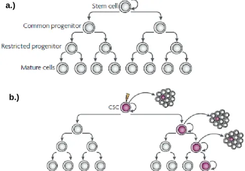
Cancer is a complex genetic disease that arises as a result of a multistep mutagenic process that disrupts the normal cellular regulatory pathways (Hahn and Weinberg, 2002). Tumor development is associated with different cellular stresses promoted by the surrounding environment (radiation and chemicals), lifestyle behaviors (diet and smoking), or by inherited predispositions (Pelengaris and Khan, 2006). These different stress inducing factors are responsible for random genetic and epigenetic mutations in multiple genes that have highly diverse biochemical functions (Weinstein and Joe, 2006). The aforementioned mutations are the driving force of cancer and their accumulation can bring forth the malignant phenotype (Evan and Vousden, 2001). The tumorigenic process is characterized by a profound cellular heterogeneity promoted not only by the ongoing mutagenesis but also by the aberrant differentiation of cancer cells (Reya et al., 2001). In an attempt to describe and account for these heterogeneous features and the tumor proliferation capacity of neoplastic cells two models have been proposed: the clonal evolution model and the cancer stem cell model (Visvader and Lindeman, 2008).
The clonal evolution model hypothesizes that cancer development can be characterized as a clonal disease in which proliferation occurs according to an evolutionary perspective in which natural selection acts upon the various somatic clones, favoring the expansion of those that carry gainful characteristics (Evan and Vousden, 2001). According to this model all of the cells have similar tumorigenic capacity and the development of a tumor begins with a single somatic cell in which an initial mutation leads to cell division and cell differentiation, culminating with the production of a genetically homogeneous clone. At this stage and although altered at the DNA level the cell is phenotypically normal (Macdonald et al., 2004).
The subsequent mutations that will occur in this neoplastic clone will be those responsible to give rise to an heterogeneous population of genetically and epigenetically unstable cells, the neoplasm, where natural selection takes place, as presented in figure 1 (Merlo et al., 2006). The selection process will benefit the clones that possess advantageous characteristics such as those that enable proliferation and autonomous survival, tissue invasion and metastasis (Stratton et al., 2009).
6
Figure 1.Cancer evolutive model. A cell that suffers a genetic mutation produces clones that in time are selected due to their growth advantages. All of the cells that make up the dominant population have similar potential to generate tumor growth (Adapted from Visvader and Linderman, 2008).
Contrariwise to the evolutionary model in which any cell can trigger tumorigenesis, the cancer stem-cell model suggests that only some cells are tumorigenic and that there is evidence of a hierarchical organization within a solid tumor (Reya et al., 2001). This hierarchy is governed by a population of stem-like cells (Visvader and Lindeman, 2008). Stem cells are unique cells that possess special characteristics like the capacity for self-renewal and potential for differentiation that distinguishes them from normal tissue cells (Rothenberg and Clarke, 2009). Cancer stem cells (CSCs) are phenotypically and functionally simillar to tissue stem cells, sharing a common set of properties with them, although with markedly differences (Table 1).
Table 1. Normal stem cell properties and cancer stem cells (Adapted from (Jordan et al., 2006, Mendelsohn et al., 2008)).
CSCs may arise from the oncogenic transformation of normal stem cells (Figure 2), a process closely related with their role in tissue homeostasis (Jordan et al., 2006). Stem cells are responsible for self-renewal of the distinctly differentiated cells that compose each tissue, however, the process of regeneration involves proliferation and
Normal Somatic Stem Cells Cancer Stem Cells
Extensive but limited self-renewal capacity Extensive and indefinite self-renewal capacity
Organogenic capacity Tumorigenic capacity
Capacity to generate differentiated lineages with limited proliferative potential, often phenotypically diverse
Capacity to generate abnormal lineages with limited proliferative potential, often
phenotypically diverse Highly regulated self-renewal and
differentiation
Highly deregulated self-renewal and differentiation
Rare in normal adult tissues Infrequent or rare within tumors
7
differentiation, the two stages where stem cells become more prone to malignant transformations (Rothenberg and Clarke, 2009).
In fact the same pathways that coordinate renewal and proliferation are likely to cause neoplasia when deregulated, among these the Wnt, Notch and Hedgehog pathways are the ones mostly responsible for CSCs generation and hyperproliferation (Visvader and Lindeman, 2008).
Figure 2. Tumor progression based on the CSC model. a.) A normal cellular hierarchy governed by stem cells generates a more restricted progeny that ultimately originates the heterogeneous mature cell types that constitute a particular tissue. b.) Cancer stem cell model where only a CSC due to its extensive proliferative capacity has the ability of triggering and sustaining tumorigenesis (Adapted from Visvader and Linderman, 2008).
It becomes therefore clear that CSCs alone can drive the continued expansion of the malignant cell population due to their substantial replicative capacity. Furthermore, to support this idea several evidences of the existence of these exceptionally tumorigenic cells have been identified in different types of malignancies, like acute myelogeneous leukemia, breast and pancreatic carcinomas, among others (Jordan et al., 2006, Visvader and Lindeman, 2008). Albeit, it is important to point out that CSCs can also arise from downstream progenitors or differentiated cells. Indeed, the existence of mutational events may confer a deregulated ability of self-renewal and other stem-like properties to these cell types, thus giving rise to CSCs (Mendelsohn et al., 2008). Even though the latter is true, it is more likely that CSCs are predominantly formed by stem cells due to their mitotic potential and preemptive activated self-renewal pathways (Mendelsohn et al., 2008).
a.)
8
In a global perspective and despite that CSC model is different from the evolutionary one they share mutual resemblances since the existence of malignant cells and genomic instability are marked characteristics of tumors (Rothenberg and Clarke, 2009). As a matter of fact CSCs themselves can undergo clonal evolution and the resulting progeny may be even more dominant and aggressive (Visvader and Lindeman, 2008).
Regardless of the model that best describes tumor development, to achieve a malignant phenotype a cell must, at some time, undergo genetic changes that are responsible for the deregulation of the normal cellular regulatory pathways (Hanahan and Weinberg, 2000). The majority of the genes that are mutated in cancer are classified as oncogenes and as tumor suppressor genes (Vogelstein and Kinzler, 2004).
Oncogenes are mutated versions of normal cellular genes (proto-oncogenes) being continuously active, or are active under the conditions where the wild type genes are dormant (Vogelstein and Kinzler, 2004). Oncogenes are responsible for accelerating cell division, growth and they are likely to play a key role in the loss of differentiation and cell motility, contributing to neoplasia (Pelengaris and Khan, 2006). On the contrary tumor suppressor genes act as guardians against DNA mutations. Tumor suppressor genes commonly supervise critical checkpoints including the mitotic cycle, transcription, differentiation and apoptosis (Pelengaris and Khan, 2006, Mendelsohn et al., 2008). Moreover, tumor suppressors also act as powerful negative regulators of oncogenes and their deactivation creates instability in proliferation and cell death, thus favoring tumorigenesis (Vogelstein and Kinzler, 2004). Furthermore and despite the controversy that surrounds the number of genetic changes required to generate malignancies (Hahn and Weinberg, 2002) point mutations in cancer genomes also need to be accounted because they foster potential to drive tumorigenesis and are responsible for an increase in cancer heterogeneity with more than 100 000 point mutations reported in some cancers (Stratton et al., 2009).
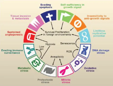
Comprehensibly the complexity of these modifications has profound impact in cancer treatment approaches. Therefore knowing the compilation of the common characteristics of cancer cells is mandatory for the development of new therapeutic approaches. At the time that a malignant transformation takes place, cancer cells collectively share a set of hallmarks, and although these are not the ones responsible for initiating tumorigenesis they are characteristic of innumerous tumor types (Figure 3) (Luo et al., 2009).
9
Figure 3. Hallmarks of cancer. Including the stress phenotypes of cancer: metabolic stress, proteo-toxic stress, mitotic stress, oxidative stress and DNA damage stress. Interrelationships between the different hallmarks promote the tumorigenic state (Adapted from Luo et al., 2009).
One of the major goals of anticancer therapy is to counteract the hallmarks depicted in figure 3, exploiting their vulnerabilities (Luo et al., 2009). Among all the phenotypic traits, the evasion of apoptosis is one of the most noteworthy, since it assures the continuous survival of the malignant cells in circumstances that otherwise would be deleterious (Evan and Vousden, 2001).
Apoptosis or programmed cell death plays a central role in the intricate balance that exists between cell survival, differentiation and death. This process is responsible for the elimination of unnecessary or dangerous cells (Melino and Vaux, 2010). The two major pathways that trigger apoptosis are the extrinsic or death-receptor mediated pathway and the intrinsic or mitochondrial mediated pathway (Melino and Vaux, 2010). The extrinsic pathway is activated whenever an extracellular signal is received by cell-surface death receptors (TNF superfamily), that in turn communicate with downstream signaling cascades. Whereas the intrinsic pathway is incited by different stresses like DNA damage, inducing the release of pro-death factors from the mitochondria (Zhivotovsky and Orrenius, 2003). Despite the pathway that is activated, both of them rely on caspases that are in charge of the effective execution of the apoptotic program (Figure 4). Caspases are proteases involved in the signaling cascade exercising their activity by cleaving several intracellular substrates that ultimately lead to cell death (Evan and Vousden, 2001).
10
Figure 4. Apoptotic pathways. The extrinsic pathway (left) is triggered by the death receptor superfamily resulting in the activation of caspases-8. The intrinsic pathway (right) is activated in response to intracellular cues like DNA damage. In the mitochondria the pro-apoptotic members of the family Bcl-2 (including Bax, Bad, Bid and Bim) compete with other anti-apoptotic Bcl-2 family members, if the pro-apoptotic cues prevail the apoptossome is formed and apoptosis continues. The extrinsic and intrinsic pathways converge at the level of caspase-3 activation. Downstream of this caspases the apoptotic program continues and results in the dismantling and removal of the cell (Adapted from Hengartner, 2000).
It is clear that apoptosis requires the combination of a multitude of cellular programs and that this process acts as a potent control mechanism. Cancer cells acquire their outstanding resistance to programmed cell death by modification of these effector pathways (Melino and Vaux, 2010). In fact malignant cells are able to rewire several key points in the apoptotic cascade and thus sustain their survival. Identified mutations in the signaling pathways of malignant cells, include deficiencies in central executioners like caspase-8, accomplished through deletions and point mutations, inactivating the caspase cascade (Zhivotovsky and Orrenius, 2003). Additional dysfunctions in the mitochondrial pathway also play a significant role in the evasion from apoptosis and among those, described the DNA damage response mediated by the protein p53 is the most critical one. The loss of function of this tumor suppressor promotes cancer proliferation (Olivier et al., 2008).
11
Taking into account that the absence of apoptosis is a key point in the origin of tumor development it is evident that there is an immense potential in its exploitation. Actually most of the therapeutic intervention in cancer aims to stimulate or restore programmed cell death and the creation of novel and effective therapeutics is becoming a reality (Luo et al., 2009). In the midst of these emerging approaches gene therapy for cancer is clearly one of the most promising, and the use expression vectors that encode genes that restart apoptosis (for instance the tumor suppressor p53) is becoming an ever growing reality (Lane et al., 2010, Tu et al., 2010).
1.2. The Tumor Suppressor p53
One of the most common dysfunctions found in cancer is the inactivation of the tumor suppressor p53, in fact, it is estimated that the p53 gene is mutated in nearly 50% of all known cancers (Melino and Vaux, 2010). P53 is a unique transcription factor and it is generally considered as the ―guardian of the genome‖ an alias that reflects its most important feature, the ability to efficiently inhibit cell proliferation (Bouchet et al., 2006). The human p53, or TP53 is a nuclear phosphoprotein composed of 393 amino acids and 5 structural and functional domains (Bouchet et al., 2006) (Figure 5). Amidst those, the DNA binding domain is the most important not only due to the fact that it is the most mutated region in cancer pathologies but also because it is important to trigger apoptosis (Zambetti, 2005, Bouchet et al., 2006)
Figure 5. Structure of the p53 protein and three dimensional structure of the DNA binding domain. The p53 protein can be divided in five functional domains, the transactivation domain, a proline rich region, the DNA-binding domain, the oligomerization domain and the regulatory domain. (Adapted from Joerger and Fersht, 2010).
12
The success of the tumor suppression mediated by p53 relies on the proper and consistent activation of the p53 pathways (Vousden and Prives, 2009). The activation of this tumor suppressor protein is triggered by a wide variety of stress events that include genotoxic stress (DNA damage), oncogene activation, loss of normal cell adhesion contacts and hypoxia (Vousden and Prives, 2009). Although this activation depends on various factors such as the intensity of the cellular stress, cell type and microenvironment (Melino and Vaux, 2010), hence it is not surprising that the loss of p53 function has such a profound influence on cancer development (Figure 6).
Figure 6. A multitude of stress signals can activate the tumor suppressor p53. The loss of function of this protein has a considerable influence in cancer development since it endows the capacity of cell survival under deleterious conditions, promotes tumor progression and metastasis (Adapted from Evan and Vousden, 2001).
Indeed the failure to activate the p53 dependent cascade in response to the stress signals that arise during the early stages of cancer development may be enough to consent the formation of pre-neoplastic lesions (Evan and Vousden, 2001). Additionally there is also the idea that the loss of p53 function may preemptively endow immediate selective disadvantages upon the modified cell in respect to normal ones, that if not overcomed may trigger proliferation (Evan and Vousden, 2001).
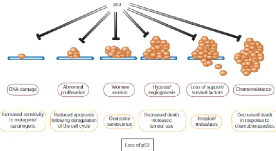
Despite these factors, once activated the tumor suppressor p53 can set off a series of pathways that ultimately lead either to cell cycle arrest, senescence or apoptosis which account for its tumor suppressor activity, as represented in figure 7 (Melino and Vaux, 2010).
13
Figure 7. p53 mediation of tumor suppression. The control of proliferation, cell survival and death by p53 is regulated by the expression of p53 target genes (blue boxes). Most of these responses contribute to tumor suppression (Adapted from Vousden and Prives, 2009).
Although p53 triggers several processes that lead to tumor suppression, it is important to underline that apoptosis is the most well established mechanism to hinder tumorigenesis, since it attacks one of the most important hallmarks of cancer (Melino and Vaux, 2010). Moreover, p53 is so versatile that it can activate both the extrinsic and intrinsic apoptotic pathways, providing a rapid response when activated, and thus effectively affect malignant cells (Melino and Vaux, 2010).
Taking into account the issues regarding cancer evasion from apoptosis and the role of p53, it seems comprehensible that targeting this unique protein as an approach to cancer therapy is a promising alternative to the generally applied treatments.
Actually several approaches that target the reactivation of p53 are showing promising results. These include the use of drugs that can influence the wild type p53 (wt-p53) or its controlling mechanisms like the case of Nutlins, small molecules that inhibit the formation of a complex between p53 and its known negative regulator MDM2 showing good results in pre-clinical models (Lane et al., 2010).
However, the most promising outcomes arise from gene therapy based therapeutics. The approaches developed so far rely on the assumption that the inclusion of a wild type p53 gene into malignant cells induces apoptosis or growth inhibition.
The majority of these approaches are based on viral delivery systems, mainly adenoviruses that are responsible for delivering the wt-p53 gene into the neoplastic cells. In fact, the first commercialized p53 cancer based therapy is an adenovirus based strategy named Gendicine, developed in China (Lane et al., 2010).
14
Nonetheless, this drug is not yet been approved by the international regulatory agencies such as the US Food and Drug Administration (FDA) due to known issues regarding the use of adenoviral vectors (Lane et al., 2010). Among those the need to deliver continuous systemic doses that may stimulate the host immune system to produce antibodies is probably the one that hinders its approval (Lane et al., 2010). Taking this into account it becomes clear that novel therapies with safer delivery vehicles, that are capable of surpassing these issues, are a requirement and their outcome will be probably responsible for the advent of a universal cancer gene based therapy.
CHAPTER II
16
Gene therapy possesses the potential to treat a wide variety of acquired or inherited diseases that nowadays are considered incurable, such as diabetes, haemophilia or cancer, as previously described in the literature (Grigsby and Leong, 2010).
The concept of this therapeutic strategy is based on the delivery of genetic material that encodes a desired gene into the cell nucleus where it will be expressed and subsequently trigger a therapeutic effect (Glover et al., 2005, Grigsby and Leong, 2010). However, this approach presents some bottlenecks regarding the sustained expression of the vector and its delivery into the intracellular environment (Grigsby and Leong, 2010). In fact, it is important to underline that often the concept of gene therapy is mostly referred as gene delivery, since the entry of the vector in the cell is the key factor that as hindered the translation of this technique into a clinical application (Grigsby and Leong, 2010).
Concerning this issue two major systems are usually used for the delivery of the genetic material into the cell: viral and non-viral systems (Glover et al., 2005). The first ones are viral systems derived from naturally occurring viruses, and they show promising results as described earlier (Glover et al., 2005). These are a consequence of the higher transfection efficiency, i.e. the ability to deliver the gene into the cell, a inherent capacity that virus possess, since to infect the host cells they need to express the virulent genome (Glover et al., 2005). Despite this fact, the use of these delivery vehicles is mostly restrained due to manufacture, structure, and safety associated limitations, such as immunogenicity and cytotoxicity, low transgene size loading and high cost (Morille et al., 2008).
Hence, as an alternative, non-viral based systems mainly cationic lipids and cationic polymers like chitosan, are now widely investigated as vehicles for gene delivery in cancer cells (Morille et al., 2008). However their outcome as the leading gene delivery vehicles is impaired due to their poor levels of transfection when compared to their viral counterparts (Morille et al., 2008, Grigsby and Leong, 2010). Nonetheless, they possess unique properties that renders them ideal carriers for DNA, as it will be further discussed. Moreover, the expansion of novel materials with nano-scale dimensions and tailored properties, could improve these carrier systems (Morille et al., 2008), making them certainly promising vehicles for use in cancer therapy in a near future.
From this viewpoint it becomes clear that non-viral gene delivery approaches are the most suitable for cancer therapy. Therefore in order to develop novel therapeutic approaches using this technique it is imperative to control and understand the whole process from the production of the expression vector and delivery system to their simultaneous application in cancer cells.
17
2.1. Plasmid DNA expression vectors for non-viral gene
therapy
Plasmids are self-sufficient replicative entities that can be found in all bacterial species possessing variable sizes that range from 1 kb to 200 kb (Schleef, 2001). One of the major advantages of these molecules is the possibility of using them as cloning vectors into which foreign DNA can be inserted and replicated (Schleef, 2001). In fact pDNA vectors are fundamental in gene delivery therapies since these double-stranded DNA (ds DNA) biomolecules encode the proteins (for example p53) that are meant to trigger the therapeutic response in the host malignant cells (Ferreira et al., 2000, Williams et al., 2009). Plasmid vectors are usually over expressed in Escherichia coli
(E.coli) bacteria before their delivery (Schleef, 2001). An integrative overview of the
process of production to application in gene therapy is presented in Fig. 8.
Figure 8. Stages from production to application of pDNA in gene therapy based treatments. In stage 1 (upstream processing) the plasmid vector is chosen and the construct designed to bear the transgene of interest a process followed by selection and optimization of the E.coli bacterial growth conditions and consequently fermentation. Stage 2 (downstream processing) comprises the final isolation of the expression vector and its purification. Stage 3 and 4 will be further addressed since there are key factors that hinder these stages before the ultimately therapeutic (Adapted from Ferreira et al, 2000; Williams et al., 2009).
As figure 8 depicts there are two key stages before the delivery of the expression vector into the malignant cells: the upstream and downstream stage. The first one can be summarized as the tailoring of conditions that allow the expression of large amounts of pDNA (Ferreira et al., 2000). Whereas the latter comprises the processing operations that aim to eliminate impurities, contaminants and recover the supercoiled plasmid DNA (sc pDNA) isoform (Ferreira et al., 2000).
18
This downstream stage is crucial since it is responsible for the removal of impurities such as endotoxins, RNA and genomic DNA that can cause serious side effects if delivered to the host (Ferreira et al., 2000). Furthermore, it is also at this stand point that the purification of the different plasmid isoforms takes place.
Plasmid DNA is considered to be a stable biomolecule, although during its production and recovery process it can undergo several stresses that may lead to its degradation (Quaak et al., 2009). This phenomenon mainly affects the sc isoform, the only plasmid isoform considered intact and undamaged (Quaak et al., 2009). Nonetheless, it is of most importance to point out that this isoform is also considered the one that possess the highest transfection efficiency and therefore it is the most appropriate one for cancer gene therapy approaches (Quaak et al., 2009). Taking this into account, it seems comprehensible that there is a growing demand for the recovery and purification of this isoform in the downstream process (Urthaler et al., 2005). In this subject, chromatography plays a central role either in the purification in large scale, or as an analytic technique used in the monitoring of the overall process. This is a key parameter, since pDNA products intended for gene therapy applications must comply to strict guidelines published by regulatory agencies such as FDA and the European Agency for the Evaluation of Medical Products (EMEA) (Diogo et al., 2005).
2.2. Amino acid-DNA affinity chromatography for sc pDNA
purification
Among the different chromatographic approaches to purify sc pDNA such as size-exclusion chromatography (SEC), hydrophobic interaction chromatography (HIC), anion-exchange (AEX) and others, affinity chromatography has shown outstanding results in the purification of pDNA intended for gene therapy (Sousa et al., 2008b). Affinity chromatography is a unique technique that relies on a specific binding ligand to purify biomolecules taking into account their biological functions and distinct chemical structures (Sousa et al., 2008a, Mondal and Gupta, 2006). The use of this methodology presents remarkable advantages since not only it abolishes unnecessary extra processing steps to purify pDNA, but also increases the yield of the recovered nucleic acids (Sousa et al., 2008a).
This purification approach separates biomolecules relying on a reversible interaction that occurs between the target molecule and the ligand (Figure 9).
19
Figure 9. Interaction between the affinity ligand and the target biomolecules. The presence of a spacer arm assures the accessibility of the ligand (Adapted from Healthcare). (Healthcare)
The specific interactions described above involve a multitude of complex physico-chemical interactions including electrostatic, hydrophobic, van der Walls forces and/or hydrogen bonds (Sousa et al., 2008a). Moreover, the selection of the matrix and the used conditions need to account not only for these interactions but also for the molecular properties of the biomolecules. (Sousa et al., 2008a). Once the sample is loaded into the column their selective capture is promoted by the ligand if the conditions are suitable, hence if that is the case the biomolecules stand bound onto the matrix (Mondal and Gupta, 2006). Subsequently, elution steps can be performed either specifically (with competitive ligands) or non-specifically by changing conditions like pH, ionic strength or polarity depend mostly on the features of the different biomolecules (Sousa et al., 2008a). In general this affinity purification strategy can purify pDNA in a single purification step that, as described earlier, is crucial to further promote the stability of the most biologically active isoform. Although it is important to underline that this unique feature is mostly dependent on the type of the affinity ligand. Regarding this issue, the use of small amino acid ligands termed pseudobio affinity ligands has proven to be a promising strategy to efficiently isolate sc pDNA with high yield and purity (Sousa et al., 2008a). In fact histidine has been used to purify the sc pDNA isoform successfully although with low yield and using high salt concentrations (Sousa et al., 2008a). To overcome the aforementioned issue, novel arginine supports have been designed, taking advantage of the fact that protein-DNA structures are mostly dependent on arginine-based interactions to purify pDNA. In fact Sousa et al., 2008, demonstrated that these supports have unmatched selectivity to the different pDNA isoforms, being capable of separating the sc pDNA isoform with high yield, stability and purity (Sousa et al., 2008b), most certainly meeting the regulatory requirements for further application in gene therapy.
20
Despite the fact that this has been a major breakthrough, efficient gene expression can only be attained if the pDNA can reach the nucleus in a structurally intact form. Hence it is crucial to assure the transport of the genetic material in a safe and efficient way.
2.3. Non-viral nanocarrier mediated gene delivery
A major challenge for cancer gene therapy is the delivery of the pDNA that contains the transgene of interest into the cell (Glover et al., 2005).
Nanoparticulated delivery systems arise nowadays as the most promising candidates for the delivery of pDNA into the intracellular compartment and ultimately into the nucleus of malignant cells (Gullotti and Yeo, 2009). These nanocarriers are exceptional delivery systems due to their unique nanoscopic size characteristics, generally situated in the range of 1 to 1000 nm (Ferrari, 2005). Furthermore, they are not only able to enter the cell but they also possess unmatched release characteristics that are responsible for the release of their payload. Such property is very important for their use as gene delivery system for cancer (Gullotti and Yeo, 2009).
However, it is important to first address the challenges of targeting these carrier systems into malignant cells. Comprehensibly, there is a pre-requisite to understand the underling concepts that the tumor biology presents to efficient gene delivery and combine them with the optimized design of nanoparticulated systems (Peer et al., 2007).
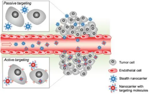
One of the most interesting features of tumors is their leaky vasculature that allows the nanoparticles to diffuse freely into tumor cells (Peer et al., 2007). This mechanism is called enhance retention and permeability effect (EPR), and is often characterized as a passive targeting strategy to deliver nanoparticles into the tumor microenvironment (Gullotti and Yeo, 2009). EPR is based on the assumption that the blood vessels surrounding tumors acquire increased permeability with fenestrations ranging from 100 to 600nm and deregulated lymphatic drainage (Gullotti and Yeo, 2009). Therefore, the passive targeting of the nanoparticulated systems is achieved due to their accumulation in the vasculature that surrounds the tumor (Figure 10).
Although, to take advantage of this effect it is crucial that the non-viral nanocarriers are able to evade the immune surveillance system and circulate for long periods of time in the bloodstream (Gullotti and Yeo, 2009). In fact, the nanoparticles must avoid the reticuloendothelial system (RES), the one responsible for their opsonization and consequent phagocytosis (Gullotti and Yeo, 2009).
21
Figure 10. Nanocarrier targeting to tumors cells. The nanocarrier systems can reach the tumor by different targeting mechanisms: passive targeting and active targeting. Upon arrival at the tumor microenvironment, nanoparticles with specifically adsorbed or conjugated molecules can bind to tumor cells, on the contrary normal carrier systems are less effective in interacting with tumor cells, although they also enter into the cellular compartment (Adapted from Gullotti and Yeo, 2009).
In addition to the EPR effect another targeting approach may also be used as depicted in figure 10. This methodology is named active targeting and involves the adsorption or chemical conjugation of specific molecules on the surface of the nanocarrier systems. In turn, these molecules are recognized by malignant cells and internalization is mediated by this specific interaction (Gullotti and Yeo, 2009). In spite of the fact that several reports describe the potential of this approach to enhance transfection efficiency and consequently the anticancer effect, it is important to underline that an ideal nanocarrier should attain delivery both relying of the EPR effect and on active targeting, thus exponentially increasing their tumor distribution (Gullotti and Yeo, 2009). At this point, another barrier to gene transfection arises since the tumor matrix that consists of collagen and other proteins obstruct the nanoparticle systems, in such a way that it limits their mobility (Morille et al., 2008).
Notwithstanding, in addition to these targeting issues, once they are located in the tumor microenvironment the non-viral nanocarriers systems face the most challenging obstacles that hinders their entry into the cell (Figure 11).
22
Figure 11. Different barriers impacting gene delivery through nanoparticulated systems before and after entering the cell (Adapted from Morille et al, 2008).
Following the arrival at the tumor site the first contact with the tumor cells is determinant, since the nanocarrier must be able to transpose the extracellular membrane (Morille et al., 2008). As figure 11 shows there are a multitude of entering pathways, although the predominant way that nanocarriers use to enter in the intracellular compartment seems to be the easier, i.e. the one that relies on non-specific endocytosis, followed by the clathrin-coated mechanism (Morille et al., 2008). Nonetheless, it is important to state that the contribution of each pathway to the internalization of the carriers is not yet defined due to their different compositions. Among the factors that may set of the entry into the cell trough one mechanism in detriment of other are the size of the carriers and their surface charge (Morille et al., 2008). Following endocytosis, the nanocarrier vectors remain captured in the vesicles, which presents itself as a major drawback to efficient transfection, since if the nanoparticle carrier stays for indeterminate time in the vesicle it will most likely be degraded by lyzossomes (Morille et al., 2008). Taking this into account, the nanocarrier
23
system must possess well defined characteristics that will ultimately allow its release into the cytoplasm a process that is thought to be mediated by a mechanism know as proton sponge effect (Pathak et al., 2009).
The proton sponge effect is a consequence of the increase in osmotic pressure promoted by the positive charge of the nanocarrier systems (Pathak et al., 2009), a crucial characteristic that determines the chosen materials for their synthesis, as it shall be seen further ahead (Mao et al., 2009).
When the nanocarrier is released into the cytoplasm two critical stages that will ultimately dictate the efficiency of the process arise, the nanocarrier unpacking of pDNA and the entry of the expression vector into the nucleus. The first one depends mostly on the properties of the nanocarrier systems and are one of those that prove most difficult to engineer (Grigsby and Leong, 2010).
It is comprehensible that the carrier systems must dissociate from their pDNA cargo so the genetic material can be translated in to the protein of interest by the cell machinery. Although the question arises, when is the best moment for this event to take place. Reports in the literature are quite opposite in what comes to this matter and some show that it occurs in the cytoplasm, well others report it happens in the nucleus (Grigsby and Leong, 2010). From this stand point it is only important to underline that these findings are only dependent on the tailoring characteristics of the carriers and they need to be carefully engineered to optimize transfection (Grigsby and Leong, 2010). Schematics of the ideal protection/release characteristics of the carriers are presented in Fig. 12.
Figure 12. DNA unpacking. After entering the cell the nanocarrier must be able to release its genetic content (Adapted from Grisby and Leong 2010).
24
As depicted in figure 12 the release of the pDNA cargo from the nanocarrier systems may occur in three distinct stages: i.) inside the endossomal vesicle; ii.) in the cytoplasm; iii.) in the nucleus (Grigsby and Leong, 2010). Ideally the carrier should protect DNA from nuclease mediated degradation, but at the same time it should be able to release it (Pathak et al., 2009). If these binding/release states will not occur in the nucleus the expression vector must also transpose this barrier until it is ultimately expressed. The nuclear uptake of the pDNA vectors may occur by two distinct routes. The first one during the cell division stage and the second one through the nuclear pore complex, a process mediated by the cell import machinery which depends on small proteins named caryopherins (Pathak et al., 2009).
From this stance it is comprehensible that at some time in the non-viral transfection process the nanocarrier intrinsic characteristics play a decisive role in the overall yield of the process, hence the type and material that composes the nano delivery system must be chosen wisely.
2.4. Non-viral polymeric nanoparticles for gene delivery
The majority of non-viral gene delivery systems comprise a wide number of polycations including cationic lipossomes, branched and linear polyethylenimine (PEI), polyamidoamine dendrimers and chitosan among others (Hart, 2010). These polycations posses unique advantages that distinguish them from viral vectors, such as low immunogenicity, easy to manufacture and no limitations in what regards the transport of genetic material (Park et al., 2006). Moreover, their positive charge allows them to condense DNA and form sub-micron ranged particles frequently referred as nanoparticles (Hart, 2010). In fact, due to their size, nanoparticles can achieve higher intracellular uptake when compared to other systems (Mosqueira et al., 2001). This structural characteristic gains special importance given the fact that until the pDNA reaches the nucleus of the cell, an adverse environment acts upon the carrier and the vector, as describer earlier.
These nanoparticulated delivery systems can be defined as solid colloids that collectively include nanospheres, nanocapsules, lipossomes and polymeric micelles as shown in figure 13.
25
Figure 13. Types of nanocarrier systems for gene delivery. A.) Lipossomes are formed by phospholipid bilayers (one or more) surrounding an aqueous core, they can be modified in their surface by PEGylation or with targeting ligands. B) Polymeric nanospheres are formed by biodegradable polymers. C.) Polymeric nanocapsules are formed by a thin polymeric membrane (with the same material as the nanospheres), that surrounds an aqueous or oil core. D.) Polymeric micelles are composed of amphiphilic polymers (Adapted from Hillaireau and Couvreur, 2009).(Hillaireau and Couvreur, 2009)
Among the delivery systems depicted in figure 13 nanocapsules are one of the most important ones. They are vesicle like polymer carriers composed of an aqueous core involved by a polymeric envelope that exhibits a typical core-shell structure in which pDNA can be entrapped (Parveen and Sahoo, 2008). Several reports exist in the literature regarding the use of nanoparticulated polymeric materials (nanoparticles and nanocapsules) for gene delivery systems such as those based on PEI (Dall'Era et al., 2008), or PEI poly (lactic-co-glycolic acid) (PLGA) polymer conjugates, polyethylene glycol (PEG) (Olga et al., 2008) or alginate (Reis et al., 2006).
Despite the diversity of materials at the disposal for synthesis of nanoparticulated systems chitosan-based nanocarriers have been gaining an exponential interest as non-viral gene delivery systems, mainly due to its undoubtedly unique properties (Mao et al., 2009).
Chitosan is synthesized from the deacetilation of chitin, which is a part of the exoskeletal structure of crustaceans (shrimp, crabs, etc). Chitosan is a biodegradable polysaccharide consisting of repeting D-glucosamine and N-acetil-D-glucosamine monomers, linked via β1-4 glycosidic bonds (Figure 14) (Mao et al., 2009). Chitosan contains a primary amine group in each deacetilated unit with a pKa of 6.5 rendering it soluble only in acidic media and insoluble in neutral or basic medium (Mao et al., 2009).
26
Figure 14. Chitosan chemical structure (Adapted from Mao et al, 2009).
In the physiological environment chitosan can be degraded by lysozymes or by chitinases which can be produced in the human intestinal flora or be present in the blood (Mao et al., 2009).
The main feature of chitosan that accounts for its potential as a gene delivery vehicle is its cationic nature (Borchard, 2001). Curiously, chitosan most important characteristic is due to the primary amine groups present in its backbone, since below the pKa they become positively charged (Mao et al., 2009). These amine groups are the ones responsible for the reaction with DNA molecules since they interact via electrostatic forces (Borchard, 2001). This contact between the positively charged chitosan molecule and the negatively charged DNA backbone (due to its phosphate groups) is responsible for the spontaneous formation of chitosan-DNA nano-complexes, also known as polyplexes, (Figure 15) which only takes place at a suitable nitrogen to phosphate ratio (N:P ratio) (Köping-Höggård et al., 2004, Mao et al., 2009).
Figure 15. Different chitosan DNA nanoparticles formulated by different synthesis mechanisms (Adapted from Mao et al., 2009).
As presented in figure 15 different nanoparticles can be formed with chitosan and DNA depending on the processing method, this is an issue of most importance since
27
the formulation of chitosan polyplexes yields rather unstable and morphologically undefined particles when compared to those obtained using cross-linking agents (Csaba et al., 2009).
Taking into account what was earlier described, chitosan as a lot of properties and some of them need to be summarized and addressed in order to understand what effect their manipulation would have in the final nanoparticle formulations for gene delivery. Chitosan can be characterized by its molecular weight, viscosity, crystallinity and most importantly its degree of deacetilation the one to be studied most thoroughly, due to the fact that it not only influences crucial nanoparticle formulation parameters but also because it plays a fundamental role in polymer degradation and transfection efficiency (Mao et al., 2009). Some of the most important formulation parameters for the synthesis of chitosan-based delivery systems are summarized in table 2.
Table 2. Summary of the most important formulation parameters in the design of
nanoparticulated systems based on chitosan.
Properties Characteristics Influences References
Deacetilation Degree Percentage of primary amine groups in the polymer backbone (determines the positive charge density) Higher degrees results in increased DNA binding, cellular uptake, longer degradation time and smaller particle size (Kiang et al., 2004); pDNA concentration Amount of pDNA that is incorporated in the cationic polymer Higher concentrations enhance transfection until a saturation point (Zhao et al., 2006)
Molecular weight Particle size, cell
uptake, transfection efficiency Decrease in size with decrease in molecular weight (Huang et al., 2005) Cross-linker agents Encapsulation of pDNA, modification of the particle physicochemical properties Higher transfection efficiency, higher stability, formation of nanocapsules (Csaba et al., 2009);(Bao et al., 2008) Polymer Concentration Amount of positive charges in solution Higher concentration increased particle size (Gan et al., 2005) pH Amount of primary protonated amines Particle size increase at pH 6.0 (Gan et al., 2005)
28
It is clear that chitosan is a polymer with immense potential for gene delivery applications. Recently some advances in respect to nano-encapsulation of pDNA (Csaba et al., 2009) inside its particle core and the results regarding gene therapy delivery systems for cancer (Hee-Dong et al., 2010) make this polymer a valuable option for gene therapy applications.
SECTION II
30
2.1. Materials
Arginine Sepharose 4B gel was obtained from Amersham Biosciences (Uppsala, Sweden). Pentasodium tripolyphosphate (TPP), ethidium bromide (purity 95%), N-acetyl-D-glucosamine, Sodium nitrate (NaNO2), Fluorescein isothiocyanate isomer I
(FITC), Anti-VE-Cadherin antibody (anti-rabbit),paraformaldehyde,Triton X-100, Tween 20, bacterial and cell culture reagents were all obtained from Sigma–Aldrich (St. Loius, MO, USA). 2-(4-aminophenyl ethylamine) was purchased from Acros Organics (Geel, Belgium). One kbp DNA ladder was obtained from Vivantis Technologies (Oceanside, CA, USA.). A549 non-small lung carcinoma cell line was purchased from ATCC (Middlesex, UK). All transfection reagents and Hoesht 33342 (trihydrochloride, trihydrate), were obtained from Invitrogen (Carlsbad, CA, USA). All used salts used in this research were of analytical grade.
2.1.1. Plasmid DNA
The 6.7 kbp pcDNA3-FLAG-p53 plasmid was purchased from Addgene (Cambridge, MA, USA). The vector encodes for the human p53 protein conjugated with a FLAG tag. The vector contains the SV40 virus mammalian expression promoter and the ampicillin resistance gene.
Figure 1. Plasmid pcDNA3-FLAG-p53 backbone (Vector drawn using the Redasoft Visual Cloning 3.2 ™ software).
2.1.2. Chitosan
Chitosan (low molecular weight (LMW)), from crab shells was purchased from Sigma-Aldrich (St. Loius, MO, USA). Chitosan molecular weight (MW) ranged between 50 and
pcDNA3-FLAG-p53 6700bp