The challe nge o f ve cto r
de ve lo pme nt in ge ne the rapy
Laboratório de Gerontogenética e Terapia Gênica, Departamento de Genética, Faculdade de Medicina de Ribeirão Preto, Universidade de São Paulo, Ribeirão Preto, SP, Brasil
S.U. Dani
Abstract
Gene therapy is the treatment of diseases based on the transfer of genetic information. Agents that carry or deliver DNA to target cells are called vectors (Latin vector: carrier, deliverer). Ideally, a vector should accommodate an unlimited amount of inserted DNA, lack the ability of autonomous replication of its own DNA, be easily manufac-tured, and be available in concentrated form. Secondly, it should have the ability to target specific cell types or to limit its gene expression to specific cell types, and to achieve sustained gene expression in the long term or in a controlled fashion. Finally, it should not be toxic or immunogenic. Such a vector does not exist and none of the DNA delivery systems so far available for in vivo gene transfer is perfect with respect to any of these points. Gene therapy and the means to promote it depend heavily on the development and improvement of new gene vector systems.
Co rre spo nde nce
S.U. Dani
Laboratório de Gerontogenética e Terapia Gênica
Departamento de Genética FMRP, USP
14049-900 Ribeirão Preto, SP Brasil
E-mail: sudani@ rgm.fmrp.usp.br
Presented at the International Symposium “The Third Revolution on Vaccines: DNA Vaccines”, Belo Horizonte, MG, Brasil, November 3-7, 1997.
Publication supported by FAPESP.
Received O ctober 15, 1998 Accepted November 9, 1998
Ke y words
·Gene therapy ·Gene transfer ·DNA delivery ·Viral vectors ·Diposomes ·Vector development
Intro ductio n
Many different ways have been devised for introducing genes into mammalian cells and tissues. The simplest technique is the inoculation of naked DNA by means of mi-croinjection, electroporation and biobalistics, the latter also known as the gene gun tech-nique. More elaborate and more efficient ways include the use of self-assembling complexes of lipid-DNA (e.g., liposomes), protein-DNA, lipid-protein-DNA, and viral vectors. Viral vectors can be fragments of viral DNA containing the DNA to be trans-ferred or the viral particle itself. The viral particle is rendered crippled or replication defective through the manipulation of the viral DNA, and the end product is a non-pathogenic viral vector carrying the genetic
information of therapeutic interest.
Five gene transfer systems most com-monly used in gene therapy trials - retroviral vectors, adenoviral vectors, liposomes, biobalistics and adeno-associated viral vec-tors - accumulate a limited clinical experi-ence of only a few hundred patients world-wide in the last eight years. Therefore, this mini-review is aimed at briefly discussing the main features of gene vector systems, rather than the clinical experience in the field of gene therapy.
Che mical me thods
less frequently, in vivo (Table 1). A substan-tial portion of the cells endocytes the DNA and is capable of transporting at least part of it into the nucleus, where the DNA is tran-siently expressed for some days. However, only a tiny fraction (usually much less than one percent) of the cells keeps the DNA permanently, incorporating it into its chro-mosomes, and continuing to express the in-troduced genes. In these methods, the vector is a purified DNA molecule (e.g., a plasmid), usually engineered by cloning methods to
contain, besides the gene of interest, regula-tory sequences such as promoters and en-hancers to facilitate the expression of the gene.
To improve efficiency and specificity, the DNA vector can be complexed to agents such as ligand proteins, to facilitate its entry into the target cells. An example is the use of conjugates of a polycation, poly-L-lysine, covalently linked to the asialo-orosomucoid glycoprotein (ASOR) (1). ASOR serves as a ligand for recognition by the asialoglycopro-tein receptor present in hepatocytes. The subsequent mixture of this conjugate ASOR-poly-L-lysine with DNA results in the for-mation of complexes stabilized by electro-static interactions, which allows the delivery of single-strand DNA or double-strand DNA specifically to hepatocytes. DNA delivered to the liver in vivo via endocytosis mediated by the asialoglycoprotein receptor is rapidly degraded, presumably in lysosomes, and can be cleared within 48 h (2). Pharmacological approaches such as the destabilization of microtubules that interfere with the translo-cation of endosomes to lysosomes have to be developed to overcome the problem of intra-cellular degradation and to augment the effi-ciency of directed gene expression.
A recent development was the in vitro
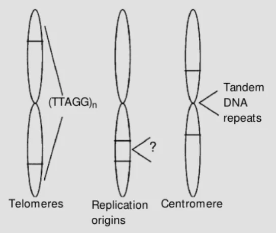
assembly of human artificial chromosomes (HACs). Chromosomes are highly structured organelles, formed by a linear DNA mole-cule wrapped and conjugated to globular hystone proteins. A chromosome has three important parts or components: a centromere (central portion), telomeres (terminal por-tions), and origins of replication (Figure 1). The functions of telomeres and centromeres are partly known and their structures are being slowly elucidated. The origins of rep-lication are involved in the initial reprep-lication of the DNA that forms the chromosome. These three components are sufficient to confer a chromosome function on a DNA fragment. Utilizing molecular biology tech-niques, it is possible to assemble the three
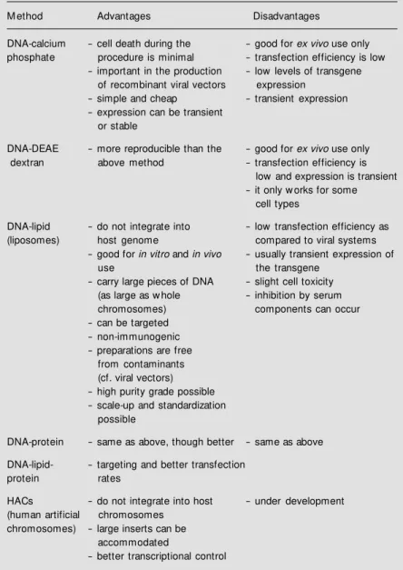
Table 1 - Chemical methods for introducing genes into mammalian cells.
M ethod Advantages Disadvantages
DNA-calcium - cell death during the - good for ex vivo use only phosphate procedure is minimal - transfection efficiency is low
- important in the production - low levels of transgene of recombinant viral vectors expression
- simple and cheap - transient expression - expression can be transient
or stable
DNA-DEAE - more reproducible than the - good for ex vivo use only
dextran above method - transfection efficiency is
low and expression is transient - it only w orks for some
cell types
DNA-lipid - do not integrate into - low transfection efficiency as
(liposomes) host genome compared to viral systems
- good for in vitro and in vivo - usually transient expression of
use the transgene
- carry large pieces of DNA - slight cell toxicity (as large as w hole - inhibition by serum
chromosomes) components can occur
- can be targeted - non-immunogenic - preparations are free
from contaminants (cf. viral vectors) - high purity grade possible - scale-up and standardization
possible
DNA-protein - same as above, though better - same as above
DNA-lipid- - targeting and better transfection
protein rates
HACs - do not integrate into host - under development (human artificial chromosomes
chromosomes) - large inserts can be accommodated
essential components to form an artificial chromosome.
Recently, a group of researchers in Cleve-land (3) described the assembly of first gen-eration human artificial minichromosomes using new assemblies of a-satellite DNA (tandem repeats of DNA sequence present in centromeres), telomeric DNA (tandem repeats of TTAGGG), and other fragments of genomic DNA. The creation of HACs is a major step forward in the studies of the functions of human chromosomes and the expression of large DNA sequences. HACs can be utilized as vectors for very large DNA sequences that in this form can be stably maintained within cells for studies of DNA sequence and gene expression, or the effect of its organization and relative position on the integrity and stability of the artificial chromosome. The cloning of genes into HACs also opens the possibility for the stable introduction and physiological expression of therapeutic genes. However, efficient HAC delivery methods have yet to be developed for the use of HACs in somatic gene therapy protocols.
Gene transfer can also be accomplished by using DNA encapsulated into agents such as cationic lipids or phospholipids, to form cationic liposomes. Liposomes are usually prepared from bipolar phospholipids and consist of a hydrophilic core delimited by an external lipid layer (4). Cationic lipids inter-act electrostatically with the strongly an-ionic DNA molecule promoting a complete encapsulation of the nucleic acid. The lipo-somes produced in this way have a broad spectrum of cellular infectivity. In a 1993 paper, Zhu and coworkers (5) reported that a single intravenous injection of cationic lipo-somes carrying a plasmid with the chloram-phenicol acetyl-transferase (CAT) gene un-der control of the strong CMV promoter was capable of transducing many different tis-sues in the mouse.
However, for in vivo use, especially for systemic applications, a series of
modifica-tions have yet to be developed both in the surface of liposomes and in the encapsulated DNA. It would be desirable, for instance, to develop modified liposomes capable of es-caping the reticuloendothelial system and tissue macrophages (e.g. Kupffer cells). This could be done through the coupling of tar-get-ligands on the liposome surface. These ligands would serve also to direct liposomes to specific targets.
An important barrier for the expression of transgenes that are introduced via lipo-somes is the transport into the nucleus. The co-encapsulation of viral envelopes with in-corporation of nucleophilic proteins that con-tain nuclear internalization signals (e.g., the high mobility group proteins, HMG1 pro-teins) can augment the efficiency of nuclear localization and gene transcription (6-8). Modifications of the encapsulated DNA in-clude the utilization of tissue-specific pro-moter-enhancer elements to achieve a tis-sue-specific expression. Tissue targeting and localization can also be improved by direct injection of liposomes into the desired re-gion.
Physical me thods
DNA vectors can be introduced into cells via a variety of physical methods (Table 2). Conceptually the most obvious of such meth-ods, direct injection, requires sophisticated techniques for injection on a micro-scale.
Tandem DNA repeats
Telomeres (TTAGG)n
?
Replication origins
Centromere
originally used in gene transfer to plant cells and tissues (9) and then applied to a variety of organisms including mammal cells and tissues (10-16) consists of coating tiny spheres of a heavy metal (e.g., gold) with DNA, and then shooting these coated par-ticles against the target cells at high speed. The accelerated particles penetrate the cells carrying the DNA. Initial studies performed with this technique have provided encourag-ing results and created the opportunity for its
in vivo application by direct shooting against a selected tissue or organ.
Biological me thods: viral ve ctors
Viruses are natural gene vectors; they are evolving in connection with virtually all kinds of organisms, from bacteria to plants and animals. The specific biomolecular systems for gene transfer, gene recombination and gene expression adopted by the viruses con-stitute powerful tools for the construction of more efficient and safe vectors, with precise indications of use. Bacteriophage, baculovi-rus, retrovibaculovi-rus, adenovibaculovi-rus, herpes simplex virus, vaccinia, human immunodeficiency virus (HIV), polyoma virus and adeno-asso-ciated virus are examples of viruses that have been successfully modified by the tech-niques of recombinant DNA. All of these viruses already have applications in research, agriculture and medicine. Conceptually, it is not surprising that animal viruses have been used as vectors for gene transfer to mammal cells (Table 3). In this article, I shall review in some depth the main properties of three of them: retrovirus, adenovirus and adeno-as-sociated virus.
Re troviral ve ctors
A murine retrovirus, the Moloney mu-rine leukemia virus (MoMuLV) was the first vector system to be developed for clinical applications of gene therapy. Utilizing re-combinant DNA techniques, the genes of the This technique, however, is limited by the
fact that only a relatively small number of cells can be injected within a given period of time. In spite of the efforts to automate the microinjection techniques, the small number of cells that can be injected each time contin-ues to be a serious limitation. This method of gene transfer could be of interest and clinical utility if a small number of purified bone marrow cells were injected and cell culture conditions were found to expand them sub-stantially. However, the problem of transient expression would still persist, since most of the injected cells would not be capable of sustaining the expression for long peri-ods. This problem requires additional im-provements in the vector sequence so as to augment its integration efficiency, for ex-ample.
There are other physical methods that can be applied to a larger number of cells. One of such methods is electroporation that can be used to introduce DNA into cells in culture. However, cell viability among re-ceptor cells varies enormously with different cell types. A physical method that has been
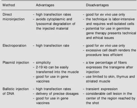
Table 2 - Physical methods for introducing genes into mammalian cells.
M ethod Advantages Disadvantages
Direct - high transfection rates - good for ex vivo use only microinjection - avoids cytoplasmic and - the technique is labor-intensive
lysosomal degradation of and requires w ell-isolated cells the injected material - potential for use in germline
gene therapy presents technical and ethical issues
Electroporation - high transfection rate - good for ex vivo use only - excessive cell death renders the
procedure less efficient
Plasmid injection - simplicity - a low percentage of fibers - 2-19 kb can be easily expresses the transgene after
transferred into the muscle injection
- good for use in gene - use limited to skin, thymus and
vaccines striated muscle
Ballistic injection - high transfection rates - transient expression of DNA - delivery of precise dosages - considerable cell lesion in the
- good for use in gene center of the region reached by
viral genome which are involved in the rep-lication of the MoMuLV - gag, pol and env
- are removed and replaced by a gene of interest. What remains from the retrovirus are its regulatory elements: the long terminal repeats (LTR) which function as provirus integration signals and transcription promot-ers, and a packaging signal, to allow the accommodation of the transcribed RNA into a viral particle. To produce retroviral vectors carrying a gene of interest, it is necessary to use a packaging cell line harboring the gag,
pol and env genes which have been previ-ously incorporated into the genome of such cells. The vectors are produced in high titers in the packaging cells, purified and injected into the patient (in vivo gene therapy) or put in contact with cells collected from the pa-tient and maintained in cell culture condi-tions (ex vivo gene therapy). Retroviral vec-tors have the ability to enter the target cells, transcribe their RNA into DNA (owing to the activity of viral reverse transcriptase), and integrate stably in a chromosome of the host cell as a result of the presence of the remaining retroviral regulatory sequences. Once integrated, the inserted gene can be expressed to produce the desired therapeutic protein. Viral vectors devoid of the genes involved in viral replication are dubbed de-fective or crippled and thus are not able to produce more replication competent virus inside the target cell. The vector acts as a final gene transfer agent, leaving a copy of its gene sequence integrated in the host cell genome.
Advantages of retroviral vectors include the fact that they are well known and well characterized, the possibility to well charac-terize them and to produce them in high titers [106
PFU/ml (plaque forming units per ml), or higher], and their high transfection effi-ciency which has been validated in various cell types. Disadvantages include an accom-modation limit of 7-8 kb of DNA, the need that the target cells be in cell division to allow for the integration of the vector
(limit-ing the use of retroviral vectors to mitotic cells), and the potential of insertional mu-tagenesis due to the fact that retroviral vec-tors integrate at random in the genome. The latter feature has the potential to interrupt important genes in the cell, with serious consequences that include oncogenesis through the activation of proto-oncogenes or inactivation of tumor suppressor genes.
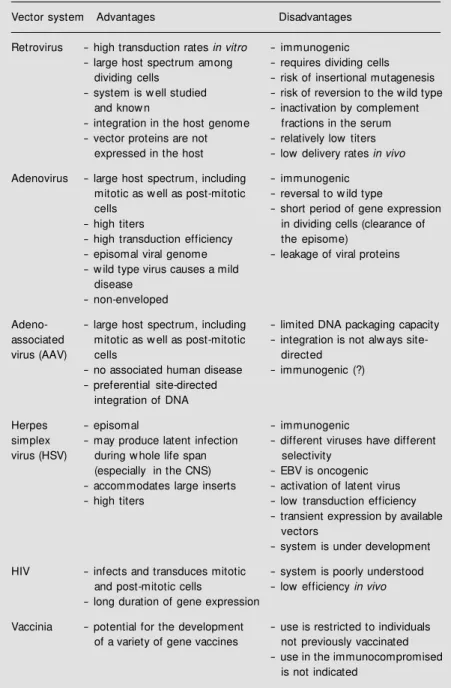
Fur-Table 3 - Biological methods for introducing genes into mammalian cells.
Vector system Advantages Disadvantages
Retrovirus - high transduction rates in vitro - immunogenic - large host spectrum among - requires dividing cells
dividing cells - risk of insertional mutagenesis - system is w ell studied - risk of reversion to the w ild type
and know n - inactivation by complement
- integration in the host genome fractions in the serum - vector proteins are not - relatively low titers
expressed in the host - low delivery rates in vivo
Adenovirus - large host spectrum, including - immunogenic mitotic as w ell as post-mitotic - reversal to w ild type
cells - short period of gene expression
- high titers in dividing cells (clearance of - high transduction efficiency the episome)
- episomal viral genome - leakage of viral proteins - w ild type virus causes a mild
disease - non-enveloped
Adeno- - large host spectrum, including - limited DNA packaging capacity associated mitotic as w ell as post-mitotic - integration is not alw ays
site-virus (AAV) cells directed
- no associated human disease - immunogenic (?) - preferential site-directed
integration of DNA
Herpes - episomal - immunogenic
simplex - may produce latent infection - different viruses have different virus (HSV) during w hole life span selectivity
(especially in the CNS) - EBV is oncogenic - accommodates large inserts - activation of latent virus - high titers - low transduction efficiency
- transient expression by available vectors
- system is under development
HIV - infects and transduces mitotic - system is poorly understood and post-mitotic cells - lowefficiency in vivo - long duration of gene expression
Vaccinia - potential for the development - use is restricted to individuals of a variety of gene vaccines not previously vaccinated
ther disadvantages of retroviral vectors that limit their in vivo use include their inactiva-tion by complement in serum, and the poorly understood spontaneous inactivation of the LTR promoter function.
Ade noviral ve ctors
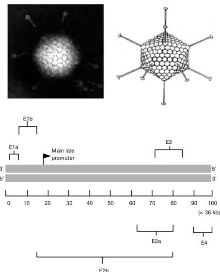
Human adenoviruses are non-enveloped DNA-viruses with a linear double-strand DNA of about 36 kb, encapsulated in an icosahedral capsid measuring 70-100 nm in diameter. The capsid has in its vertices rod-like structures that interact with cell recep-tors (Figure 2). In the wild, adenoviruses are capable to infect cells in the gastrointestinal
tract causing common cold, gastroenteritis in children or epidemic conjunctivitis. The adenoid glands are one of its targets, from which the name adenovirus has originated. Infections involving the urinary tract, he-patic pathways and the central nervous sys-tem can occur sporadically. The majority if not the totality of adults has been exposed to adenoviruses and has antibodies.
Differently from retrovirus, adenovirus does not depend on host cell division for its replication, and its chromosome rarely inte-grates into the cell genome, remaining episo-mal in most cases. Integration seems to oc-cur only in the presence of high levels of infection in dividing cells, but this event does not contribute significantly to the util-ity of these viruses as vectors. Adenoviral vectors have a broad spectrum of cell infec-tivity that includes virtually all post-mitotic and mitotic cells, and also can be produced in high titers.
The genomic structure of adenovirus is more complex than that of retrovirus. The adenoviral genome codes for approximately 15 proteins. Viral gene expression occurs in a coordinated fashion and is mainly directed by the E1A and E1B genes, localized in the 5' portion of the adenoviral genome (Figure 2). These genes have transactivation func-tions for the transcription of various viral and host cell genes. Since E1 genes are involved in adenoviral replication, their re-moval renders the virus replication incom-petent or defective. The removal also cre-ates room for the insertion of a gene of therapeutic interest. An exogenous DNA can also replace the E3 region, whose product is involved in the ability of the virus to escape the host immune system. Some properties of the adenovirus E1 proteins can be relevant in some forms of gene therapy, namely cancer gene therapy. E1A is the first viral transcrip-tion unit to be expressed after the viral chro-mosome has reached the nucleus. A consti-tutively active promoter with duplicated en-hancer elements controls transcription of the
E2b
Figure 2 - Adenoviral particles (top); representation of the adenoviral genome, w ith impor-tant genes indicated (bottom). 100 = 36 kb. See Ref. 31.
E1b
3’
5’
0 10 20 30 40 50 60 70 80 90 100
E2a E4
(= 36 kb) M ain late
promoter U
E3 E1a
E1A unit. E1A transcripts also are capable to induce apoptosis through the induction of the p53 gene. An E1A protein appears to stabilize the p53 protein, resulting in the accumulation of this protein in the nucleus, and high levels of the p53 protein can block the progression of the cell cycle and induce apoptosis. Similarly to the E1A protein, a 55-kDa E1B protein of type 5 adenovirus also modulates the progression into the cell cycle. This E1B protein has the p53 protein as a ligation target, and has the ability to block E1A-induced apoptosis.
Packaging cells carrying adenoviral genes that provide transcomplementation functions are also required to produce defective aden-oviral vectors. Packaging cells of the NIH-293 cell lineage are human embryo kidney (HEK) cells that have been previously trans-formed with type 5 adenovirus. These cells retain the E1A and E1B regions of the viral genome covalently linked to their genomic DNA. The construction of an adenoviral vector begins with the production of a bacte-rial plasmid carrying the adenovirus genome deleted in the E1 and E3 regions. E1 deletion renders the virus defective, whereas E3 dele-tion does not affect viral replicadele-tion. The genes of interest can be cloned in these deleted regions, and the plasmid can be growth-amplified in a bacterial cell culture. The purified plasmid is then transfected to 293 cells, where it is packaged into defective adenoviral particles. The virions are isolated from the cell culture medium and purified by ultracentrifugation in cesium chloride gradi-ents to achieve suspensions with high viral titers (higher than 1013
PFU/ml). The puri-fied viral vector is stable in a variety of aqueous buffers, and can be frozen for long periods without loss of activity.
An alternative strategy for the production of adenoviral vectors begins with the prepa-ration of a plasmid in which the gene of interest is flanked by adenoviral DNA se-quences. These sequences serve as control regions and contain packaging signals and
sites for recombination with the genomic adenoviral DNA that will be used to recon-stitute defective adenovirions inside the pack-aging cell. The cotransfection of this plas-mid together with the adenoviral genomic DNA with selected deletions, e.g., E1 and E3, to 293 cells leads to the formation of adenoviral particles, through homologous recombination, with the gene of interest re-placing the E1 and E3 genes. Thus, both direct cloning and homologous recombina-tion can be used to produce a defective aden-ovirus. Further development of new packag-ing cell lines carrypackag-ing a larger number of adenoviral genes for transcomplementation, or helper adenovirus, shall enable the pro-duction of adenoviral vectors containing a small number of adenoviral sequences, and a great deal of heterologous sequences. An all deleted adenoviral vector has been re-cently produced (17). This vector carries the full-length human dystrophin cDNA.
The disadvantages of adenoviral vectors include: i) short duration of transgene ex-pression since the vector usually does not integrate stably into the host cell genome; ii) limits to the size of the DNA sequence of interest to be packaged into the adenoviral particle, and iii) humoral and cellular im-mune responses triggered against the aden-oviral particles or against the host cell that eventually expresses adenoviral proteins, in addition to the transgene expression. The latter is called viral protein leakage. Fur-ther problems include the potential of viral replication through transcomplementation of the vector by cell proteins with functions homologous to the E1A protein, or through a superinfection with wild adenovirus. Actu-ally, this concerns not only adenoviral vec-tors, but also any kind of defective viral vector.
Ade no-associate d ve ctors
in-clude the lack of integration of adenoviral DNA and the random integration of retroviral vectors into the host cell genome. Besides, these vectors can trigger immune responses of the humoral and cell types, and they can be pathogenic.
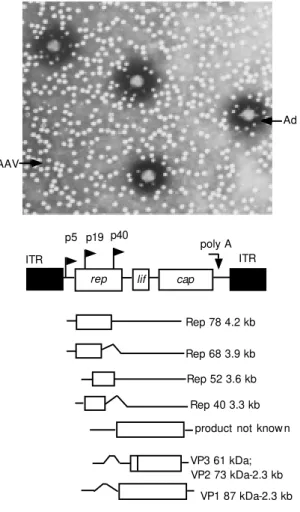
Vectors alternatively indicated to circum-vent these problems are based on the adeno-associated virus (AAV), a small non-envel-oped, non-pathogenic DNA virus belonging to the Parvoviridae family. The AAV ge-nome is a single-strand DNA molecule, 4681 bases long including two inverted terminal repeats (ITRs). ITRs are 145 bases long palyndromic sequences involved in the regu-lation of the AAV cell cycle. They are lo-cated in the 5' and 3' terminal portions of the viral genome, and serve as origins and initia-tors for DNA replication. Flanked by the ITRs, two large open reading frames code
for a regulatory protein and a structural pro-tein, rep and cap, respectively (Figure 3). The reading region located in the 5' region (rep gene) encodes four non-structural pro-teins involved in the genomic replication. The 3' portion contains the cap gene, which encodes three structural proteins for the for-mation of the viral capsid.
AAV is considered to be a dependovirus because it is capable of replication in a cell only in the presence of a helper virus (aden-ovirus or herpes virus), which provides by transcomplementation the helper factors that are essential for its replication. In the ab-sence of a helper virus, the AAV genome preferentially integrates into a specific site on the short arm of chromosome 19, be-tween q13.3 and qter, called AAVS1. The ITRs as well as a rep transcript (18) play an important role in this process, resulting in a latent infection in mitotic as well as in post-mitotic cells. Recently, episomal forms of the virus have been identified, and the inte-gration into non-specific sites has been docu-mented; however, there is no report of inser-tional oncogenesis. The latent provirus can be rescued and replicated following a super-infection with the helper virus.
There is a great interest in using AAV as a potential gene vector in human gene therapy trials (19). Among its most favorable proper-ties are i) no relation to human diseases; ii) broad infectivity spectrum, and iii) ability to stably integrate into the host genome. This kind of integration can occur in cells that are not dividing, although at a lower frequency than in dividing cells. The site-directed inte-gration is also a most favorable property of AAV.
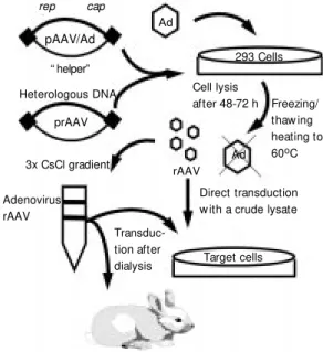
Recombinant adeno-associated viral vec-tors (rAAV) are derived from plasmids that carry ITRs flanking the exogenous gene of interest. These vectors can be packaged into the AAV capsid through co-transfection with the following elements in packaging cells: i) adenovirus and ii) a second packaging plas-mid carrying the rep and cap genes. The
Figure 3 - Adenovirus (Ad) and adeno-associated virus (AAV), AAV genome, differ-ent transcripts. ITR, Inverted terminal repeats. See Ref. 32.
AAV
Ad
p5 p19
poly A ITR
rep lif cap
U U U U
U
U
Rep 78 4.2 kb
Rep 68 3.9 kb
Rep 52 3.6 kb
Rep 40 3.3 kb
product not know n
VP3 61 kDa;
VP1 87 kDa-2.3 kb ITR
rAAV is rescued from cell lysates, and the helper virus is removed or inactivated (Fig-ure 4).
The disadvantages of rAAV particles as gene vectors include their small genome size (5 kb is the upper limit to AAV encapsida-tion), and the difficulty to produce large amounts of the vector. However, several improvements have been obtained in the production and use of AAV-based vectors. Richard J. Samulski and coworkers (20) have constructed a plasmid, pSub201, which con-tains the full-length AAV genome. Since then, the difficulty to produce rAAV DNA on a large scale has been minimized by the utilization of recombinant plasmids that can be growth-amplified in bacterial cultures.
The size limit has been circumvented in the sense that the rAAV DNA can be introduced into cells by lipofection. A summary of the advantages and disadvantages of the main viral vectors is provided in Table 3.
Combination of viral and non-viral e le m e nts
The combination of viral and non-viral elements can be used to increase the effi-ciency of gene transfer to cells. The incorpo-ration of adenoviral lysosomal degradation escape functions to encapsulated DNA is an example of such strategy (21,22). Complexes are prepared with adenoviral particles and poly-L-lysine linked to DNA, or to DNA encapsulated into liposomes. These com-plexes enable the simultaneous delivery of its components to each lysosome in the cell, diminishing lysosomal degradation of the DNA, and increasing the efficiency of trans-fection. Since adenoviruses used in this strat-egy can be irradiated to become non-infec-tious, the method has the potential to elimi-nate completely the use of any infectious viral DNA. However, there are unresolved questions concerning the duration of gene expression using such complexes, in vivo
efficiency, etc.
Another example of combination of viral and non-viral elements was mentioned in the AAV section and consists of the construc-tion of plasmids carrying the AAV ITR se-quences flanking the gene of interest to pro-mote the transformation of cells. AAV can be replaced for this kind of construction encapsulated into liposomes. The level of gene expression and transgene retention is similar to that observed with AAV alone. In 1994, Philip and coworkers (23) reported that the presence of the ITRs of AAV in plasmids could confer prolonged persistence of the transfected DNA if compared to the transfection mediated by conventional plas-mids lacking ITRs. In 1995, Vieweg and colleagues (24) demonstrated that the utili-zation of plasmids containing the ITR se-quences of AAV, complexed to cationic li-posomes can produce gene transfer and in vitro interleukin-2 (IL-2) gene expression that are 3 to 10 times higher than the levels obtained with plasmids lacking the ITRs. The levels of IL-2 expression achieved with these plasmids in an in vivo model of anti-tumor induction were comparable to the se-cretion levels obtained with the use of retroviral vectors. Expression levels are pos-sibly dose-dependent, according to results
Figure 4 - Steps in the pro-duction of recombinant ad-eno-associat ed part icles (rAAV). Ad, Adenovirus. From ref erence 33, w it h modifications.
rep cap
Ad
293 Cells
pAAV/Ad
“ helper”
Heterologous DNA
prAAV
3x CsCl gradient
Adenovirus rAAV
rAAV Cell lysis
after 48-72 h Freezing/ thaw ing heating to 60oC
Direct transduction w ith a crude lysate
Target cells
Transduc-tion after dialysis
reported by Malik and coworkers (25) show-ing that the frequency of AAV integration can jump from 2 to 49% of transfected cells, only by changing the AAV titer from 1.3 multiplicity of infection (MOI) to 130 MOI, respectively, representing a 25-fold increase in integration efficiency. This suggests that a higher number of rAAV genomic particles increases the likelihood of a copy being inte-grated into the specific chromosomal site.
A further improvement involves chang-ing the structure of the encapsulatchang-ing lipo-some. Hong and coworkers (26) combined adenoviral capsid proteins or adenoviral fi-ber proteins with liposomes, obtaining a con-siderable increase in the efficiency of trans-fection of the reporter gene LacZ cloned in an AAV-derived vector. Taken together, these results allow us to reach the following conclusions: i) plasmids carrying ITR se-quences produce high and stable levels of expression, and ii) cationic liposomes are an excellent alternative to carry rAAV into cells without triggering any immune response by the host.
The pote ntial use of mitochondria as D NA ve ctors
Mitochondria are the only cytoplasmic organelles that possess their own DNA (mtDNA). In this respect, mitochondria look
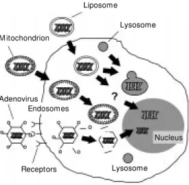
like liposomes that are used in gene transfer, constituted by lipid membranes enveloping DNA molecules (Figure 5). Since mitochon-dria can be purified by ultracentrifugation from cell homogenates, they could be used as gene transfer vectors. Mitochondria iso-lated from donor blood can be fused to re-ceptor cells, generating viable cytoplasmic hybrids (cybrids) (27,28).
The use of mitochondria as gene vectors has potential application in the replacement of mitocondrial DNA (mtDNA) in cells with oxidative phosphorylation defects caused by mutations of the mtDNA. mtDNA mutations are linked to a series of maternally inherited degenerative neuromuscular syndromes in-cluding mitochondrial encephalopathy, lac-tic acidosis, vomiting and stroke (MELAS), myoclonic epilepsy and ragged-red fiber (MERRF), neurogenic muscular weakness, and ataxia and retinitis pigmentosa (NARP), among others. mtDNA mutations also occur in cells of the somatic lineage and accumu-late with aging and in conditions of oxida-tive stress (29,30), and may account for a great deal of the phenotypes of advanced age, including muscle weakness, Alzheimer disease, and Parkinson disease.
Technically, the in vivo use of mitochon-dria as gene vectors is cumbersome. Target-ing the mtDNA to mitochondria in vivo of-fers a more viable approach to somatic gene therapy of diseases of the mitochondrial ge-nome.
In se arch of the ide al ve ctor: the future of ge ne the rapy
Ideally, a vector should accommodate an unlimited amount of inserted DNA, lack the ability of autonomous replication of its own DNA, be easily manufactured, and be available in concentrated form. Secondly, it should have the ability to target specific cell types or to limit its gene expression to specific cell types, and to achieve sustained gene expression in the long term or in a
Figure 5 - Potential use of mito-chondria as gene vectors for stable gene transfer to the cy-toplasm, as compared to lipo-somes and adenoviral vectors. This figure show s w hat hap-pens to liposomes (lysosomal degradation or nuclear incorpo-ration as an episome) and ad-enovirus (episomal incorpora-tion) used in gene transfer. M i-tochondria apparently possess membrane properties that en-dow them w ith cytoplasm sta-bility.
Lysosome
Nucleus Lysosome Liposome
M itochondrion
Adenovirus Endosomes
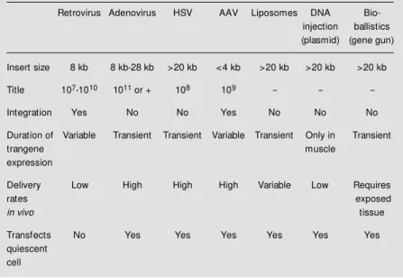
controlled fashion. Finally, it should not be toxic or immunogenic. Table 4 shows a comparison of seven main vector systems available to date. The analysis of the proper-ties of the main vector types reviewed in Tables 1-3 shows that none of these vector systems conforms to the notion of the ideal vector (Table 5).
There still exist serious limitations re-garding the efficiency and cell specificity of the available gene vector systems. Recombi-nant viral vectors are the most potent gene transfer vectors, but host immune response, safety issues, and the difficulty of scaling up their production constitute important barri-ers for their clinical use. The delivery of DNA via liposomes or via cell receptor tar-geting offers alternatives for virus-mediated gene transfer, but the levels of gene expres-sion obtained with these methods need to be significantly improved before they can be used for the treatment of human diseases. Furthermore, our knowledge about the tran-scriptional control is incomplete. All these aspects are under intense investigation; meth-ods for ex vivo and in vivo gene transfer are under rapid development, and there is the hope that considerable progress will be at-tained within the next decade.
The development of highly efficient sys-tems for viral packaging and the refinement of the purification and concentration pro-cesses could improve the titers of viral vec-tors to values that would allow gene transfer through systemic administration. Targeted delivery could be made possible through the incorporation of single chain antibodies raised against cell surface antigens or ligands for transmembrane receptors incorporated into the retrovirus envelope or adenovirus penton proteins, and there are encouraging signs that this strategy could reach success. Design vectors are expected to be avail-able in the short term. These vectors shall incorporate the most useful elements of pres-ently available viral and synthetic systems, with variations depending on the
applica-Table 5 - The ideal vector for gene transfer in gene therapy protocols.
Insert size - can accommodate one or more genes
Targeting - delivery to specific cell types or gene expression limited to target cells
Regulation - transgene expression levels can be up- or dow n-regulated
Safety and stability - safe for the patient and the environment - devoid of the risk of insertional mutagenesis
Immune response - none
Titles - high concentrations, final product stable
M anufacture - easy, reproducible, scale-up and standardization possible
tion. For example, the ITR of AAV, that promote stable chromosomal integration, can be combined to sequences of other vectors with more insert accommodation capacity. Integration efficiency can be increased through the packaging of a functional recombinase together with the gene expres-sion construction in the form of liposome complexes directed at a specific cell type by antibodies or receptor-ligand proteins.
In the long term, new delivery methods could be developed for the intranuclear trans-fer of HACs carrying whole gene blocks
Table 4 - Vector comparison.
HSV, Herpes simplex virus; AAV, adeno-associated virus.
Retrovirus Adenovirus HSV AAV Liposomes DNA Bio-injection ballistics (plasmid) (gene gun)
Insert size 8 kb 8 kb-28 kb >20 kb <4 kb >20 kb >20 kb >20 kb
Title 107-1010 1011 or + 108 109 - -
-Integration Yes No No Yes No No No
Duration of Variable Transient Transient Variable Transient Only in Transient
trangene muscle
expression
Delivery Low High High High Variable Low Requires
rates exposed
in vivo tissue
Transfects No Yes Yes Yes Yes Yes Yes quiescent
with natural control elements. In the future, basic research shall try to define which ge-nomic elements are involved in the temporal and spatial control of gene expression during the whole lifespan of an organism. Advances in these fields will require parallel develop-ments in vector design before the knowledge can be utilized in clinical practice.
From the wide variety and creativity of vector systems and methods of gene transfer under development it seems clear that in the
future there will be a wide choice of different gene transfer methods for different clinical applications. This situation certainly will broaden the opportunities for the use of gene transfer technology in the clinical context. Gene therapy promises to be a fruitful area of scientific and clinical research for many years to come, and there is no doubt that it will become an important clinical practice in the next century.
Re fe re nce s
1. Findeis M A, Wu CH & Wu GY (1994). Ligand-based carrier systems for delivery of DNA to hepatocytes. M ethods in Enzy-mology, 247: 341-351.
2. Stankovics J, Crane AM , Andrew s E, Wu CH, Wu GY & Ledley FD (1994). Overex-pression of human methylmalonyl CoA mutase in mice after in vivo gene transfer w ith asialoglycoprotein/polylysine/DNA complexes. Human Gene Therapy, 5: 1095-1104.
3. Harrington JJ, Van Bokkelen G, M ays RW, Gustashaw K & Willard HF (1997). Forma-tion of de novo centromeres and con-struction of first-generation human artifi-cial microchromosomes. Nature Genetics, 15: 345-355.
4. Lasic DD & Papahadjopoulos D (1995). Liposomes revisited. Science, 267: 1275-1276.
5. Zhu N, Liggitt D, Liu Y & Debs R (1993). Systemic gene expression after intrave-nous DNA delivery into adult mice. Sci-ence, 261: 209-211.
6. Hangai M , Kaneda Y, Tanihara H & Honda Y (1996). In vivo gene transfer into the retina mediated by a novel liposome sys-tem. Investigative Ophthalmology and Visual Science, 37: 2678-2685.
7. Kato K, Nakanishi M , Kaneda Y, Uchida T & Okada K (1991). Expression of hepatitis B virus surface antigen in adult rat liver: co-introduction of DNA and nuclear pro-tein by a simplified liposome method. Journal of Biological Chem istry, 266: 3361-3364.
8. Takeda S, M iyoshi S, Omori K, Utsumi T, Kogaki S, Saw a Y, Yanagisaw a M & M atsuda H (1998). Pulmonary disease models induced by in vivo hemagglutinat-ing virus of Japan liposome-mediated en-dothelin-1 gene transfer. Journal of
Car-diovascular Pharmacology, 31 (Suppl 1): S336-S338.
9. Sanford JC, Klein TM , Wolf ED & Allen N (1987). Delivery of substances into cell tissues using a particle bombardment pro-cess. Particulate Science and Technology, 5: 27-37.
10. Klein TM , Arent zen R, Lew is PA & Fitzpatrick-M cElligott S (1992). Transfor-mation of microbes, plants and animals by particle bombardment. Bio/Technol-ogy, 10: 286-291.
11. Klein TM & Fit zpat rick-M cElligot t SF (1993). Particle bombardment: a universal approach for gene transfer to cells and tissues. Current Opinion in Biotechnol-ogy, 4: 583-590.
12. Sanford JC, Smith FD & Russell JA (1993). Optimizing the biolistic process for differ-ent biological applications. M ethods in Enzymology, 217: 483-510.
13. Vainstein M H, Alves SA, Lima BD, Aragão FJL & Rech EL (1994). Stable DNA trans-fection in a flagellate trypanosomatid by microparticle bombardment. Nucleic Ac-ids Research, 22: 3263-3264.
14. Aragão FJL, Barros LM G, Brasileiro ACM , Ribeiro SG, Smith FD, Sanford JC, Faria JC & Rech EL (1996). Inheritance of for-eign genes in transgenic bean (Phaseolus vulgaris L.) co-transformed via particle bombardment. Theoretical and Applied Genetics, 93: 142-150.
15. Tang D, De Vit M & Johnston SA (1992). Genetic immunization is a simple method for eliciting an immune response. Nature, 356: 152-154.
16. Rech EL, De Bem R & Aragão FJL (1996). Biolistic-m ediated gene expression in guinea pigs and cattle tissues in vivo. Bra-zilian Journal of M edical and Biological Research, 29: 1265-1267.
17. Haecker SE, Stedman HH, Balice-Gordon RJ, Smith DB, Greelish JP, M itchell M A, W ells A, Sw eeney HL & W ilson JM (1996). In vivo expression of full-length human dystrophin from adenoviral vec-tors deleted of all viral genes. Human Gene Therapy, 7: 1907-1914.
18. Balague C, Kalla M & Zhang WW (1997). Adeno-associated virus Rep78 protein and terminal repeats enhance integration of DNA sequences into the cellular genome. Journal of Virology, 71: 3299-3306. 19. Page KW & Lebkow ski JS (1997).
Adeno-associated virus: a vector for high-effi-ciency gene transduction. In: Bertino JR (Editor), Encyclopedia of Cancer. Vol. II. Academic Press, San Diego, CA, 1-9. 20. Samulski RJ, Chang LS & Shenk T (1987).
A recombinant plasmid from w hich an in-fectious adeno-associated virus genome can be excised in vitro and its use to study viral replication. Journal of Virology, 61: 3096-3101.
21. Curiel DT (1994). High-efficiency gene transfer employing adenovirus-polylysine-DNA complexes. Natural Immunity, 13: 141-164.
22. Fasbender A, Zabner J, Chillon M , M oninger TO, Puga AP, Davidson BL & Welsh M J (1997). Complexes of adenovi-rus w ith polycationic polymers and cat-ionic lipids increase the efficiency of gene transfer in vitro and in vivo. Journal of Biological Chemistry, 272: 6479-6489. 23. Philip R, Brunette E, Kilinski L, M urugesh
and Cellular Biology, 14: 2411-2418. 24. View eg J, Boczkow ski D, Roberson KM ,
Edw ards DW, Philip M , Philip R, Rudoll T, Smith C, Robertson C & Gilboa E (1995). Efficient gene transfer w ith adeno-associ-ated virus-based plasmids complexed to cationic liposomes for gene therapy of human prostate cancer. Cancer Research, 55: 2366-2372.
25. M alik P, M cQuiston SA, Yu X-J, Pepper KA, Krall WJ, Podsakoff GM , Kurtzman GJ & Kohn DB (1997). Recombinant adeno-associated virus mediates a high level of gene transfer but less efficient integra-tion in the K562 human hematopoietic cell line. Journal of Virology, 71: 1776-1783.
26. Hong Z, Guangbin Z, Xiaojun Z, Jian T, Guanghui C, Qianhui H, Tongcai P & Bingquan H (1995). Enhanced adeno-as-sociated virus vector expression by
aden-ovirus protein-cationic liposome complex. A novel and high efficient w ay to intro-duce foreign DNA into endothelial cells. Chinese M edical Journal, 108: 332-337. 27. Fukami M H & Salganicoff L (1973).
Isola-tion and properties of human platelet mi-tochondria. Blood, 42: 913-918.
28. Chomyn A, Lai ST, Shakeley R, Bresolin N, Scarlato G & Attardi G (1994). Platelet-mediated transformation of mtDNA-less human cells: analysis of phenotypic vari-ability among clones from normal individu-als and complementation behavior of the tRNALys mutation causing myoclonic epi-lepsy and ragged red fibers. American Journal of Human Genetics, 54: 966-974. 29. M erril CR, Zullo S, Ghanbari H, Herman M M , Kleinman JE, Bigelow LB, Bartko JJ & Sabourin DJ (1996). Possible relation-ship betw een conditions associated w ith chronic hypoxia and brain mitochondrial
DNA deletions. Archives of Biochemistry and Biophysics, 326: 172-177.
30. Wallace DC, Brow n M D, M elov S, Gra-ham B & Lott M (1998). M itochondrial biology, degenerative diseases and aging. Biofactors, 7: 187-190.
31. Stannard LM (1995). Electron micrograph image of adenovirus. WWW site at: http:// w w w .uct .ac.za/dept s/m m i/st annard/ emimages.html
32. M ayor HD & Kurstak E (1974). Viruses w ith separately encapsidated comple-mentary DNA strands. In: Kurstak E & M aramorosch K (Editors), Viruses, Evolu-tion and Cancer. Academic Press, Inc., New York.