ACTA RADIOLÓGICA PORTUGUESA September-December 2019 Vol 31 nº 3 13-18
Accessing Non-Atherosclerotic Vasculopathies Using
Ultrasound-Doppler Imaging
Avaliação das Vasculopatias Não Ateroscleróticas por Ecografia-Doppler
Carolina Ferreira Pinheiro, Teresa Morais, Catarina Perry da Câmara, Cristina Rios, Mariana Cardoso Diogo, Madalena Patrício Ferreira
Departament of Neurorradiology, Centro Hospitalar e Universitário de Lisboa Central, Lisboa, Portugal
Address
Carolina Ferreira Pinheiro Departamento de Neurorradiologia Centro Hospitalar e Universitário de Lisboa Central
Rua José António Serrano 1150-199 Lisboa, Portugal
e-mail: carolina.pinheiro@chlc.min-saude.pt
Resumo
As vasculopatias não ateroscleróticas são um grupo de doenças pouco frequentes, de diversa etiopatogenia, que envolvem grupos etários mais jovens, comparativamente à doença aterosclerótica, e que se manifestam clinicamente por eventos vasculares agudos- isquémicos ou hemorrágicos - ou quadros neurológicos incaracterísticos.
Sendo a angiografia cerebral diagnóstica o método gold standard para o diagnóstico destas patologias, devido à sua elevada sensibilidade, a sua especificidade na avaliação da parede vascular é menor quando comparada com a avaliação por ecografia-Doppler carotídeo e vertebral. Salientando-se o carácter não invasivo da técnica, e o facto de não usar radiação ionizante, o eco-Doppler permite, não só o diagnóstico, como a monitorização repetida e a longo prazo destes jovens pacientes.
Palavras-chave
Ecografia-doppler; Ecografia dos vasos do pescoço; Vasculopatias não-ateroscleróticas; Imagem da parede vascular; Doença de Moyamoya; Disseção arterial; Displasia fibromuscular; Arterite de Takayasu; Arterite de células gigantes; Drepanocitose; Vasculopatia induzida pela radiação.
Abstract
The non-atherosclerotic vasculopathies are an uncommon group of disorders, with diverse etiopathogenesis, involving younger patients when compared with atherosclerotic disease. Clinical presentation varies from acute vascular events - ischemic or hemorrhagic- to uncharacteristic neurologic syndromes. Although the cerebral angiography is the gold standard diagnostic method due to its high sensibility, its specificity is low mainly when compared to imaging evaluation of the arterial wall with carotid and vertebral ultrasound-doppler. We reinforce the importance of this non-invasive and radiation-free exam, not only in the diagnosis but also in the monitoring of these patients - young patients who need a regular and extended evaluation.
Keywords
Ultrasound-doppler; Carotid ultrasound; Non-atherosclerotic vasculopathies; Vessel-wall imaging; Moyamoya disease; Vertebral artery dissection; Fibromuscular dysplasia; Takayasu arteritis; Giant cells arteritis; Sickle cell disease; Radiation-induced vasculopathy.
Review Article / Artigo Revisão
Introduction
The non-atherosclerotic vasculopathies are an uncommon group of disorders, with diverse etiopathogenesis, involving younger patients when compared with atherosclerotic disease. Clinical presentation varies from acute vascular events - ischemic or hemorrhagic- to uncharacteristic neurologic syndromes.
Cerebral angiography is considered the gold standard diagnostic method for imaging blood vessels, but carotid and vertebral ultrasound (US)-Doppler is an excellent alternative and complementary technique to evaluate the arterial wall. The use of color and/or power Doppler US in the assessment of non-atherosclerotic vasculopathies is of great importance due to different reasons. Not only is this a helpful diagnostic tool, but also an inexpensive imaging technique, radiation-free and non-invasive technique. Also, it allows a frequent and feasible monitorization of different diseases, which is especially important in younger patients who need a lifetime follow-up.
It is also important to note that there are some disadvantages: it is highly operator-dependent and it must be performed or validated by experienced and trained operators and, due to artifacts, it does not allow a precise study of the arterial wall in the distal internal carotid arteries and intracranial vessels.
We present six different examples of non-atherosclerotic vasculopathies, namely Moyamoya disease, vertebral artery dissection in a patient with fibromuscular dysplasia, Takayasu arteritis, giant cells arteritis, sickle cell disease and radiation-induced vasculopathy.
Moyamoya Disease
Described in the English literature by Japanese neurosurgeons in the 1960’s, Moyamoya disease (MMD) is a chronic, occlusive cerebrovascular disease characterized by progressive stenosis at the terminal portion of the internal carotid artery (ICA) and an abnormal vascular network at the base of the brain.1 The term “moyamoya”
is derived from a Japanese expression for something hazy, like a puff of cigarette smoke drifting in the air.
The diagnostic criteria,1 from 1997, proposed by the
Research Committee on Spontaneous Occlusion of the Circle of Willis suggested three principal factors:
-Stenosis or occlusion at the terminal portion of the internal carotid artery (ICA) and/or at the proximal portion of the anterior and/or middle cerebral arteries; -Abnormal vascular networks in the vicinity of the
occlusive or stenotic lesions in the arterial phase; -Lesion bilaterality.
Recently, in 2015, a statement by the Research Committee of MMD of the Japanese Ministry of Health, Labour and Welfare,2 proposed the key modification that the
“bilaterality” should be omitted. If unilateral disease is observed, a cerebral angiography is needed to confirm the diagnosis, whereas bilateral cases can be promptly diagnosed by either catheter angiography or magnetic resonance imaging/angiography (MRI/MRA).2
The US-Doppler technique, particularly power Doppler, is of great value as it is more sensitive to detect low-velocity blood flow signals in arteries that appeared occluded on cerebral angiography.3
Case 1
A 11-year-old boy who had a right hemispheric ischemic stroke when he was only 10-months-old. By the age of 14-month-old, he had another ischemic event, involving his left hemisphere, resulting in a right-sided hemiparesis. During the investigation, MMD was confirmed on cerebral angiography (not shown).
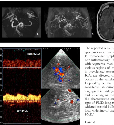
Due to lack of cerebral collateralization (Figure 1.A), he was submitted to revascularization surgery with good results (Figure 1.B).
The transcranial Doppler US (TCD) (Figure 2) confirmed left middle cerebral artery (MCA) patency after surgery and, in some patients, this method may be more sensitive in detecting arterial flow than MRA.
Vertebral Artery Dissection in a Patient with
Fibromuscular Dysplasia
A dissection is produced by subintimal penetration of blood in a vessel with subsequent longitudinal extension of the intramural hematoma for a varying distance between its layers, and it is usually associated with an intimal tear.4 Although vertebrobasilar and intracranial
carotid dissections are less common than the extracranial carotid artery, with the advance in imaging techniques asymptomatic multiple vessel involvement can be detected.5
Cervicocephalic arterial dissections have been reported after trivial trauma,4 but it also can occur spontaneously
particularly in association with fibromuscular dysplasia, connective tissue disease such as Marfan’s syndrome and Ehlers-Danlos syndrome,5 and MMD.
The traditional method for visualization of arterial dissection is cerebral angiography. MR images can show a periarterial rim of intramural hematoma typically with hyperintense signal on both T1 and T2 weighted images.
Figure 1 – A. TOF MRA
before revascularization surgery. B. TOF MRA after revascularization surgery. C. Axial T2/FLAIR, encephalomalacia involving the left frontal and parietal lobes - consequence of ischemic stroke.
Figure 2 – TCD reveals patency of both MCAs.
The reported sensitivity of neurovascular US for detecting spontaneous arterial dissection varies from 80 to 96%.5
Fibromuscular dysplasia (FMD) is a non-atheromatous, non-inflammatory arteriopathy of unknown etiology with segmental manifestation on medium-sized arteries in various regions of the body.6 Second to the renal arteries
in prevalence,7 extracranial cervical arteries, including the
ICAs are affected, often bilaterally but manifestation also occurs on the vertebral artery (VA).6
Depending on the type of FMD (intimal, medial, and subadventitial-perimedial), US findings correlate with the angiographic findings, and may show segmental narrowing and widening or the color flow in cervical arteries, with the characteristic string of beads appearance in medial type of FMD, long tubular stenosis, usually distally from a widened carotid bulb in intimal type of FMD, or irregular local widening of the arterial wall in subadventitial type of FMD.5
Case 2
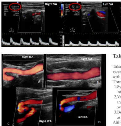
A 44-year-old female patient presenting cervical pain and right hemiparesis, after physical exercise.
An emergent CT-angiography (Figure 2) and MRI (Figure 3), showed an irregular right VA (segment V4) and an acute ischemic stroke on the right dorsolateral medulla oblongata. During her hospitalization, a color Doppler US was performed (Figure 4 and 5) showing different blood flow velocities in the vertebral arteries (V2 segments), with lower velocity on the right (Figure 4). The carotid examination (Figure 5) revealed wall irregularities, with bilateral kinking of both ICAs.
Figure 3 – A+B: CT- angiography revealed an irregular filling of the right vertebral artery (V4 segment) (green arrow). C+D:
Axial DWI+ADC map showed a restricted diffusion area (red arrow) in the right dorsolateral medulla oblongata compatible with acute ischemic stroke.
Figure 4 – Color Doppler ultrasound demonstrating VAs
with different blood flows velocities.
Figure 5 – A+B: Power Doppler US imaging, revealing wall irregularities
in the right ICA. C+D: Color Doppler US imaging showing kinking of both ICAs.
Takayasu’s Arteritis
Takayasu’s arteritis (TA) is an idiopathic granulomatous vasculitis that affects the aortic arch and its main branches with unknown etiology.8 Most patients are young women.9
Three stages have been described:5
1.Systemic stage, where symptoms and signs of an active inflammatory illness dominate;
2.Vascular inflammatory stage, when stenosis, aneurysms, and vascular pain tend to occur. This stage sometimes overlaps with the systemic stage;
3.Burned-out stage develops when fibrosis sets in, and it is usually associated with remission.
Although it is difficult to establish the diagnosis in the early stages, US can help identify early TA. Cerebral angiography is the gold standard,5 and it usually demonstrates changes/
narrowing in the lumen of multiple arteries. Sonography shows changes mainly of the arterial wall in a small number of important arteries, as delineated characteristic long segments with homogeneous, mid echoic, circumferential arterial wall thickening, which has been described as the ‘macaroni sign’.9
Sonography and cerebral angiography are complementary methods in the diagnosis of Takayasu arteritis.
Case 3
A 15-year-old female patient, diagnosed with TA at 11-years-old, after episodes of severe headache and multiple transient ischemic accidents with left sided hemiparesis and dysarthria. During the hospital stay she underwent several complementary diagnostic examinations: carotid Doppler US discovered mural thickening on both common carotid arteries with partial luminal obliteration and low flow velocities (Figure 7).
A diagnostic cerebral angiography (Figure 6) confirmed the CT-angiography and US findings: an irregular right vertebral, with a stenosis followed by a post stenosis dilation compatible with V4 segment dissection (B+C orange arrow) and marked wall irregularities, with areas of stenosis followed by dilatation in the more distal right ICA (A) - compatible with FMD.
Figure 6 – Cerebral angiography: A. contrast injection in the
brachiocephalic trunk - right carotid axis. B: Anterior-Posterior view of a right VA injection. C: Lateral view of a right VA injection – vessel irregularity (orange arrow) compatible with dissection.
These US findings were confirmed by MRA (Figure 8), with marked stenosis and occlusion of large vessels (right and left CCA and left VA).
Giant Cell Arteritis
Giant cell arteritis (GCA), also known as temporal arteritis or cranial arteritis, is the most common form of vasculitis that occurs in adults older than the age of 50. It is a granulomatous arteritis affecting large or medium sized artery, usually temporal or ophthalmic artery.5 It
can be considered an ophthalmological emergency, its most serious complication being irreversible visual acuity loss secondary to ischemic optic neuropathy, that may become bilateral within a few days or weeks if a prompt diagnosis and treatment are not established.10 Standard test
for diagnosing GCA is biopsy of the temporal artery.5 In
recent years, non-invasive imaging techniques such as color Doppler US have been applied to overcome the limitations of the biopsy. Temporal artery color Doppler US can identify three characteristic US features:10
1.A periluminal hypoechogenic halo reflecting arterial wall edema;
2.Segmental arterial stenosis;
3.Arterial luminal occlusion in severe cases.
A positive halo sign strongly supports a diagnosis of GCA in the presence of compatible clinical manifestations, but the absence of a halo does not rule it out.11 This sign is
Figure 7 – A+C - Marked wall thickening on both common carotid
arteries (CCA) - blue asterisk. Using power Doppler (B+D) note the weak luminal filling, indicating stenosis.
Figure 8 – A: Gadolinium-enhanced MRA (3D) B: TOF-MRA: circle of
willis (3D). Important vessels asymmetry. Intracranial circulation is mainly dependent on the right vertebral artery (blue arrow).
due to vessel wall edema and can be distinguished from the focal hyperechoic wall thickening seen in atherosclerosis.12
During healing, regression of the dark halo will be visible parallel with the restitution of the color-coded flow.5
Case 4
A 72-year-old female patient, with jaw claudication, was admitted in the emergency room due to prostration, walk imbalance and nausea, related with an acute posterior circulation ischemic stroke confirmed with an emergent MRI (Figure 9).
During the hospital stay, the US (Figure 10) revealed thickening / “halo sign” on both superficial temporal arteries.
The CT-angiography revealed irregularities on both VA (Figure 11) supporting the diagnostic hypothesis of vasculitis. Her erythrocyte sedimentation rate was elevated and a GCA was considered. She started methylprednisolone with improvement of symptoms.
Figure 9 – Axial DWI (A), ADC map (B) confirmed the bilateral acute
ischemic lesions involving the pontine tegmentum, the right middle cerebellar peduncle and the right cerebellar hemisphere, also visible on Axial T2/FLAIR (C) showing the same hyperintense lesions (blue arrows).
A B
C
Figure 10 – A:”Halo” sign (red circle) of the right TSA, corresponding
to wall artery thickening (White arrows) due to wall edema on both STA (B+C).
Sickle Cell Disease
Children with sickle cell disease (SCD) are at high risk for stroke. The risk is highest in children with elevated blood flow velocity in the distal/ terminal internal carotid or proximal middle cerebral artery (MCA), as measured with transcranial Doppler US (TCD).13
Chronic blood transfusions, if implemented in a timely fashion in those with flow velocity >200 cm/s, can reduce the risk of stroke by as much as 92%. It is also suggested that unilateral high-flow velocity indicates stenosis, whereas bilateral high-velocity represents bilateral stenosis, hyperemia, or both.14 It is important to emphasize that the
STOP velocity criteria apply only to children with SCD who have not had a stroke. Those with abnormal velocity should undergo repeated screening within the next few weeks and if the second study is also abnormal they should be offered transfusion therapy.15
Figure 11 – CT-
angiography confirmed irregular VAs, in the context of vasculitis.
Case 5
A 17-year-old female patient diagnosed at 1 year of age with sickle cell disease after several vaso-occlusive crises. She was under a regular regime of blood transfusions since the age of 14. Due to important cerebral vasculopathy (bilateral ischemic lesions and a right ICA stenosis), she was submitted to an indirect brain revascularization (encephalo-duro-arterio-synangiosis) by the age of 15, with good development of collateral circulation (Figure 12). The TCD (Figure 13) confirmed normalization of velocities on the left MCA after surgery.
Table 1 – Adapted from: Nichols, F. et al; American Society of
Neuroimaging (2001)
Figure 12 – MRA-TOF (3D) before (A) and after (B) the revascularization
surgery. Note the anatomic variant: left VA fenestration.
Figure 13 – TCD images: MCA velocities, before (A+C) and after (B+D) the indirect
revascularization surgery. Note the regularization of arterial velocity flow on the left MCA.
Radiation Induced Vasculopathy
Cerebral arteriopathy may result from therapeutic irradiation of neck or intracranial malignancies. The vascular complications usually develop 6 months to 10 years after irradiation.8 It is an underrecognized precursor
to ischemic stroke in these patients.16 In children, and
rarely in adults, it may be associated with the formation of netlike vessels and transdural anastomoses (Moyamoya disease like). The exact pathogenesis of radiation-induced carotid artery disease is still unclear.17 Some authors have
argued that chronic occlusive radiation vasculopathy is primarily an accelerated form of atherosclerosis; others have described it as a distinct disease entity shaped by the initial radiation insult to the vasa vasorum.16 A hallmark of
radiation vasculopathy is its occurrence in patients who lack traditional vascular risk factors. The diagnosis of carotid stenosis still relies principally on the imaging approaches.17
Case 6
A 65-year-old man patient, with a history of larynx cancer, treated with surgery and radiation, had a wake-up stroke with left sided hemiparesis. Brain-CT (Figure 14. A.) showed a right insular and external capsule ischemic lesion.
Figure 14 – A: Brain-CT: right insular and
external capsule infarct lesion (red arrow) Note no color mode filling nor flux registered either in the right common carotid artery (B) and vertebral artery (C).
Figure 15 – A: cerebral angiography: contrast injection on the
brachiocephalic trunk. No right CCA or right VA are seen. Thyrocervical trunk (green arrow); divides into inferior thyroid artery (blue arrow) and ascending cervical artery (red arrow). Also note the deep cervical artery (yellow arrow). B: CT-angiography - 3D reconstruction.
During the hospital stay, he underwent vascular study with carotid US that showed hyperechoic infiltration on both carotid axes, with an occluded right CCA and VA, and a > 50% stenosis on left ICA, most likely related with neck radiation performed 12 years ago. Both the cerebral angiography and CT-angiography confirmed US findings (Figure 15).
Conclusion
The US-Doppler technique is an alternative exam to the cerebral angiography, with a high diagnostic capacity (sensibility and specificity), being the method of excellence in monitoring the arterial wall disease.
Received / Recebido 29/08/2018 Acceptance / Aceite 29/07/2019 Ethical disclosures / Divulgações Éticas
Conflicts of interest: The authors have no conflicts of interest to declare. Conflitos de interesse: Os autores declaram não possuir conflitos de interesse. Financing Support: This work has not received any contribution, grant or scholarship.
Suporte financeiro: O presente trabalho não foi suportado por nenhum subsídio ou bolsa.
Confidentiality of data: The authors declare that they have followed the protocols of their work center on the publication of data from patients. Confidencialidade dos dados: Os autores declaram ter seguido os protocolos do seu centro de trabalho acerca da publicação dos dados de doentes. Protection of human and animal subjects: The authors declare that the procedures followed were in accordance with the regulations of the relevant clinical research ethics committee and with those of the Code of Ethics of the World Medical Association (Declaration of Helsinki). Protecção de pessoas e animais: Os autores declaram que os procedimentos seguidos estavam de acordo com os regulamentos estabelecidos pelos responsáveis da Comissão de Investigação Clínica e Ética e de acordo com a Declaração de Helsínquia da Associação Médica Mundial.
Bibliography
1. Hishikawa T, et al. Moyamoya disease: a review of clinical research. Acta Med Okayama. 2016;70:229-36.
2. Kim JS. Moyamoya disease: epidemiology, clinical features, and diagnosis. Journal of Stroke. 2016;18:2-11.
3. Ruan L,et al. Color and power doppler sonography of extracranial and intracranial arteries in moyamoya disease. Journal of Clinical Ultrasound. 2006;34:60-9.
4. Biller J. Non-atherosclerotic cerebral vasculopathies in the young. Journal of Stroke and Cerebrovascular Diseases. 1997;6:173-7.
5. Huzjan A. Diagnosis of non-atherosclerotic carotid disease. Perspectives in Medicine. 2012;1:244-9.
6. Arning C, et al. Color doppler imaging of cervicocephalic fibromuscular dysplasia. Cardiovascular Ultrasound. 2004;2:7
7. Southerland A, et al. Shared associations of nonatherosclerotic, largevessel, cerebrovascular arteriopathies: considering intracranial aneurysms, cervical artery dissection, moyamoya disease and fibromuscular dysplasia. Curr Opin Neurol. 2013;26:13-28.
8. Kim J, et al. Non-atherosclerotic intracranial arterial diseases. Front Neurol Neurosci. 2016;40:179-203.
9. Schmidt W, et al. Diagnosis of early takayasu arteritis with sonography. Rheumatology. 2002:41:496-502.
10. Suelves A, et al. Doppler ultrasound and giant cell arteritis. Clinical Ophthalmology. 2010;4:1383-4.
11. Pipitone N, et al. Role of imaging studies in the diagnosis and follow-up of large-vessel vasculitis: an follow-update. Rheumatology. 2008;47:403-8. 12. Ninan J, et al. Giant cell arteritis – beyond temporal artery biopsy and steroids. Intern Med J. 2017;47:1228-40.
13. Krejza J, et al. Sickle cell disease and transcranial doppler imaging inter-hemispheric differences in blood flow doppler parameters. Stroke. 2011;42:81-6.
14. Adams R, et al. Prevention of a first stroke by transfusions in children with sickle cell anemia and abnormal results on transcranial doppler ultrasonography. N Engl J Med. 1998;339:5-11.
15. Zétola V. Role of TCD in sickle cell disease: a review. Perspectives in Medicine. 2012;1:265-8.
16. Plummer C, et al. Ischemic stroke and transient ischemic attack after head and neck radiotherapy. Stroke. 2011;42:2410-8.
17. Xu J, et al. Radiation-induced carotid artery stenosis: a comprehensive review of the literature. Intervent Neurol. 2013;2:183-92.