Antioxidant status of
Penaeus monodon
fed
with
Dunaliella salina
supplemented diet
and resistance against WSSV
M. MADHUMATHI*
Research scholar, Center for advanced studies in Botany University of Madras, Maraimalai campus, Chennai-65
Tamil Nadu, India. Mobile No: +91 9841111534
madhumathi83@gmail.com
R.RENGASAMY
Director &Professor, Center for advanced studies in Botany University of Madras, Maraimalai campus, Chennai-65
Tamil Nadu, India. Mobile No: +91 9176295052 profrrengasamy@yahoo.com
Abstract
The present study investigates the protection of shrimp Penaeus monodon against white spot syndrome virus (WSSV) using Dunaliella salina algal cells which contains an antioxidant betacarotene for the shrimp non-specific immunity. To determine the antioxidant activity, the shrimp were treated in vivo (orally with feed) methods at the concentration of 0.5 %, 1.0 % and 2 % D. salina incorporated with pellet feed, respectively. In the present study, anti-WSSV activity of D. salina incorporated diet by in vivo methods showed strong antioxidant activity and the immunological parameters such as proPO, SOD,catalase were higher in the WSSV-infected shrimp treated with D. salina incorporated diet when compared to control groups. These results strongly indicate that in vivo of D. salina incorporated diet enhances immunity of the shrimp. Based on the present data and the advantages of harvesting D. salina at low price, we believe that oral administration of D. salina live cells along with the pellet feed is a potential prophylactic agent against WSSV infection of shrimp to some extent.
Keywords:Penaeus monodon; Dunaliella salina; antioxidant activity.
1. Introduction
studies have been carried out to obtain the performance of immunostimulants for enhancing immune response and reduction of disease impacts. Several diseases were controlled by oral administration of immunostimulation.
Immunostimulants are suitable for boosting immature immune system and effective against a number of opportunistic and secondary pathogens (Felix et al., 2004). Thus modern shrimp hatchers are now trying to resolve this problem of WSSV infection by using immunostimulant incorporated shrimp feed. Studies have been carried out to obtain the performance of immunostimulants for enhancing immune response and reduction of disease impacts. Certain carotenoids obtained from algal sources like astaxanthin and β-carotene induces immune system in shrimp and will certainly continue to play an important role in disease control in intensive shrimp culture (Karin van de, 2002). In order to know the role of this immunostimulant of carotenoids obtained from algae on resistance of shrimp against WSSV and its consequences on the immune condition of shrimp, the present study was carried out to determine the effect of betacarotene obtained from Dunaliella salina incorporated with different concentrations on prophenol oxidase, superoxide dismutase, catalase (SOD) production during the course of the WSSV challenge to assess the non-specific immune status of the shrimp.
2. Materials and Methods
2.1. Preparation of betacarotene from D. salina
Ten litre of Dewalnes medium were prepared with 35 ppt and inoculated with 100 ml of pure Dunaliella salina culture. The cultures were kept in out door condition for a period of 20 days. On 15th day orange stage is attained. Then the cultures were harvested and centrifuged at 10,000 rpm for 10 min. The obtained biomass were lyophilised and stored in the refrigerator until they were used for the bioassay test.
2.2. Preparation of test diets
One litre of fresh water was taken in a vessel and boiled on a heater. The ingredients were cooked as described below. The rice bran was added at first and mixed thoroughly, followed by the addition of fishmeal, coconut cake, squid meal, wheat flour powder and mixed thoroughly, until the feed became a paste and cooled to room temperature and added the ingredients such as fish oil, vitamin mix and tapioca. To this different percentages of harvested biomass of D. salina cells (dry wt) such as 0.5 %, 1.0 % and 2 % were added separately and mixed thoroughly. Then the paste was taken pressed and squeezed through 2 mm2 pores and collected the shreds on a polythene sheet. The shreads without D. salina served as control. The shreds were allowed to dry at room temperature for 2 days kept in plastic bags and sealed properly until given to shrimp.
2.3. Collection and maintenance of experimental animals
used for the experiment. Temperature, pH and salinity (Aqua fauna, Japan) were recorded on the spot. This experiment was carried out for a period of 30 days.
2.4.Feeding trial on Penaeus monodon
The experimental shrimp, Penaeus monodon was fed with normal diet (control) for a period of 2 days and then taken for the experimental study. The animals were divided into four groups of each 10 shrimps in 100 L seawater in fibre tanks at room temperature (27-30°C) with salinity between 20 and 25 ppt. Group I animals were fed with the normal pellet feed without D. salina (control). In Group II, the shrimp were fed with 0.5 % D. salina incorporated feed, Group III fed with 1.0 % D. salina incorporated feed, and Group IV fed with 2 % D. salina incorporated feed for a period of one month. The animals were fed twice daily at 09:00: and 17:00 h for a period of 30 days. The amount of feed given was 5 % of animal body weight. Every alternate days, 50 % volume of the water along with the excreta of animals and other waste were removed and compensated with fresh seawater.
2.5.Determination of immunological and antioxidant activity
The uninfected shrimp fed with different concentrations of D. salina incorporated diet were sacrificed weekly for a period of one month for immunological assay such as prophenoloxidase assay (proPO), superoxide anion assay, superoxide dismutase activity and catalase assay were analyzed for different groups.
2.6.Haemolymph collection
Haemolymph of 300 µl per shrimp was collected directly from the heart of animal every week for a period of one month. The haemolymph was collected by using a 23-guage needle and 1.0 mL syringe contained 300 µl (4°C) precooled 10 % sodium citrate solution as anticoagulant in glass distilled water.
2.7.Estimation of protein
The estimation of protein was carried out by following the method of Lowery et al., 1951. One hundred µL of haemolymph samples were made up to 1 mL separately with glass distilled water. To this 4.5 mL of alkaline copper reagent was added shaken well and allowed to stand for 10 minutes. To this 0.5 mL of Folin’s reagent was added and kept at room temperature at 37°C for 20 minutes. The absorbance was read at 660 nm using UV visible spectrophotometer (UV- 1601 Shimadzu). Total protein expressed as µg/mL in haemolymph.
2.8.Phenoloxidase activity
L-DOPA (Leonard et al., 1985). The data were recorded every one minute interval for a period of 5 minutes at 490 nm.
2.9.Assay of Super oxide dismutase
SOD assay was carried out in Haemolymph protein samples of the experimental animals as described by Misra and Fridovich , 1972. To 0.1 mL of haemolymph protein 0.75 mL ethanol and 0.15 mL chloroform (chilled in ice) were added separately and centrifuged at 10,000 rpm at 4°C for 10 minutes. To 0.5 mL of supernatant 0.5 mL of EDTA (0.6 mM) solution and 1mL of carbonate bicarbonate buffer (0.1 M) pH 10.2 were added. The reaction was initiated by the addition of 0.5 mL of substrate (Epinephrine 1.8 mM) and the increase in absorbance was recorded at 480 nm at every 30 seconds for 3 minutes. The values are expressed as 50 % inhibition of epinephrine auto oxidation /min /mg protein.
2.10.Catalase Assay
The Catalase assay was carried out by following the method of Takaharat et al., 1960. To 1.2 mL of phosphate buffer (0.05 M, pH 7), 0.2 mL haemolymph of protein samples was added separately and the catalase enzyme reaction was initiated by addition of 1 mL of substrate H2O2 (0.03 M in phosphate buffer). The decrease
in OD at 240 nm was recorded at every 30 seconds for 3 minutes. The enzyme blank was run simultaneously with 1mL of glass distilled water instead of H2O2. A standard contained catalase was carried out simultaneously
and expressed as µmoles of H2O2 decomposed /min/mg protein.
2.11. In vivo determination of antioxidant activity of healthy shrimp challenged with WSSV infected shrimp
parameters, samples of haemolymph were taken from live infected animals on 1, 3, 6 and 9 days. The procedures for the estimation of the assays were described earlier. The immunological results were compared with the experimental animals. All the live and dead animal samples were analyzed for PCR using the primer meant for WSSV.
Statistical analyses
The experimental data were tabulated and analyzed using one-way ANOVA by the Agres statistical software package (Agres, 1994). The least significant difference (LSD) analysis was performed to group the treatment mean values.
3. Results
3.1. Immunostimulant and antioxidant activity of healthy P. monodon fed with D. salina test diets 3.1.1 Prophenoloxidase assay
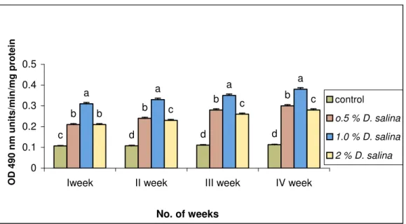
The shrimp fed with the diet without D. salina (control) showed the proPO levels of 0.11, 0.11, 0.11, and 0.11 U/min/mg of protein on 1st, 2nd, 3rd and 4th weeks (Fig. 1), respectively. The above values were less than 64 %, 66 %, 68 % and 70 % to the animal fed with 1.0 % D. salina incorporated diet. Among the three different concentrations of D. salina incorporated diets, the animal fed with 1.0 % diet showed maximum proPO of 0.38 U/min/mg of protein on 4th week. Whereas the shrimp fed with 0.5 % and 2.0 % D. salina incorporated diet showed 0.3 and 0.28 U/min/mg of protein, respectively on 4th week.
c d d d
b b
b b
a a
a a
b c
c c
0 0.1 0.2 0.3 0.4 0.5
Iweek II week III week IV week
No. of weeks
O
D
490 nm units/min/mg pr
o
tein
control
o.5 % D. salina
1.0 % D. salina
2 % D. salina
Fig. 1. Prophenol oxidase assay of P. monodon fed with D. salina incorporated diets
3.2. Superoxide dismutase assay
3.2.1. Superoxide dismutase assay of Haemolymph
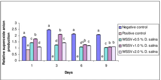
with 0.5 % D. salina incorporated diet showed 1.2, 1.32, 1.2 and 1.04 (RAP) and 2 % D. salina incorporated diet showed 1.6, 1.21, 1.2 and 1.07 (RAP) on 1st, 2nd, 3rd and 4th weeks, respectively.
a b c c d c b
d a b
a a c c d b 0 0.5 1 1.5 2 2.5
I week II week III week IV week
No. of weeks
R e la ti ve super o xi de ani on pr oducti on Control
0.5 % D. salina
1.0 % D. salina
2 % D. salina
Fig. 2. Superoxide dismutase assay of hemolymph of P. monodon fed with D. salina incorporated diets
3.3. Catalase assay
3.3.1. Catalase assay of haemolymph
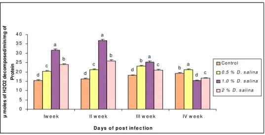
The catalase activity of haemolymph in control ranged from 15.46 to 19.23 µmoles H2O2 decomposed
-/min/mg of protein. The shrimp fed with 1.0 % D. salina incorporated diet showed the maximum catalase activity of 31.69, 36.69, 25.31 and 15.34 µmoles H2O2 decomposed /min/mg of protein (Fig. 3) which were
more than 51 %, 55 %, 28 % and 20 % to that of the control on 1st, 2nd, 3rd and 4th weeks respectively.
b d d d a b c c d a a a c c b b 0 5 1 0 1 5 2 0 2 5 3 0 3 5 4 0
Iw e e k II w e e k III w e e k IV w e e k
Da ys o f p o s t infe c tio n
µ m ole s of H 2 O 2 de c o m p os e d /m in/ m g of Pr o te
in Co n tr o l
0 .5 % D . s a l i n a 1 .0 % D . s a l i n a 2 % D . s a l i n a
Fig. 3. Catalase assay of hemolymph of P. monodon fed with D. salina incorporated diets
3.4. Challenge test
The shrimp, P. monodon fed with three different percentages (0.5 %, 1.0 % and 2.0 %) D. salina
incorporated diets for a period of 30 days were challenged with WSSV. The animal P. monodon fed with D. salina incorporated diet without any challenge towards WSSV (negative control) showed 100 % survival till
the end of the study period (up to 10th day). Whereas the animal challenged with WSSV infection (positive
the animals fed with 0.5 % D. salina incorporated diet showed 50 % mortality on 5th day and 100 % mortality on 10th day and 2 % D. salina incorporated diet showed 55 % mortality on 5th day and 100 % mortality on 10th day.
3.5. PCR amplification of genomic DNA of WSSV P. monodon
The live animals in treated and negative control groups were WSSV negative by PCR whereas moribund animals from positive group showed WSSV positive by PCR. The animals kept in the tank without WSSV survived during the study period (Data not shown).
3.6. Immunostimulant and antioxidant activity of WSSV infected P. monodon fed with D. salina incorporated diets
3.6.1. Prophenoloxidase assay
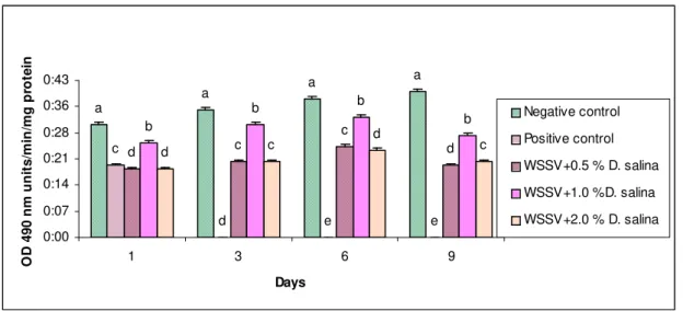
The WSSV infected shrimp fed with 1.0 % D. salina incorporated diet showed proPO levels of 0.26, 0.31, 0.33 and 0.28 (unit/min/mg of protein) on 1st, 3rd, 6th and 9th day of post infection (Fig. 4) respectively. The above values were less than 16 %, 6 %, 5 % and 26 % respectively, to that of the negative control (without infection). Whereas the infected animals fed with 2 % showed less than 26 %, 36 % and 35 % compared to the animals fed with 1.0 % D. salina incorporated diets.
a
a a
a
c
d e e
c d
c
d b
b b
b
c d
d
c
0:00 0:07 0:14 0:21 0:28 0:36 0:43
1 3 6 9
Days
O
D
4
9
0
nm
uni
ts
/m
in/
m
g
pr
ot
e
in
Negative control
Positive control
WSSV+0.5 % D. salina
WSSV+1.0 %D. salina
WSSV+2.0 % D. salina
Fig. 4. Prophenol oxidase assay of WSSV infected P. monodon fed with D. salina incorporated diets
3.7. Superoxide dismutase assay
3.7.1. Superoxide dismutase assay of haemolymph
a
a
a a
e
e e e
c d d c b b b b d c c b 0 0.5 1 1.5 2 2.5 3
1 3 6 9
Days R elat ive su p er o x id e an io n pr odu c ti o n Negative control Positive control
WSSV+0.5 % D. salina
WSSV+1.0 % D. salina
WSSV+2.0 % D. salina c
Fig. 5. Superoxide dismutase assay of haemolymph of WSSV infected P. monodon fed with D. salina incorporated diet
3.8. Catalase assay
3.8.1. Catalase assay of haemolymph
The catalase activity in haemolymph of WSSV infected shrimp fed with 1.0 % D. salina incorporated diet showed the catalase level of 25.46 µmoles H2O2 decomposed /min/mg of protein on 6th day (Fig. 6) which was
more than 35 % and 43 % of the animals fed with 0.5 % and 2 % D. salina incorporated diets, respectively. Whereas the animals without WSSV infection (negative control) fed with D. salina incorporated diet showed 31.69, 36.69, 25.31 and 15.34 µmoles H2O2 decomposed /min/mg of protein on 1st, 3rd, 6th and 9th day,
respectively. a a a a e
e e e
d d
d
d
b b b
b
c c c
c 0 5 10 15 20 25 30 35 40
1 3 6 9
Days µ m o les of H 2 O 2 decom p o sed/ m in /m g of P rot ei n Negative control Positive control
WSSV+0.5 % D. salina
WSSV+1.0 % D. salina
WSSV+2.0 % D. salina
Fig. 6. Catalase assay of haemolymph of WSSV infected P. monodon fed with D. salina incorporated diet
4.Discussion
different concentrations viz: 0.5 %, 1.0 % and 2.0 % when incorporated in the diet and fed to shrimp showed that the growth of P. monodon was found similar among the three concentrations. When compared to the control (without D. salina diet) 1.0 % diet showed better growth. Similarly, Boonyaratpalin et al., 2001 reported a higher survival rate and growth in P. japonicus fed with astaxanthin-supplemented diets than supplement of β -carotene or algal meal.
Carotenoids pigments are involved in antioxidants activities in aquatic animals and moreover carotenoids are known to enhance immune function and disease resistance in higher animals as stated by (Hunter, 2000, Supamattaya et al., 2005). Phenoloxidase (PO), the key enzyme in the synthesis of melanin, occurs in haemolymph as an inactive proenzyme prophenoloxidase (proPO). ProPO is activated to form PO (phenol oxidase) when it reacts with zymosan (carbohydrates from yeast cell walls), bacterial lipopolysaccharide (LPS), urea, calcium ions, trypsin, or heat (Soderhall et al., 1986). Hence an attempt was made on the present study with P. monodon fed with β-carotene producing D. salina incorporated diets for a period of 30 days, and also challenged against WSSV infection. The animal without WSSV infection but fed with D. salina incorporated diet showed 100 % survival till the experimental study indicated its non-toxic effects. It is worthwhile to mention that the control animals (fed without D. salina but with WSSV infection) showed 100 % mortality within 48 h. However, the animal fed with 1.0 % D. salina incorporated diet challenged with WSSV showed 40 % mortality on 5th day followed by 100 % mortality at the end of 10th day. Thus indicating that the possible role of D. salina for protecting the animals to WSSV to certain extent.
Screening of Penaeus monodon shrimp for WSSV is one of the effective ways to check the vertical transmission of disease through the hatcheries (Hsu et al., 1999). Henceforth, efforts were initiated to check the presence of WSSV through PCR amplification in P. monodon. In the present study all the dead animals showed amplification with the primer corresponded to WSSV confirmed that their mortality was due to WSSV infection. Instead the animals kept in the tank without WSSV survived well during the study period.
postponing the dead of WSSV Shrimp to some extent. Similar findings were observed by Felix et al., 2004 noticed highest proPO activity was observed in WSSV infected P. monodon fed with Sargassum wightii (seaweed). A gradual increase of proPO activity was observed up to 6th day after challenging with Vibrio parahemolyticus thereafter there was a gradual decrease in the activity. Balasubramanian et al., (2008) reported that increased levels of proPO assay were observed in shrimp fed with plant derived antiviral compound from Cyanodon dactylon.
Super oxide dismutase (SOD) is one of the main antioxidant defence enzymes generated in response to oxidative stress. Sarathi et al., (2007) and Mohankumar and Ramasamy (2007) observed the activity of SOD was significantly lowered in WSSV-infected F. indicus. Also in the present study, the activity of SOD was significantly lowered in the WSSV-infected hemolymph of P. monodon, whereas D. salina incorporated diet -treated in vivo shrimp significantly recovered when compared with control animals. These results concur with the findings of Lin and Chang et al., 2003 who have found that SOD decreases in WSSV infected P. monodon.
In the present study, SOD level in the Haemolymph of shrimp fed with three different concentrations of D. salina incorporated diet showed high levels of SOD when compared to control (without D. salina). Nakano
et al., (1999) observed for the first time that astaxanthin (algal pigment) supplementation in diet fed to rainbow trout influenced liver function and increased SOD defensive potential against oxidative stress. Similarly, Chang et al., 2003 observed that the shrimp fed with b-glucan (BG) diets showed significantly higher levels of O2
concentration than the BG free group as observed in shrimp treated with C. dactylon plant extract. Holmblad
and Soderhall (1999) observed that SOD is related to immunity in crustacean. The high level of O2 in
P. monodon fed with D. salina incorporated diets indicated that the alga may be the potential immunostimulant. Mohankumar and Ramasamy, (2007) observed that hydrogen peroxide is toxic to cells and catalase is a major primary antioxidant defense component that catalyses the decomposition of H2O2 which is produced by
the action of superoxide dismutase to H2O The present study revealed that the catalase assay of haemolymph of
P. monodon fed with three different concentrations of D. salina incorporated diets showed increased levels of catalase when compared to control. In the present study, the activity of catalase in haemolymph of WSSV infected P. monodon fed with 1.0 % D. salina incorporated diet showed less than 30 % to that of the negative control.
In conclusion, this study suggests that increasing proPO activity, the superoxide anion production and catalase production of WSSV infected P. monodon showed lower activities than the healthy shrimp (without WSSV infection). Whereas the SOD of Haemolymph of WSSV infected P. monodon fed with D. salina incorporated diet showed a slightly less activity when compared to the shrimp fed with D. salina without WSSV infection. The above results indicated that the shrimp fed with D. salina incorporated diet showed a slightly enhancement in immune resistance towards WSSV infected P. monodon
References
[1] Albores V.F, Guzman-Murillo A, Ochoa J.L. (1993) A lipopolysaccharide-binding agglutinin isolated from brown shrimp Penaeus
californiensis Holmes haemolymph. Comp Biochem Physiol, 104A:407–13.
[2] Balasubramanian, G., Sarathi, M., Venkatesan, C., John Thomas, and Sahul Hameed, A.S. (2008) Studies on the immunomodulatory effect of extract of Cyanodon dactylon in shrimp, Penaeus monodon, and its efficacy to protect the shrimp from white spot syndrome virus (WSSV). Fish Shellfish Immunol, 25, 820-828.
[3] Boonyaratpalin, M., S. Thongrod, K., Supamattaya, G., Britton. and Schlipaulis, L.E. (2001) Effect of β- carotene source, Dunaliella
salina, and astaxanthin on pigmentation, growth, survival and health of Penaeus monodon. Aquaculture research, 32, 182- 190.
[4] Chang C.F, Su MS, Chen HY, Liao IC. (2003) Dietary-1,3-glucan effectively improves immunity and survival of Penaeus monodon
[5] Chang P.S, Chen H.C, Wand Y.C. (1998) Detection of white spot syndrome associated baculovirus WSBV in experimentally infected wild shrimps, crabs and lobsters by in situ hybridization. Aquaculture, 164, 23–43.
[6] Felix, S., Herald Robins, P. and Rajeev, A. (2004) Immune enhancement assessment of dietary incorporated marine alga Sargassum
wightii (Phaeophyceae/Punctarials) in tiger shrimp Penaeus monodon (Crustacia/Penaeidae) through Phenoloxidase (proPO) systems.
Indian Journal of marine Sciences, 33, (4), 361-364.
[7] Flegel, T.W. (1997) Major viral diseases of the black tiger prawn (Penaeus monodon) in Thailand. In: Inui, Y. (eds.), New approaches to viral diseases of aquatic animals. NRIA international workshop proceedings. National Research Institute of Aquaculture, Nansei, p 167-187.
[8] Holmblad, T. and Soderhall, K. (1999) Cell adhesion molecules and antioxidative enzymes in a crustacean; possible role in immunity.
Aquaculture, 172, 111-123.
[9] Hsu, H.C., Lo, C.F., Liu, K.F., Su, M.S. and Kou, G.H. (1999) Studies on effective PCR screening strategies for white spot syndrome virus (WSSV) detection in Penaeus monodon brooders. Dis. Aquat. Organ, 39, 13-19.
[10] Hunter, B. (2000) Physiological functions of astaxanthine and other carotenoids in marine organisms. In: Sungpuang, P. (eds.), First South East Asia and Pacific Regional meeting on carotenoids, Mahidol University, Bangkok, 2-5, 19 p.
[11] Johanson, M.W., Keyser, P., Sritunyalucksana, K. and So¨derha¨ll, K. (2000) Crustacean hemocytes and haematopoiesis. Aquaculture, 191, 45-52.
[12] Karin van de Braak. (2002) Haemocytic defence in black tiger shrimp (Penaeus monodon) PhD thesis, Wageningen University - with ref. - with summary in Dutch ISBN 90-5808-651-8. Institute of Animal Sciences. Wageningen
[13] Karunasagar, I., Otta, S.K. and Karunasagar, I. (1997) Histopathological and bacteriological study of white spot syndrome of Penaeus
monodon along the west coast of India. Aquaculture, 153, 9- 13.
[14] Lowry, O.H., Rosenbrough, N.J., Farr, A.L. and Randall, R.J. (1951) Protein measurement with the Folin phenol reagent. J. Biol.
Chem, 193, 265-275.
[15] Misra, H.P. and Fridovich, I. (1972) The role of superoxide anion in the auto oxidation of epinephrine and a simple assay of superoxide dismutase. J. Biol. Chem 247, 3170-3175.
[16] Mohankumar K, and Ramasamy P. (2007) White spot syndrome virus infection decreases the activity of antioxidant enzymes in
Fenneropenaeus indicus. Virus Res, 115, 69–75.
[17] Nakano, T., Kanmuri, T. Sato, M. and Takeuchi, M. (1999) Effect of astaxanthin rich red yeast (Phaffia rhodozyma) on oxidative stress in rainbow trout. Biochimica et Biophysica Acta, 1426, 119-125.
[18] Ourth D.D, Renis H.E. (1993) Antiviral melanization reaction of Heliothis virescens hemolymph against DNA and RNA viruses in vitro. Comp Biochem Physiol, 105, 719–23.
[19] Pascual, C., Sa´nchez, A., Sa´nchez, A., Vargas-Albores, F., LeMoullac, G. and Rosas, C. (2003) Haemolymph metabolic variables and immune response in Litopenaeus setiferus adult males: the effect of an extreme temperature. Aquaculture , 218, 637- 650. [20] Sahul Hameed A.S, Balasubramanian G, Syed Musthaq S and Yoganandhan K. (2003) Experimental infection of twenty species of
Indian marine crabs with white spot syndrome virus (WSSV). Dis Aquat Organ, 57, 157–61.
[21] Sarathi M, Ishaq Ahmed VP, Venkatesan C, Balasubramanian G, Prabavathy J and Sahul Hameed A.S. (2007) Comparative study on immune response of Fenneropenaeus indicus to Vibrio alginolyticus and white spot syndrome virus. Aquaculture; 271, 8–20. [22] Soderhall, K., Smith, V.J. and Johansson, M. (1986) Exocystosis and uptake of bacteria by isolated haemocyte populations of two
crustaceans: evidence for cellular co-operation in the defense reactions of arthropods. Cell Tissue Res, 243, 43-49.