Surgery-first approach with 3D customized
passive self-ligating brackets and 3D surgical
planning: Case report
Juan Fernando Aristizábal1, Rosana Martínez-Smit2,3, Cristian Díaz1, Valfrido Antonio Pereira Filho4
1 Universidad del Valle, Departamento de Ortodoncia (Cali, Colombia). 2 CES University, Departamento de Ortodoncia (Medellín, Colombia). 3 Universidade Estadual Paulista, Departamento de Ortodontia e Pediatria,
Faculdade de Odontologia de Araraquara (Araraquara/SP, Brazil). 4 Universidade Estadual Paulista, Departamento de Diagnóstico e Cirurgia
Bucomaxilofacial, Faculdade de Odontologia de Araraquara (Araraquara/SP, Brazil).
» Patients displayed in this article previously approved the use of their facial and in-traoral photographs.
How to cite: Aristizábal JF, Martínez-Smit R, Díaz C, Pereira Filho VA. Sur-gery-first approach with 3D customized passive self-ligating brackets and 3D sur-gical planning: Case report. Dental Press J Orthod. 2018 May-June;23(3):47-57. DOI: https://doi.org/10.1590/2177-6709.23.3.047-057.oar
Submitted: March 21, 2017 - Revised and accepted: July 02, 2017
» The authors report no commercial, proprietary or financial interest in the products or companies described in this article.
Contact address: Dr. Rosana Martínez-Smit Cra 48 # 12 sur – 70 office 603, Medellin, Colombia E-mail: [email protected]
It is possible to unify three-dimensional customized orthodontic techniques and three-dimensional surgical technology. In this case report, it is introduced a treatment scheme consisting of passive self-ligation customized brackets and virtual surgical planning combined with the orthognathic surgery-first approach in a Class III malocclusion patient. Excellent facial and occlusal outcomes were obtained in a reduced treatment time of five months.
Keywords:Angle Class III malocclusion. Orthodontic surgery. Orthodontics. Three-dimensional image.
DOI: https://doi.org/10.1590/2177-6709.23.3.047-057.oar
É possível unificar técnicas ortodônticas personalizadas e tecnologia de planejamento cirúrgico 3D. No presente relato de caso, apresenta-se um plano de tratamento envolvendo o uso de braquetes autoligáveis passivos personalizados e planeja-mento cirúrgico virtual, combinado com cirurgia ortognática de benefício antecipado, em um paciente com má oclusão de Classe III. Foram obtidos excelentes resultados faciais e oclusais em um tempo reduzido de tratamento, de 5 meses.
INTRODUCTION
Accurate surgical treatment starts with precise diagnosis, by evaluating all dimensions and deter-mining the nature of deformity, because it might be a combination of hard and soft tissue components.1
The main limitation of conventional surgical planning is its two-dimensional approach that in-volves clinical examination, extraoral and intraoral photographs, lateral and posteroanterior cephalo-grams, and plaster dental models.2,3 To overcome
these deficiencies, cone-beam computed tomog-raphy (CBCT) for imaging the craniofacial region brought a true paradigm shift from a two-dimen-sional to a three-dimentwo-dimen-sional (3D) approach.4
Computer-aided surgical simulation (CASS) utilizing three-dimensional images obtained from multislice computed tomography (MSCT)/cone beam computer tomography (CBCT) has been successfully performed previously to plan cranio-facial surgery.5-8 Also, CASS has been combined
with the surgery-first approach (SFA) to demon-strate two useful and practical methods for plan-ning these cases.9
Furthermore, the patient can be virtually visual-ized by generating a fusion model with digital den-tal casts, a CBCT reconstructed bony volume and textured facial soft tissue image.10,11 Additionally,
with this fusion model the clinicians can accurately create surgical splints using the computer-aided de-sign/computer-aided manufacturing (CAD/CAM) system for successful surgical treatments.11,12
Recently, significant technological advancements have been made in computer-aided orthodontic treatment. In the Insignia system (Ormco Corpora-tion, Orange County, CA), polyvinyl siloxane (PVS) impressions are digitized with computed tomogra-phy to produce highly detailed digital models, or an intraoral dental scanner is used to generate 3D digital models. The orthodontist adjusts the digital setup using a real-time 3D interface, while referring to the patient’s intra and extraoral photographs and radiographs for consideration of esthetic treatment goals. After the clinician approves the final setup, the customized brackets, tubes, and arch-wires are fabri-cated and bracket-positioning jigs are provided, for accurate indirect transfer.13
In the present case report, 3D virtual custom-ized bracket design (Insignia, Ormco Corporation, Orange County, CA) was integrated with 3D vir-tual surgical planning along with fabrication of digi-tal surgical splints using a CAD/CAM technique. This article aims to report how the use of 3D digital technology, self-ligating brackets and the SFA can drastically reduce treatment time.
CASE REPORT
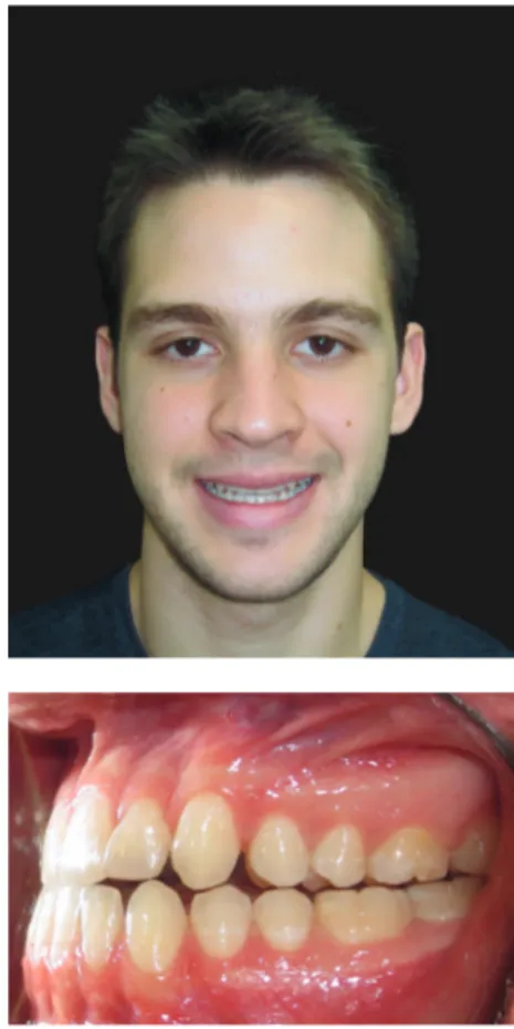
A 21-year-old Hispanic male reported to the or-thodontist office with the primary complaint of not feeling comfortable with the bite and chin projection (Fig 1). A subsequent clinical examination showed that the profile had worsened since a previous orth-odontic treatment.
Systemically, he referred controlled Diabetes Mellitus Type I. The extraoral examination showed concave facial profile, with a slight maxillary hypo-plasia, significant chin projection, upper lip retrusion and adequate nasolabial angle (Fig 1). Dentally, the patient presented a Class III malocclusion with pro-clined upper incisors and retropro-clined lower incisors, edge to edge bite, lower proper alignment and spac-ing of 2mm in the upper arch (Figs 1, 2, and 3A). The panoramic radiograph showed mild different ramus lengths (Fig 3B). Skeletally, Class III pattern with mandibular prognathism and macrognathism was observed (Fig 3A, 3C).
The treatment objectives were to correct the Class III skeletal pattern, to improve profile, to increase overjet and to improve facial aesthetics. The treatment options pre-sented were presurgical orthodontic treatment followed by mandibular setback surgery and SFA with mandibular setback followed by fixed appliances to align, level and sta-bilize the occlusion. Considering that the patient’s chief concern was his facial esthetics, it was decided to proceed with SFA, because the patient wanted immediate facial change. This approach would avoid deterioration in his profile and malocclusion during presurgical orthodontics, and would also take advantage of the biological potential of the regional acceleratory phenomenon (RAP).
A computed tomography (CT) (Bright Speed Elite, General Electric, and Fairfield, Connecticut, USA) was taken for the construction of a model of the skull8
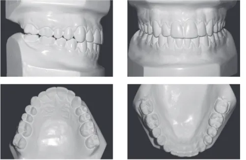
Figure 1 - Pre-treatment photographs showing skeletal and dental Class III malocclusion.
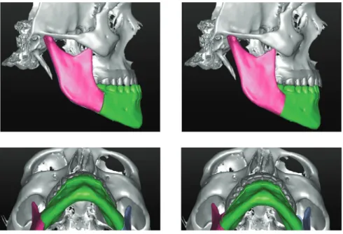
The surgical plan was mandibular setback (Fig 4). The virtual design was transferred to the CAD/CAM software for production of surgical splints. The inter-mediate splint was physically generated by a 3D printer (Fortus 250mc, Stratasys, Eden Prairie, MN, USA) with hybrid epoxy-acrylate polymer.
The first step in the Insignia system (Ormco Corporation, Orange, CA) for custom-designed or-thodontics is to send precise polyvinyl siloxane im-pressions as well as photographic and radiographic information to the manufacturer. The brackets cho-sen were Insignia self-ligating (SL) brackets, which
are the customized version of Damon Q SL brackets (Ormco Corporation, Orange, CA).14 The final
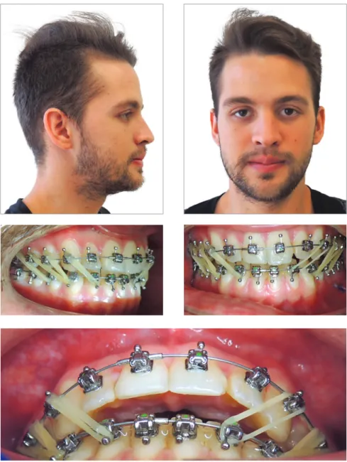
set-up for the patient was approved with an overcorrec-tion of lower incisors positive torque, ensuring opti-mal expression of the lower incisors decompensation exploiting the massive RAP after orthognathic sur-gery (Fig 5). The selected sequence of wires was Cu-NiTi 0.014-in, CuNiTi 0.014 x 0.025-in, CuNiTi 0.018 x 0.025-in, TMA 0.019 x 0.025-in and stain-less steel 0.019 x 0.025-in (Ormco Corporation, Orange, CA). The brackets were bonded three days before surgery and no archwire was placed.
Figure 3 - A) Pre-treatment lateral cephalometric
ra-diograph. B) Pre-treatment panoramic radiograph. C) Pre-treatment lateral cephalometric tracing.
A
B
Figure 4 - Surgical planning of mandibular set-back.
Figure 5 - Custom designed orthodontics, with Insignia.
Figure 7 - Class III intermaxillary elastics.
In the day of the surgery, immediately before intu-bation assisted by a fiber optic probe, CuNiTi 0.014-in (Ormco Corporation, Orange, CA) archwires were placed (Fig 6). After mandibular setback surgery by sagittal osteotomy, under brain activity monitoring,
and once a suitable rigid fixation and postoperative occlusion were established, ¼ 3.5 oz intermaxillary elastics were applied with Class III vector.
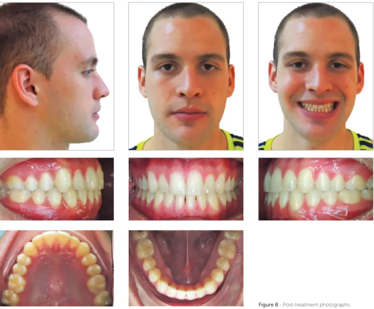
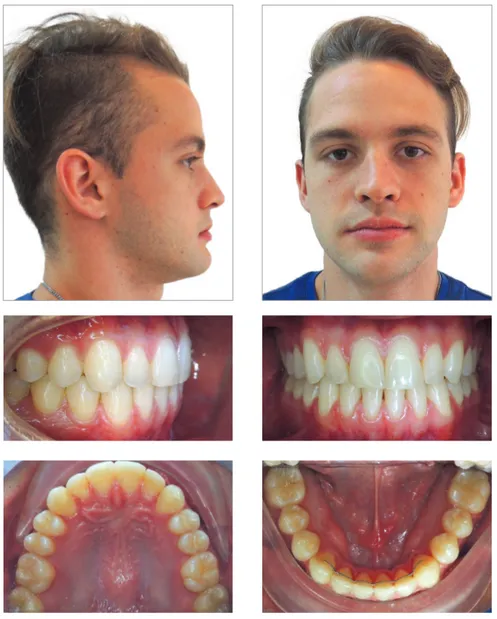
Figure 8 - Post-treatment photographs.
0.014 x 0.025-in CuNiTi (Ormco Corporation, Or-ange, CA). One month after surgery 0.018 x 0.025-in CuNiTi archwires (Ormco Corporation, Orange, CA) were placed and Class III intermaxillary elastics were continued. Then, 0.019 x 0.025-in TMA arches (Ormco Corporation, Orange, CA) were placed six
Figure 10 - A) Post-treatment lateral cephalomet-ric radiograph. B) Post-treatment cephalometric tracing. C) Superimposition of pre and post-treat-ment cephalometric tracings. D) Post-treatment panoramic radiograph.
A
D
B C
Figure 11 - Follow-up photographs (24 months).
DISCUSSION
Surgery-first approach (SFA) was first proposed by Nagasaka et al,15 in 2009. With the orthognathic
surgery performed before the orthodontic correc-tion, total treatment time could be reduced to even less than the average period for presurgical ortho-dontics.16-19 Considering the number of patients who
want orthognathic surgery mainly for esthetic reasons and would appreciate a shorter treatment time, SFA offers an attractive alternative for managing skeletal malocclusions while improving patients’ self-esteem and function at the beginning of treatment.20,21
and surrounding soft tissues; and (6) stability of results equal to, or in some cases superior to, those achieved using the traditional orthodontics-first approach.22
Most articles recommended that orthodontic ap-pliances should be placed prior to surgery, even when using a surgery-first approach. Studies reported bond-ing the orthodontic brackets immediately before,15,23
1 week before,24-261 month before27-29 or 1-2 months
before30surgery. Only one of the papers reported the
total elimination of preoperative orthodontic treat-ment and the fitting of orthodontic brackets 10-14 days after surgery20 Studies described that active
orth-odontic force can be applied before26-29 or shortly
af-ter15,23-25,30 surgery. Preoperative orthodontic
prepara-tion can, therefore, be started immediately before or approximately 1-2 months before surgery. Occasion-ally, it might be completely eliminated.
The shortest reported treatment time for postop-erative orthodontic treatment was 4 months for cor-rection of a skeletal Class III malocclusion with ante-rior open bite and dental crowding26 and 4.5 months
in the management of unilateral condylar hyperpla-sia,11 similar to this case report, with total treatment
time of 5 months. Most studies described completing postoperative orthodontic treatment within approxi-mately 1 year15,27,28,30 or in 6-9 months.20,23,25
Treat-ment time was approximately 6-12 months shorter using the SFA, compared to using a conventional or-thodontics-first approach. Only one study described similar treatment time (approximately 1.5 years) for both approaches.29
There is no doubt that SFA requires precise and ac-curate diagnosis and planning. Post-surgical orthodon-tic movements must be carefully executed according to the surgical plan, which implies constant communica-tion between orthodontist and oral surgeon.
To expedite post-surgical orthodontics, Insignia System (Ormco Corporation, Orange, CA) is an
important tool for offering customized brackets and archwires, also diminishing errors from appliance po-sitioning. Customized devices in orthodontics have been reported before. Subjects treated with SureS-mile (OraMetrix, Richardson, Tex) were compared with those undergoing conventional orthodontic treatment, concluding that treatment time was 7 months shorter in patients treated with SureSmile.31
Saxe et al32 obtained comparable results. However,
SureSmile technology (OraMetrix, Richardson, Tex) customizes only the archwires, using roboti-cally-assisted archwire bending technology.32,33
In-signia (Ormco Corporation, Orange County, CA) customizes bracket prescription, bonding and arch-wires.14 Besides, the light forces produced by the
passive self-ligating system with high-tech archwires will control the transverse dimension in coordina-tion with post-surgical sagittal changes.19
With two-dimensional (2D) imaging, the most usual problems are landmark identification, image distortion and magnification.34,35 However 2D
imag-ing remains as the gold standard for the craniofacial region. The 3D computer-assisted surgical planning benefits the specialists because it can predict surgical movements including translations in anteroposterior, lateral, and vertical directions, and rotations around the x-, y-, and z-axes, the so-called pitch, roll, and yaw rotations36 and this is an undisputed advantage
in determining the best treatment option.
CONCLUSIONS
» The 3D diagnostics, digital surgical planning and CAD/CAM customized bracket systems with passive self-ligation offer a more accurate alternative to im-prove the efficiency of orthodontic-surgical treatment.
1. Cheong YW, Lo LJ. Facial asymmetry: etiology, evaluation, and management. Chang Gung Med J. 2011 July-Aug;34(4):341-51.
2. Schwartz HC. Efficient surgical management of mandibular asymmetry. J
Oral MaxillofacSurg 2011;69:645-54.
3. Baek SH, Cho IS, Chang YI, Kim MJ. Skeletodental factors affecting chin point deviation in female patients with Class III malocclusion and facial asymmetry: a three-dimensional analysis using computed tomography. Oral Surg Oral Med Oral Pathol Oral Radiol Endod. 2007 Nov;104(5):628-39.
4. Uribe F, Janakiraman N, Shafer D, Nanda R. Three-dimensional cone-beam
computed tomography-based virtual treatment planning and fabrication of a surgical splint for asymmetric patients: surgery first approach. Am J Orthod Dentofacial Orthop. 2013 Nov;144(5):748-58.
5. Gateno J, Xia JJ, Teichgraeber JF, et al. Clinical feasibility of computer-aided surgical simulation (CASS) in the treatment of complex cranio- maxillofacial deformities. J Oral MaxillofacSurg 2007; 65:728-734.
6. Xia JJ, Gateno J, Teichgraeber JF, Christensen AM, Lasky RE, Lemoine JJ, et al. Accuracy of the computer-aided surgical simulation (CASS) system in the treatment of patients with complex craniomaxillofacial deformity: a pilot study. J Oral Maxillofac Surg. 2007 Feb;65(2):248-54.
7. Xia JJ, Shevchenko L, Gateno J, Teichgraeber JF, Taylor TD, Lasky RE, et al. Outcome study of computer-aided surgical simulation in the treatment of patients with craniomaxillofacial deformities. J Oral Maxillofac Surg. 2011 July;69(7):2014-24.
8. Xia JJ, McGrory JK, Gateno J, Teichgraeber JF, Dawson BC, Kennedy KA, et al. A new method to orient 3-dimensional computed tomography models to the natural head position: a clinical feasibility study. J Oral Maxillofac Surg. 2011 Mar;69(3):584-91.
9. Hsu SS, Singhal D, Xia JJ, Gateno J, Lin C-H, Huang C-S, et al. Planning the Surgery-first approach in Surgical- orthodontic treatment with a computer aided Surgical Simulation (CASS) Planning Protocol. J Taiwan Assoc Orthod. 2012;24(2):24-37.
10. Plooij JM, Maal TJ, Haers P, Borstlap WA, Kuijpers-Jagtman AM, Berge SJ. Digital three-dimensional image fusion processes for planning and evaluating orthodontics and orthognathic surgery. Int J Oral Maxillofac Surg. 2011 Apr;40(4):341-52.
11. Janakiraman N, Feinberg M, Vishwanath M, NalakaJayaratne YS, Steinbacher DM, et al. Integration of 3-dimensional surgical and orthodontic
technologies with orthognathic “surgery-first” approach in the management of unilateral condylar hyperplasia. Am J Orthod Dentofacial Orthop. 2015 Dec;148(6):1054-66
12. Gateno J, Xia J, Teichgraeber JF, Rosen A, Hultgren B, Vadnais T. The precision of computer-generated surgical splints. J Oral Maxillofac Surg. 2003 July;61(7):814-7.
13. Breuning KH. Efficient tooth movement with new technologies for customized treatment. J Clin Orthod. 2011 May;45(5):257-62; quiz 287. 14. Gracco A, Stellini E, Parenti SI, Bonetti GA. Individualized orthodontic
treatment: the Insignia system. Orthod (Chic.). 2013;14(1):e88-94.
15. Nagasaka H, Sugawara J, Kawamura H, Nanda R. “Surgery first” skeletal Class III correction using the Skeletal Anchorage System. J Clin Orthod. 2009 Feb;43(2):97-105.
16. Luther F, Morris DO, Hart C. Orthodontic preparation for orthognathic surgery: how long does it take and why? A retrospective study. Br J Oral Maxillofac Surg. 2003 Dec;41(6):401-6.
17. Dowling PA, Espeland L, Kroonstad O, Stenvik A, Kelly A. Duration of orthodontic treatment involving orthognathic surgery. Int J Adult Orthod Orthog Surg. 1999;14(2):146-52.
18. Luther F, Morris DO, Karnezi K. Orthodontic treatment following
orthognathic surgery: How long does it take and why? A retrospective study J Oral Maxillofac Surg. 2007 Oct;65(10):1969-76.
REFERENCES
19. Aristizábal JF, Martínez Smit R, Villegas C. The “surgery first” approach with passive self-ligating brackets for expedited treatment of skeletal Class III malocclusion. J Clin Orthod. 2015 June;49(6):361-70.
20. Hernández-Alfaro F, Guijarro-Martínez R, Molina-Coral A, Badía-Escriche C. “Surgery first” in bimaxillary orthognathic surgery. J Oral Maxillofac Surg. 2011 June;69(6):e201-7.
21. Peiró-Guijarro MA, Guijarro-Martínez R, Hernández-Alfaro F. Surgery first in orthognathicsurgery: a systematicreview of theliterature. Am J Orthod Dentofacial Orthop. 2016 Apr;149(4):448-62.
22. Huang CS, Hsu SS, Chen YR. Systematic review of the surgery-first approach in orthognathic surgery. Biomed J. 2014 July-Aug;37(4):184-90.
23. Sugawara J, Aymach Z, Nagasaka DH, Kawamura H, Nanda R. “Surgery first” orthognathics to correct a skeletal class II malocclusion with an impinging bite. J Clin Orthod. 2010 July;44(7):429-38.
24. Liou EJ, Chen PH, Wang YC, Yu CC, Huang CS, Chen YR. Surgery-first accelerated orthognathic surgery: Orthodontic guidelines and setup for model surgery. J Oral Maxillofac Surg. 2011 Mar;69(3):771-80.
25. Villegas C, Uribe F, Sugawara J, Nanda R. Expedited correction of significant dentofacial asymmetry using a “surgery first” approach. J Clin Orthod. 2010 Feb;44(2):97-103; quiz 105.
26. Yu CC, Chen PH, Liou EJ, Huang CS, Chen YR. A Surgery-first approach in surgical-orthodontic treatment of mandibular prognathism- a case report. Chang Gung Med J. 2010 Nov-Dec;33(6):699-705.
27. Wang YC, Ko EW, Huang CS, Chen YR, Takano-Yamamoto T. Comparison of transverse dimensional changes in surgical skeletal Class III patients with and without presurgical orthodontics. J Oral Maxillofac Surg. 2010 Aug;68(8):1807-12.
28. Liao YF, Chiu YT, Huang CS, Ko EW, Chen YR. Presurgical orthodontics versus no presurgical orthodontics: Treatment outcome of surgical-orthodontic correction for skeletal class III open bite. Plast Reconstr Surg. 2010 Dec;126(6):2074-83.
29. Ko EW, Hsu SS, Hsieh HY, Wang YC, Huang CS, Chen YR. Comparison of progressive cephalometric changes and postsurgical stability of skeletal Class III correction with and without presurgical orthodontic treatment. J Oral Maxillofac Surg. 2011 May;69(5):1469-77.
30. Baek SH, Ahn HW, Kwon YH, Choi JY. Surgery-first approach in skeletal class III malocclusion treated with 2-jaw surgery: evaluation of surgical movement and postoperative orthodontic treatment. J Craniofac Surg. 2010 Mar;21(2):332-8.
31. Alford TJ, Roberts WE, Hartsfield JK Jr, Eckert GJ, Snyder RJ. Clinical outcomes for patients finished with the SureSmile! Method compared with conventional fixed orthodontic therapy. Angle Orthod. 2011 May;81(3):383-8.
32. Saxe AK, Louie LJ, Mah J. Efficiency and effectiveness of SureSmile. World J Orthod. 2010 Spring;11(1):16-22.
33. Mah J, Sachdeva R. Computer-assisted orthodontic treatment: the SureSmile process. Am J Orthod Dentofacial Orthop. 2001 July;120(1):85-7. 34. Trpkova B, Major P, Prasad N, Nebbe B. Cephalometric landmarks
identification and reproducibility: a meta-analysis. Am J Orthod Dentofacial Orthop. 1997 Aug;112(2):165-70.
35. Timock AM, Cook V, McDonald T, Leo MC, Crowe J, Benninger BL, et al. Accuracy and reliability of buccal bone height and thickness measurements from cone-beam computed tomography imaging. Am J Orthod Dentofacial Orthop. 2011 Nov;140(5):734-44.