Advances in early biomarkers of diabetic nephropathy
JIN ZHANG1, JIANHUA LIU2, XIAOSONG QIN3*
1Masters Student, Department of Laboratory Medicine, Shengjing Hospital of China Medical University, Shenyang, Liaoning, China
2MD, PhD. Associate Professor of Laboratory Medicine, Department of Laboratory Medicine, Shengjing Hospital of China Medical University, Shenyang, Liaoning, China 3MD, PhD. Professor of Laboratory Medicine, Department of Laboratory Medicine, Shengjing Hospital of China Medical University, Shenyang, Liaoning, China
S
UMMARYStudy conducted at Shengjing Hospital of China Medical University, Shenyang, Liaoning, China
Article received: 8/9/2017
Accepted for publication: 9/9/2017
*Correspondence:
Address: No 36, Sanhao Street, Heping District Shenyang, Liaoning – China Postal code: 110004 qinxs@sj-hospital.org
http://dx.doi.org/10.1590/1806-9282.64.01.85
Diabetic nephropathy is the main cause of chronic kidney disease, and represents the most common and serious complication of diabetes. The exact pathogenesis is complex and not elucidated. Several factors and mechanisms contribute to the development and outcome of diabetic nephropathy. An early diagnosis and intervention may slow down disease progression. A variety of biological markers associated with diabetic nephropathy were found in recent years, which was important for predicting the occurrence and development of the disease. Therefore, this article provides an overview of early biomarkers that are associated with diabetic nephropathy.
Keywords: Diabetes Mellitus. Diabetic Nephropathies. Biomarkers.
I
NTRODUCTIONDiabetes mellitus (DM) is an endocrine and metabolic disease that has serious impact on human health. The morbidity and mortality of DM have risen continually at an alarming rate in recent years, and the population with diabetes mellitus is predicted to be about 439 million worldwide by 2030.1 The complications of DM include
diabetic retinopathy, diabetic cardiovascular diseases and diabetic nephropathy (DN), which is the most com-mon and serious complication of DM. DN has become the leading cause of chronic kidney failure, starting with normoalbuminuria, microalbuminuria, macroalbumin-uria and ultimately leading to end stage renal disease (ESRD).2 For a long time, proteinuria has been considered
the gold standard for evaluation and monitoring of renal function. However, renal function declines in about one-third of the patients before the occurrence of proteinuria,3
which makes it inadequate to detect proteinuria alone to monitor the incidence and progression of DN. Therefore, we need to look for laboratory biomarkers that are ear-lier than microalbuminuria or those appearing at the same time. This review focuses on the early biomarkers associated with the pathogenesis and pathology of DN and changes in renal function.
B
IOMARKERSASSOCIATED WITHDN
PATHOGENESISA large number of prospective studies confirm that hyper-glycemia is the most important risk factor for DN.4,5
Hy-perglycemia promotes mitochondrial electron transport chain to generate excessive reactive oxygen species (ROS) through formation of the advanced glycation end products (AGEs) and activation of the polyol pathway, hexosamine pathway, protein kinase C (PKC) and angiotensin II. Then, the ROS initiate or enhance the oxidative stress and even-tually cause the inflammatory response and formation of fibrosis.6,7 In addition, lipid metabolism abnormality,
renin-angiotensin-aldosterone system (RAAS) activation, sys-temic and glomerular hypertension, insulin signaling im-pairment, increased growth factors and pro-inflammatory cytokines, and intracellular signaling pathway activation also play a role in the occurrence and progression of DN.6,8
Biomarkers of oxidative stress
The occurrence and progression of DN is closely related with oxidative stress. Excessive ROS, which are induced by hyperglycemia, are involved in oxidative stress causing direct oxidation and damage of deoxyribonucleic acid (DNA), proteins and lipids.8,9
Biomarkers of DNA injury
8-hydroxy-2’-deoxyguanine (8-OHdG) is a sensitive bio-marker of DNA damage to assess oxidative stress in the human body. In 1994, Ha et al.10 found that the 8-OHdG
suggested that DN might be associated with oxidative stress and the formation of 8-OHdG.The following study by Hinokio et al.11 showed that urinary 8-OHdG excretion
in patients suffering from type 2 diabetes mellitus com-plicated by nephropathy was higher than in patients with-out complications or in healthy control subjects. Moreover, there was a correlation between urinary 8-OHdG level and glycosylated hemoglobin (HbA1c). In this report,
8-OHdG was speculated to be a useful biomarker associ-ated with complications secondary to DM. Zhao et al.12
measured the serum concentration of 8-OHdG using enzyme-linked immunosorbent assay (ELISA) and drew a similar conclusion. However, Serdar et al. demonstrat-ed that there was no difference in urinary 8-OHdG levels between the groups with and without diabetic nephrop-athy on liquid chromatography-mass spectrometry, sug-gesting that 8-OHdG in urine was not a sensitive bio-marker regarding albumin to creatinine ratio (UACR) for distinguishing DN patients from DM patients.13
Differ-ent biological fluids and methods might contribute to the lack of consistency in these studies, so that the predic-tive value of 8-OHdG in the early stages of DN needs further research to be determined.
Biomarkers of protein and lipid injury
Biomarkers associated with protein injury comprise pen-tosidine, 2,4-dinitrophenylhydrazine (DNPH) and ad-vanced oxidation protein product (AOPP). F2-isoprosta-glandin and 4-hydroxy-nonenal (HNE) are related to lipid injury. Calabrese et al. found that both urinary and serum levels of pentosidine, DNPH, F2-isoprostaglandin and HNE of DN patients were higher than those of con-trol subjects.14 Tabak et al. showed that the level of AOPP
in type 2 diabetes mellitus patients with complications such as DN and diabetic retinopathy was significantly higher than in patients without complications.15 These
two studies have confirmed that oxidative stress damage is involved in the development of diabetic nephropathy.
Biomarkers of glutathione antioxidant system and lipid peroxidation
A growing number of studies reported that DM and its complications were closely related to oxidative stress, so we supposed that the biomarkers related to antioxidant defense system and lipid peroxidation (LPO) induced by free radicals may be potential biomarkers of kidney dam-age in diabetic patients.8 Glutathione s-transferase (GST),
a kind of enzyme involved in cell detoxification, promotes inactivation and excretion of toxins by combining toxic drophobic compounds with glutathione.8
Experimental data from a study by Jiang et al. showed that the expression level of GST in diabetic rats induced by streptozotocin was remarkably higher than in control rats, suggesting that hyperglycemia may be the major cause for elevated GST. Eight weeks after treatment with resveratrol, the GST expression decreased and several indicators suggesting the occurrence of DN such as uri-nary protein excretion, creatinine, cellular apoptosis and renal hypertrophy were all improved, leading researchers to suppose that resveratrol likely played a role in reno-protection by lowering the expression level of GST.16 In
agreement with GST, animal experiments on LPO have yielded the same results.17,18 In addition, genetic
investi-gation also found that knockout of GST coding genes can lead to decreased GST levels and increased malo-ndialdehyde (MDA) levels, an important biomarker of LPO, demonstrating that GST has an effect against oxidative stress.19
Human research was consistent with the experimen-tal studies above. Compared with healthy subjects, in-creased activity of GST and inin-creased level of MDA were found in type 2 diabetes mellitus patients. These results suggested that oxidative stress was involved in the occur-rence of DM and GST was likely to play an important role in antioxidation.20,21 In the study about GST and DN,
Noce et al. reported that GST activity in type 2 diabetes mellitus patients with and without nephropathy were both significantly higher than that of control subjects, appearing to be closely related with the stages of DN and indicating that GST was likely to be a potential biomark-er in early stage DN.22
Biomarkers of inflammation
the control subjects. Besides, the levels of MCP-1 and IP-10 were positively correlated with proteinuria and HbA1c, while negatively correlated with the estimated
glomerular filtration rate (eGFR).23 These outcomes
sug-gest that urinary inflammation-related factors may con-tribute to the diagnosis in early stages of DN.
In addition, some studies have shown that serum interleukin-18 (IL-18) level was elevated in DN patients and associated with HbA1c or UACR, thus being
specu-lated as a potential biomarker of diabetic nephropathy.24
On the other hand, the value of interleukin-6 (IL-6) in early diagnosis of diabetic nephropathy remains to be further confirmed. A number of studies have found that serum IL-6 levels of patients with normoalbuminuria or microalbuminuria were higher than those of control subjects and showed a positive correlation with UACR.25-28
However, some other studies have found that serum IL-6 level was elevated in patients with macroalbuminuria alone, and its early diagnosis value was not as good as that of urinary albumin excretion.24,29
Some studies demonstrated that an increase in both urinary and serum levels of TNF-α in patients with ne-phropathy secondary to DM was found compared to those with normoalbuminuria and control subjects. Besides, levels of TNF-α in urine and serum were both signifi-cantly associated with urinary albumin excretion. These results revealed that TNF-α might be an early biomarker of kidney damage in diabetic patients.30,31 Soluble CD40
ligand (sCD40L) is a transmembrane protein of the tumor necrosis factor superfamily and regulates inflammatory response by binding with CD40. A study by El-Asrar et al.32 showed that serum sCD40L level in type 1 diabetes
mellitus patients with microangiopathy such as diabetic nephropathy, retinopathy or neuropathy was significant-ly higher than that of patients without complications and healthy control subjects, and diabetic patients without any of these complications presented higher sCD40L concentration as compared to healthy subjects. The re-searchers also found that serum sCD40L was signifi-cantly associated with the severity of kidney damage and the level of glycemic control.33
In addition to the biomarkers cited above, glycosyl hydrolase family of 18 members, including chitotriosidase (CHIT1) and cartilage glycoprotein 40 (YKL-40), com-monly activated by macrophages cells and neutrophils, were also involved in the inflammatory response.34,35
Sev-eral studies showed that both CHIT1 activity and YKL-40 level of type 2 diabetes mellitus patients in all subgroups were higher than that of control subjects. CHIT1 activity and YKL-40 level increased gradually along with the
stag-es of DN according to UACR, which was correlated with activity of CHIT1 and level of YKL-40 even after adjustment for clinical parameters, suggesting that they were both associated with kidney damage of DN patients. However, because of the higher sensitivity and specificity, CHIT1 activity was better in the diagnosis of persistent microal-buminuria compared with serum level of YKL-40.36,37
Biomarkers of RAAS activation
Renin-angiotensin-aldosterone system (RAAS) plays an important role in regulating blood pressure by producing aldosterone in human body. Angiotensinogen, produced by liver, was reported in patients with chronic glomeru-lonephritis in a previous study.38 The following study
found that urinary angiotensinogen excretion of type 2 diabetes mellitus patients with microalbuminuria and macroalbuminuria were both significantly increased com-pared to control subjects, as well as to normoalbuminuric patients, suggesting that angiotensinogen appeared pri-or to the establishment of albuminuria. Also, angioten-sinogen level shows a strong association with urinary albumin excretion, which is an indicator of the severity of kidney damage in diabetic patients. Angiotensinogen may be a promising biomarker in the early stages of DN due to its high sensitivity and specificity in diagnostic analysis of diabetic nephropathy.39
These biomarkers were summarized in Figure 1.
B
IOMARKERS ASSOCIATED WITHDN
PATHOLOGYBiomarkers of damage of glomerular filtration membrane
Under normal circumstances, podocyte and foot process, glomerular basement membrane and capillary endothe-lial cells constitute the glomerular filtration barrier. The damage of this filtration barrier can affect the glomerular filtration function. Markers such as podocytes, basement membrane and endothelial cell damage may have poten-tial to indicate kidney damage in DN patients.
Biomarkers of podocytes injury
Studies have shown that a decline in the number of podo-cytes and disappearance of foot processes often occur in the early stages of DN due to apoptosis or shedding of podocytes. Therefore, urinary podocytes and their spe-cific protein products may be regarded as potential bio-markers of podocyte injury.40 Currently, the studies
fo-cused on the podocyte-specific protein products because it was difficult to detect urinary podocytes directly. One study by Wang et al.41 showed that urinary mRNA levels
by real-time quantitative PCR. These results were also proved by renal biopsy. Also, synaptopodin level was positively correlated with urinary albumin excretion and serum creatinine concentration while negatively corre-lated with GFR. Patients, however, were not divided into different subgroups according to their average level of urinary protein. The validation of these podocyte-specif-ic protein products in early stages of DN was not con-firmed in this study.41 Further research performed by Hara
et al. revealed that urinary synaptopodin level of type 2 diabetes mellitus patients complicated by nephropathy was higher when compared to control subjects, even before the occurrence of proteinuria and associated with the level of urinary albumin and HbA1c, indicating that
syn-aptopodin was a biomarker with high sensitivity to podo-cyte injury in diabetic patients.42 Another report by Jim
et al. revealed that nephrin level in urine was elevated in all DN patients and 54% of normoalbuminuric subjects. In addition, urinary level of nephrin showed a strong as-sociation with UACR so that it might be a useful bio-marker for nephropathic patients in preclinical stage.43
Biomarkers of basement membrane injury
Type IV collagen is the main component of the glomeru-lar basement membrane and extracelluglomeru-lar matrix, and does not pass through glomerular filtration barrier under normal circumstances. Therefore, type IV collagen could be used as a biomarker of basement membrane injury.
The study found that urinary type IV collagen levels were higher before microalbuminuria and associated with urinary albumin and serum creatinine, suggesting that urinary type IV collagen may be a promising biomarker for early diagnosis of DN.44
Biomarkers of endothelial cells injury
Endothelial cells injury can directly affect the permeabil-ity of the glomerular filtration membrane. Generally, von Willebrand factor (vWF) is mostly synthesized by endo-thelial cells. Plasma vWF levels increase when endothe-lial cells are stimulated or damaged. Jensen45 first
discov-ered that plasma levels of vWF are higher in type 1 diabetes mellitus patients, indicating that there is endo-thelial cell dysfunction in diabetic patients. Subsequent-ly, a number of studies have shown that plasma vWF levels in patients with DN are significantly higher than those in patients without kidney disease and control subjects, indicating that plasma vWF may contribute to the early diagnosis of diabetic nephropathy.46-48
Hyperglycemia does aggravate vascular endothelial injury by up-regulating the expression of adhesion mol-ecules by endothelial cells.49 The study about type 2
dia-betes mellitus patients from Malaysia discovered that plasma levels of intercellular adhesion molecule-1 (ICAM-1) are elevated in DN patients.50
Vascular endothelial growth factor (VEGF) can affect the filtration of large molecular weight proteins through FIGURE 1 Summary of biomarkers associated with DN pathogenesis.
DN: diabetic nephropathy; RAAS: renin-angiotensin-aldosterone system; DNA: deoxyribonucleic acid; IL-8: interleukin-8; IL-18: interleukin-18; IL-6: interleukin-6; TNF-α: tumor necrosis factor-α; MCP-1: monocyte chemoattractant protein-1; IP-10: interferon-inducible protein-10; 8-OHdG: 8-dihydro-2’-deoxyguanosine; AOPP: advanced oxidation protein product; GST: glutathione s-trans-ferase; sCD40L: soluble CD40 ligand; CHIT1: chitotriosidase; DNPH: 2,4-dinitrophenylhydrazine; HNE: 4-hydroxy-nonenal; MDA: malondialdehyde; YKL-40: cartilage glycoprotein 40.
Biomarkers associated with DN pathogenesis
Inflammation
Angiotensinogen DNA
injury
Protein injury
Lipid
injury MCP-1
IP-10
CHIT1
YKL-40
DNPH HNE MDA
sCD40L 8-OHdG Pentosidine AOPP
Antioxidant and lipid peroxidation
RAAS activation Oxidative stress
IL-8, IL-18, IL-6, TNF-α
glomerular filtration barrier by promoting endothelial cell proliferation and increasing vascular permeability. Researchers have found that plasma and urinary levels of VEGF in DN patients were both elevated. Especially in type 2 diabetes mellitus subjects, urinary VEGF level was higher in normoalbuminuric patients than in control subjects and gradually increased along with the DN stag-es. These findings suggested that VEGF may be an effec-tive biomarker for early diagnosis in DN patients.51,52
Biomarkers of mesangial expansion and fibrosis
Fibrosis is one of the pathological features of diabetic complications caused by extracellular matrix alterations and mesangial expansion. Hyperglycemia up-regulates the expression of transforming growth factor-β1 (TGF-β1), which is considered to be the most crucial cytokine in glomerulosclerosis and tubulointerstitial fibrosis.53 Data
by Xie showed that serum TGF-β1 level of patients with microalbuminuria was significantly higher than that of patients with normoalbuminuria and control subjects. Interestingly, urinary levels of TGF-β1 are already ele-vated in normoalbuminuria subjects and gradually increase along with DN progression, so that TGF-β1 was considered a sensitive biomarker in the early phase of diabetic nephropathy.54
Pigment epithelial-derived factor (PEDF) is a mem-ber of the serine protease superfamily and is involved in the formation of extracellular matrix and vascular en-dothelial growth factor. PEDF levels were found to be decreased in the kidney of diabetic mice, suggesting that it may have a protective effect in diabetic microvascular lesions.55 Researchers also found that urinary PEDF
levels in DN patients are significantly higher than in control patients, indicating that PEDF is probably an effective biomarker of DN.56
These biomarkers were summarized in Table 1.
B
IOMARKERS ASSOCIATED WITH RENALFUNCTION CHANGES
The level of proteinuria in the early stages of DN can tell us whether there is glomerular damage or not and the extent of the damage. Investigation of proteinuria con-tinues to be the gold standard for diagnosis and staging of DN.57 In addition, albumin, transferrin (TRF),
cerulo-plasmin (CER) and immunoglobulin G (IgG) in urine can also reflect functional changes in glomerular filtration. Narita et al. found that urinary levels of TRF, CER and IgG in normoalbuminuric patients were significantly higher than those in control subjects and they strongly correlated with each other, indicating that TRF, CER and
IgG may be more sensitive makers for changes in filtration function than albuminuria in the early stages of DN.58
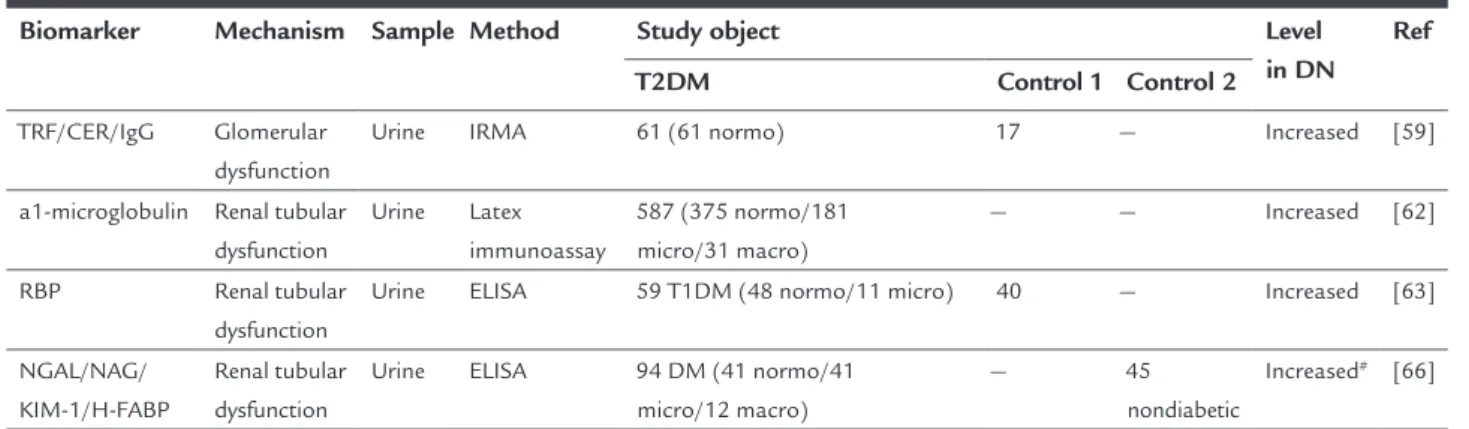
Biomarkers of renal tubular dysfunction
Tubulointerstitial injury plays an important role in DN development process and even prior to glomerular injury. In addition, about one third of the patients with diabetes mellitus have decreased renal function prior to proteinuria. Therefore, we should pay more attention to the biomark-ers of tubulointbiomark-erstitial injury, which can contribute to the early diagnosis and treatment of DN patients.59,60
a1-Microglobulin, retinol-binding protein 4 (RBP4) and other low molecular weight proteins can freely pass through the glomerular filtration membrane and then be reabsorbed in the tubules. These were early biomarkers of tubular injury because of their increase in urine after renal tubular damage. Researchers found that urinary levels of a1-microglobulin and RBP4 in patients with normoalbuminuria were significantly higher than those in control subjects and were both associated with the levels of HbA1c, so that detection of two biomarkers may
be helpful for early diagnosis of diabetic nephropathy.61,62
Some other biomarkers of tubular injury, such as neutrophil gelatinase-associated lipocalin (NGAL), N-acetyl-β-D-glucosidase (NAG), kidney injury mole-cule-1 (KIM-1) and heart-type fatty acid binding protein (H-FABP) applied only to predict acute kidney injury.63,64
A recent study discovered that urinary levels of NGAL, NAG, KIM-1, H-FABP of patients with normoalbumin-uria were significantly higher than those of control sub-jects and increased gradually along with the DN stages. In addition, they all significantly correlated with urinary albumin levels, indicating that they might be early bio-markers for DN diagnosis.65
These biomarkers were summarized in Table 2.
C
ONCLUSIONTABLE 1 Summary of biomarkers associated with DN pathology.
Biomarker Mechanism Sample Method Study object Level
in DN Ref
T2DM Control 1 Control 2
Podocin Podocytes injury Urine RT-QPCR 21 DN patients by biopsy 9 — Increased [42]
Synaptopodin Podocytes injury Urine RT-QPCR 21 DN patients by biopsy 9 — Increased [42]
Urine ELISA 71 (39 normo/17 micro/15 macro) 69 — Increased# [43]
Nephrin Podocytes injury Urine RT-QPCR 21 DN patients by biopsy 9 — Increased [42]
Urine ELISA 66 (26 normo/11 micro/29 macro) 10 — Increased# [44]
Type IV collagen
Basement membrane injury
Urine ELISA 698 DM (264 normo/169 micro/181
macro/84 renal failure)
191 — Increased [45]
vWF Endothelial
cells injury
Plasma ELISA 109 (66 normo/26 micro/17 macro) — 31
nondiabetic
Increased [47]
Plasma ELISA 24 (12 normo/12 micro) 12 — Increased [48]
Serum ELISA 60 (30 DN/30 without DN) 60 — Increased [49]
VEGF Endothelial
cells injury
plasma ELISA 387 T1DM (188 normo/199 DN) — — Increased [52]
Urine ELISA 107 (37 normo/37 micro/
33 proteinuria)
47 — Increased# [53]
TGF-β1 Mesangial
expansion/fibrosis Serum/ urine
ELISA 54 (20 normo/34 micro) 30 — Increased [55]
PEDF Mesangial
expansion/fibrosis
Urine ELISA 228 (59 normo/130 micro/
39 macro)
46 — Increased [57]
DN: diabetic nephropathy; T2DM: type 2 diabetes mellitus; Ref: reference; RT-QPCR: real-time quantitative polymerase chain reaction; ELISA: enzyme-linked immunesorbent assay; normo: nor-moalbuminuria; micro: microalbuminuria; macro: macroalbuminuria; DM: diabetes mellitus; vWF: von Willebrand factor; VEGF: vascular endothelial growth factor; T1DM: type 1 diabetes melli-tus; TGF-β1: transforming growth factor-β1; PEDF: pigment epithelial-derived factor.
Control 1: healthy subjects; Control 2: not healthy subjects.
#: increased prior to albuminuria.
TABLE 2 Summary of biomarkers associated with renal function changes.
Biomarker Mechanism Sample Method Study object Level
in DN Ref
T2DM Control 1 Control 2
TRF/CER/IgG Glomerular
dysfunction
Urine IRMA 61 (61 normo) 17 — Increased [59]
a1-microglobulin Renal tubular dysfunction
Urine Latex immunoassay
587 (375 normo/181 micro/31 macro)
— — Increased [62]
RBP Renal tubular
dysfunction
Urine ELISA 59 T1DM (48 normo/11 micro) 40 — Increased [63]
NGAL/NAG/ KIM-1/H-FABP
Renal tubular dysfunction
Urine ELISA 94 DM (41 normo/41
micro/12 macro)
— 45
nondiabetic
Increased# [66]
T2DM: type 2 diabetes mellitus; Ref: reference; TRF: transferrin; CER: ceruloplasmin; IgG: immunoglobulin G; IRMA: immunoradiometric assay; normo: normoalbuminuria; micro: microalbumi-nuria; macro: macroalbumimicroalbumi-nuria; RBP: retinol-binding protein; ELISA: enzyme-linked immunosorbent assay; T1DM: type 1 diabetes mellitus; NGAL: neutrophil gelatinase-associated lipocalin; NAG: N-acetyl-β-D-glucosidase; KIM-1: kidney injury molecule-1; H-FABP: heart-type fatty acid binding protein; DM: diabetes mellitus.
Control 1: healthy subjects; control 2: not healthy subjects.
#: increased prior to albuminuria.
R
EFERENCES1. Shaw JE, Sicree RA, Zimmet PZ. Global estimates of the prevalence of diabetes for 2010 and 2030. Diabetes Res Clin Pract. 2010; 87(1):4-14.
2. Gross JL, Azevedo MJ, Silveiro SP, Canani LH, Caramori ML, Zelmanovitz T. Diabetic nephropathy: diagnosis, prevention, and treatment. Diabetes Care. 2005; 28(1):164-76.
3. Tabaei BP, Al-Kassab AS, Ilag LL, Zawacki CM, Herman WH. Does microalbuminuria predict diabetic nephropathy? Diabetes Care. 2001; 24(9):1560-6.
5. Intensive blood-glucose control with sulphonylureas or insulin compared with conventional treatment and risk of complications in patients with type 2 diabetes (UKPDS 33). UK Prospective Diabetes Study (UKPDS) Group. The Lancet. 1998; 352(9131):837-53.
6. Brownlee M. Biochemistry and molecular cell biology of diabetic complications. Nature. 2001; 414(6865):813-20.
7. Cooper ME. Pathogenesis, prevention, and treatment of diabetic nephropathy. Lancet. 1998; 352(9123):213-9.
8. Forbes JM, Coughlan MT, Cooper ME. Oxidative stress as a major culprit in kidney disease in diabetes. Diabetes. 2008; 57(6):1446-54.
9. Giacco F, Brownlee M. Oxidative stress and diabetic complications. Circ Res. 2010; 107(9):1058-70.
10. Ha H, Kim C, Son Y, Chung MH, Kim KH. DNA damage in the kidneys of diabetic rats exhibiting microalbuminuria. Free Radic Biol Med. 1994; 16(2):271-4.
11. Hinokio Y, Suzuki S, Hirai M, Chiba M, Hirai A, Toyota T. Oxidative DNA damage in diabetes mellitus: its association with diabetic complications. Diabetologia. 1999; 42(8):995-8.
12. Zhao L, Xiang G, Yang L, Sun H, Le L, Liu Y. Relationship of serum 8-OHdG and VEGF with diabetic nephropathy in diabetics. Chin J Diabetes. 2012; 20(9):667-70.
13. Serdar M, Sertoglu E, Uyanik M, Tapan S, Akin K, Bilgi C, et al. Comparison of 8-hydroxy-2’-deoxyguanosine (8-OHdG) levels using mass spectrometer and urine albumin creatinine ratio as a predictor of development of diabetic nephropathy. Free Radic Res. 2012; 46(10):1291-5.
14. Calabrese V, Mancuso C, Sapienza M, Puleo E, Calafato S, Cornelius C, et al. Oxidative stress and cellular stress response in diabetic nephropathy. Cell Stress Chaperones. 2007; 12(4):299-306.
15. Tabak O, Gelisgen R, Erman H, Erdenen F, Muderrisoglu C, Aral H, et al. Oxidative lipid, protein, and DNA damage as oxidative stress markers in vascular complications of diabetes mellitus. Clin Invest Med. 2011; 34(3):E163-71. 16. Jiang B, Guo L, Li BY, Zhen JH, Song J, Peng T, et al. Resveratrol attenuates
early diabetic nephropathy by down-regulating glutathione s-transferases Mu in diabetic rats. J Med Food. 2013; 16(6):484-6.
17. Ahmed S, Mundhe N, Borgohain M, Chowdhury L, Kwatra M, Bolshette N, et al. Diosmin modulates the NF-kB signal transduction pathways and downregulation of various oxidative stress markers in alloxan-induced diabetic nephropathy. Inflammation. 2016; 39(5):1783-97.
18. Kishore L, Kaur N, Singh R. Renoprotective effect of Bacopa monnieri via inhibition of advanced glycation end products and oxidative stress in STZ-nicotinamide-induced diabetic nephropathy. Ren Fail. 2016; 38(9):1528-44. 19. Datta SK, Kumar V, Ahmed RS, Tripathi AK, Kalra OP, Banerjee BD. Effect of GSTM1 and GSTT1 double deletions in the development of oxidative stress in diabetic nephropathy patients. Indian J Biochem Biophys. 2010; 47(2):100-3. 20. Annadurai T, Vasanthakumar A, Geraldine P, Thomas PA. Variations in
erythrocyte antioxidant levels and lipid peroxidation status and in serum lipid profile parameters in relation to blood haemoglobin A1c values in individuals with type 2 diabetes mellitus. Diabetes Res Clin Pract. 2014; 105(1):58-69.
21. Giebułtowicz J, Sołobodowska S, Bobilewicz D, Wroczyński P. Blood ALDH1 and GST activity in diabetes type 2 and its correlation with glycated hemoglobin. Exp Clin Endocrinol Diabetes. 2014; 122(1):55-9.
22. Noce A, Fabrini R, Dessi M, Bocedi A, Santini S, Rovella V, et al. Erythrocyte glutathione transferase activity: a possible early biomarker for blood toxicity in uremic diabetic patients. Acta Diabetol. 2014; 51(2):219-24.
23. Liu J, Zhao Z, Willcox MD, Xu B, Shi B. Multiplex bead analysis of urinary cytokines of type 2 diabetic patients with normo- and microalbuminuria. J Immunoassay Immunochem. 2010; 31(4):279-89.
24. Moriwaki Y, Yamamoto T, Shibutani Y, Aoki E, Tsutsumi Z, Takahashi S, et al. Elevated levels of interleukin-18 and tumor necrosis factor-alpha in serum of patients with type 2 diabetes mellitus: relationship with diabetic nephropathy. Metabolism. 2003; 52(5):605-8.
25. Navarro JF, Mora C, Gomez M, Muros M, Lopez-Aguilar C, García J. Influence of renal involvement on peripheral blood mononuclear cell expression behavior of tumour necrosis factor-alpha and interleukin-6 in type 2 diabetic patients. Nephrol Dial Transplant. 2008; 23(3):919-26.
26. Shikano M, Sobajima H, Yoshikawa H, Toba T, Kushimoto H, Katsumata H, et al. Usefulness of a highly sensitive urinary and serum IL-6 assay in patients with diabetic nephropathy. Nephron. 2000; 85(1):81-5. 27. Dimas G, Iliadis F, Tegos T, Spiroglou S, Kanellos I, Karamouzis I, et al.
4B.08: serum levels of TIMP-1 and IL-6 are associated with hypertension
and atherosclerosis in patients with early stages of chronic kidney disease and type 2 diabetic nephropathy. J Hypertens. 2015; 33(Suppl 1):e55. 28. Zhang C, Xiao C, Wang P, Xu W, Zhang A, Li Q, et al. The alteration of Th1/
Th2/Th17/Treg paradigm in patients with type 2 diabetes mellitus: Relationship with diabetic nephropathy. Hum Immunol. 2014; 75(4):289-96. 29. Dalla Vestra M, Mussap M, Gallina P, Bruseghin M, Cernigoi AM, Saller A,
et al. Acute-phase markers of inflammation and glomerular structure in patients with type 2 diabetes. J Am Soc Nephrol. 2005; 16(Suppl 1):S78-82. 30. Navarro JF, Mora C, Muros M, García J. Urinary tumour necrosis factor-alpha excretion independently correlates with clinical markers of glomerular and tubulointerstitial injury in type 2 diabetic patients. Nephrol Dial Transplant. 2006; 21(12):3428-34.
31. Wu CC, Chen JS, Lu KC, Chen CC, Lin SH, Chu P, et al. Aberrant cytokines/ chemokines production correlate with proteinuria in patients with overt diabetic nephropathy. Clin Chim Acta. 2010; 411(9-10):700-4.
32. El-Asrar MA, Adly AA, Ismail EA. Soluble CD40L in children and adolescents with type 1 diabetes: relation to microvascular complications and glycemic control. Pediatr Diabetes. 2012; 13(8):616-24.
33. Chiarelli F, Giannini C, Verrotti A, Mezzetti A, Mohn A. Increased concentrations of soluble CD40 ligand may help to identify type 1 diabetic adolescents and young adults at risk for developing persistent microalbuminuria. Diabetes Metab Res Rev. 2008; 24(7):570-6.
34. Kanneganti M, Kamba A, Mizoguchi E. Role of chitotriosidase (chitinase 1) under normal and disease conditions. J Epithel Biol Pharmacol. 2012; 5:1-9. 35. Rathcke CN, Vestergaard H. YKL-40, a new inflammatory marker with relation to insulin resistance and with a role in endothelial dysfunction and atherosclerosis. Inflamm Res. 2006; 55(6):221-7.
36. Røndbjerg AK, Omerovic E, Vestergaard H. YKL-40 levels are independently associated with albuminuria in type 2 diabetes. Cardiovasc Diabetol. 2011; 10:54.
37. Żurawska-Płaksej E, Ługowska A, Hetmańczyk K, Knapik-Kordecka M,
Adamiec R, Piwowar A. Proteins from the 18 glycosyl hydrolase family are associated with kidney dysfunction in patients with diabetes type 2. Biomarkers. 2015; 20(1):52-7.
38. Urushihara M, Kondo S, Kagami S, Kobori H. Urinary angiotensinogen accurately reflects intrarenal renin-angiotensin system activity. Am J Nephrol. 2010; 31(4):318-25.
39. Satirapoj B, Siritaweesuk N, Supasyndh O. Urinary angiotensinogen as a potential biomarker of diabetic nephropathy. Clin Kidney J. 2014; 7(4):354-60. 40. Reddy GR, Kotlyarevska K, Ransom RF, Menon RK. The podocyte and diabetes mellitus: is the podocyte the key to the origins of diabetic nephropathy? Curr Opin Nephrol Hypertens. 2008; 17(1):32-6. 41. Wang G, Lai FM, Lai KB, Chow KM, Li KT, Szeto CC. Messenger RNA expression
of podocyte-associated molecules in the urinary sediment of patients with diabetic nephropathy. Nephron Clin Pract. 2007; 106(4):c169-79.
42. Hara M, Yamagata K, Tomino Y, Saito A, Hirayama Y, Ogasawara S, et al. Urinary podocalyxin is an early marker for podocyte injury in patients with diabetes: establishment of a highly sensitive ELISA to detect urinary podocalyxin. Diabetologia. 2012; 55(11):2913-9.
43. Jim B, Ghanta M, Qipo A, Fan Y, Chuang PY, Cohen HW, et al. Dysregulated nephrin in diabetic nephropathy of type 2 diabetes: a cross sectional study. PLoS One. 2012; 7(5):e36041.
44. Tomino Y, Suzuki S, Azushima C, Shou I, Iijima T, Yagame M, et al. Asian multicenter trials on urinary type IV collagen in patients with diabetic nephropathy. J Clin Lab Anal. 2001; 15(4):188-92.
45. Jensen T. Increased plasma concentration of von Willebrand factor in insulin dependent diabetics with incipient nephropathy. BMJ. 1989; 298(6665):27-8. 46. Yu Y, Suo L, Yu H, Wang C, Tang H. Insulin resistance and endothelial dysfunction in type 2 diabetes patients with or without microalbuminuria. Diabetes Res Clin Pract. 2004; 65(2):95-104.
47. Hirano T, Ookubo K, Kashiwazaki K, Tajima H, Yoshino G, Adachi M. Vascular endothelial markers, von Willebrand factor and thrombomodulin index, are specifically elevated in type 2 diabetic patients with nephropathy: comparison of primary renal disease. Clin Chim Acta. 2000; 299(1-2):65-75. 48. Fang YH, Zhang JP, Zhou SX, Zheng JF, Yu YW, Yan SG, et al. [Relationship between serum vWF and PAF in type 2 diabetic patients and diabetic nephropathy]. Di Yi Jun Yi Da Xue Xue Bao. 2005; 25(6):729-31. 49. Nong S, Ke L, Zhang X, Huang X, Man Y, Wang S, et al. Mechanism underlying
up-regulation of ICAM-1 and VCAM-1 expressions induced by high glucose in endothelial cells. Chinese J Cardiovasc Med. 2010; 15(3):219-22. 50. Abu Seman N, Anderstam B, Wan Mohamud WN, Östenson CG, Brismar
molecule 1 in Malaysian subjects with type 2 diabetes and diabetic nephropathy. J Diabetes Complications. 2015; 29(8):1234-9.
51. Hovind P, Tarnow L, Oestergaard PB, Parving HH. Elevated vascular endothelial growth factor in type 1 diabetic patients with diabetic nephropathy. Kidney Int Suppl. 2000; 75:S56-61.
52. Kim NH, Kim KB, Kim DL, Kim SG, Choi KM, Baik SH, et al. Plasma and urinary vascular endothelial growth factor and diabetic nephropathy in Type 2 diabetes mellitus. Diabet Med. 2004; 21(6):545-51.
53. Tamaki K, Okuda S. Role of TGF-beta in the progression of renal fibrosis. Contrib Nephrol. 2003; 139:44-65.
54. Xie F. Significance of serum and urinary TGF-β1 to the early diagnosis of diabetic nephropathy. Strait Pharmaceutical J. 2009; 21(5):145-6. 55. Wang JJ, Zhang SX, Lu K, Chen Y, Mott R, Sato S, et al. Decreased expression
of pigment epithelium-derived factor is involved in the pathogenesis of diabetic nephropathy. Diabetes. 2005; 54(1):243-50.
56. Chen H, Zheng Z, Li R, Lu J, Bao Y, Ying X, et al. Urinary pigment epithelium-derived factor as a marker of diabetic nephropathy. Am J Nephrol. 2010; 32(1):47-56.
57. Cohen-Bucay A, Viswanathan G. Urinary markers of glomerular injury in diabetic nephropathy. Int J Nephrol. 2012; 2012:146987.
58. Narita T, Sasaki H, Hosoba M, Miura T, Yoshioka N, Morii T, et al. Parallel increase in urinary excretion rates of immunoglobulin G, ceruloplasmin,
transferrin, and orosomucoid in normoalbuminuric type 2 diabetic patients. Diabetes Care. 2004; 27(5):1176-81.
59. Gewin L, Zent R, Pozzi A. Progression of chronic kidney disease: too much cellular talk causes damage. Kidney Int. 2017; 91(3):552-60.
60. Perkins BA, Ficociello LH, Silva KH, Finkelstein DM, Warram JH, Krolewski AS. Regression of microalbuminuria in type 1 diabetes. N Engl J Med. 2003; 348(23):2285-93.
61. Hong CY, Hughes K, Chia KS, Ng V, Ling SL. Urinary alpha1-microglobulin as a marker of nephropathy in type 2 diabetic Asian subjects in Singapore. Diabetes Care. 2003; 26(2):338-42.
62. Salem MA, el-Habashy SA, Saeid OM, el-Tawil MM, Tawfik PH. Urinary excretion of n-acetyl-beta-D-glucosaminidase and retinol binding protein as alternative indicators of nephropathy in patients with type 1 diabetes mellitus. Pediatr Diabetes. 2002; 3(1):37-41.
63. Bagshaw SM, Bellomo R. Early diagnosis of acute kidney injury. Curr Opin Crit Care. 2007; 13(6):638-44.
64. Han WK, Wagener G, Zhu Y, Wang S, Lee HT. Urinary biomarkers in the early detection of acute kidney injury after cardiac surgery. Clin J Am Soc Nephrol. 2009; 4(5):873-82.