Hydrocortisone reduces serum concentrations of inflammatory
biomarkers in patients subjected to carotid endarterectomy
Hidrocortisona reduz as concentrações séricas dos biomarcadores inflamatórios séricos
em pacientes submetidos a endarterectomia de carótida
Sthefano Atique Gabriel1
*
, Leila Antonangelo2, Vera Luiza Capelozzi2, Camila Baumann Beteli3, Otacílio de Camargo Júnior1, José Luis Braga de Aquino1, Roberto Augusto Cafaro4
Abstract
Background: Hydrocortisone may reduce serum and tissue concentrations of inlammatory biomarkers. Objective: To analyze the inlammatory activity of serum and tissue high-sensitivity C-reactive protein (hsCRP), tumor necrosis factor (TNF)-α and vascular endothelial growth factor (VEGF) after intraoperative administration of hydrocortisone, after carotid endarterectomy (CEA). Method: Twenty-two patients were allocated to a Control Group (5 asymptomatic and 6 symptomatic patients) and were not administered hydrocortisone or to Group 1 (4 asymptomatic and 7 symptomatic patients) and were administered 500 mg intravenous hydrocortisone. Serum levels of hsCRP, TNF-α and VEGF were tested for the preoperative period and at 1 hour, 6 hours and 24 hours after CEA. Levels of TNF-α and VEGF were also measured in carotid plaques. Results: Group 1 exhibited lower concentrations of serum TNF-α at 1 hour (p=0.031), 6 hours (p=0.015) and 24 hours (p=0.017) after CEA and lower concentrations of serum VEGF at 1 hour (p=0.006) and 6 hours (p=0.005) after CEA, relative to controls. Symptomatic patients in group 1 exhibited lower concentrations than controls for serum TNF-α at 1 hour and 6 hours after CEA and lower concentrations than controls for serum VEGF at 1 hour after CEA. here were no statistical diferences in tissue concentrations of TNF-α or VEGF between the control group and group 1. Conclusion: Hydrocortisone reduces postoperative concentrations of serum TNF-α and VEGF, especially in symptomatic patients; but does not reduce tissue levels of these biomarkers.
Keywords: hydrocortisone; inlammation; carotid stenosis.
Resumo
Contexto: A hidrocortisona pode reduzir a concentração dos biomarcadores inlamatórios séricos e teciduais.
Objetivo: Analisar a atividade inlamatória da proteína C-reativa ultrassensível (PCR-US), do fator de necrose tumoral (FNT)-alfa e do fator de crescimento do endotélio vascular (FCEV) séricos e teciduais, mediante administração intraoperatória de hidrocortisona, após endarterectomia de artéria carótida (EAC). Método: Vinte e dois pacientes foram divididos em Grupo Controle (5 assintomáticos e 6 sintomáticos) – não foi administrada hidrocortisona – e Grupo 1 (4 assintomáticos e 7 sintomáticos) – foram administrados 500 mg intravenoso de hidrocortisona. O PCR-US, o FNT-alfa e o FCEV séricos foram dosados no pré-operatório e em 1 hora, 6 horas e 24 horas após a EAC. Na placa carotídea, mensuramos os níveis de FNT-alfa e FCEV. Resultados: O grupo 1 exibiu menor concentração sérica de FNT-alfa em 1 hora (p=0,031), 6 horas (p=0,015) e 24 horas (p=0,017) após a EAC, e menor concentração de FCEV em 1 hora (p=0,006) e 6 horas (p=0,005) após a EAC, em relação ao grupo controle. Os pacientes sintomáticos do grupo 1 exibiram menor concentração de FNT-alfa em 1 hora e 6 horas após a EAC, e menor concentração de FCEV em 1 hora após a EAC, em relação ao grupo controle. Não houve diferença estatística entre as concentrações teciduais de FNT-alfa e FCEV entre o grupo controle e o grupo 1. Conclusão: A hidrocortisona reduz as concentrações séricas pós-operatórias de FNT-alfa e FCEV, em especial nos sintomáticos; porém, não reduz os níveis teciduais destes biomarcadores.
Palavras-chave: hidrocortisona; inlamação; estenose das carótidas.
1Pontifícia Universidade Católica de Campinas - PUC-Campinas, Campinas, SP, Brazil. 2Universidade de São Paulo - USP, São Paulo, SP, Brazil.
3Instituto Dante Pazzanese de Cardiologia, São Paulo, SP, Brazil.
4Faculdade de Ciências Médicas da Santa Casa de Misericórdia de São Paulo, São Paulo, SP, Brazil.
Financial support: None.
Conlicts of interest: No conlicts of interest declared concerning the publication of this article. Submitted: March 18, 2015. Accepted: June 11, 2015.
INTRODUCTION
Inlammatory biomarkers, which are produced by immunocompetent cells in atheromatous plaques, can orchestrate both systemic and tissue inlammatory responses, contributing to formation of complex atherosclerotic plaques, which are responsible for manifestation of ischemic neurological events, such as transitory ischemic attacks (TIA) and cerebral
vascular accidents (CVA).1-4
Elevated expression of inlammatory biomarkers in patients with unstable carotid plaques suggests that immunomodulatory treatments focused on the inlammatory process within the carotid plaque should be tested with a view to reducing progression of the
disease and the risk of TIA and CVA.5-7
Glucocorticoids have anti-inflammatory and immunosuppressive properties, reducing secretion of inflammatory biomarkers by monocytes and
macrophages.8,9 Elenkov10 points out that glucocorticoids
are immunomodulatory medications capable of modulating the systemic inlammatory response. The objective of this study was to analyze the inlammatory activity of hsCRP, of TNF-alpha and of VEGF in serum and tissue in response to intraoperative administration of hydrocortisone.
METHODS
Population
A total of 22 patients were treated with EAC from October of 2012 to September of 2013. Their ages ranged from 50 to 84, with a mean of 69.50 ± 9.09 years. In terms of sex distribution, 68.18% (15) were male and 31.82% (7) were female. With regard to neurological symptoms, 50% (11) were symptomatic and 50% (11) asymptomatic. Contralateral carotid stenosis percentages were as follows: 54.54% (12) of the patients had < 50% stenosis; 36.36% (8) had from 50% to 69% stenosis, and 9.1% (2) had stenosis ≥ 70%.
Symptomatic and asymptomatic patients were enrolled on the study if they had carotid stenosis ≥ 70% with indications for EAC. Patients were excluded in they exhibited carotid stenosis < 70% or occlusion of this arterial segment; indications for carotid angioplasty; autoimmune or systemic diseases; recent infections (< 1 month); or a recent CVA (< 1 month); hypersensitivity to and/or contraindications to use of hydrocortisone, or were taking glucocorticoids at the time of enrolment.
The study was approved by the Research Ethics Committee (Hearing No. 108.870) and patients signed free and informed consent forms.
Preoperative analysis
Carotid stenosis was investigated using color Doppler ultrasonography and stenosis ≥ 70% was conirmed with angiography of cerebral and supra-aortic trunks, since we do not have access to angiotomography equipment.
A data collection form covering epidemiological data and cardiovascular risk factors was designed for the study and completed for all patients, as shown in Table 1. Systemic arterial hypertension (SAH) was deined as systolic pressure ≥ 140 mmHg, diastolic pressure ≥ 90 mmHg and/or use of antihypertensive medications. Diabetes mellitus (DM) was deined as fasting glycemia level ≥ 126 mg/dL or use of hypoglycemic medications. Smoking was assessed in terms of mean consumption at the time of enrollment. Hypercholesterolemia was deined as total cholesterol > 200 mg/dL or use of hypolipidemic medications . Obesity was deined as body mass index
(BMI) ≥ 30 Kg/m2.
Patients were deined as symptomatic if they had suffered a TIA or CVA less than six months prior
to EAC.11
Patients who were to undergo EAC were allocated to one of two groups: either a Control Group (n=11, 5 asymptomatic and 6 symptomatic patients), containing patients who would not be administered hydrocortisone, or Group 1 (n=11, 4 asymptomatic and 7 symptomatic patients) comprising patients who would be given 500 mg of intravenous hydrocortisone during induction of anesthesia. Randomization of patients was achieved by randomly distributing 22 sequential numbers into 22 envelopes numbered from 1 to 22. Patients who were drawn even numbers were allocated to the control group and patients drawn uneven numbers were allocated to Group 1. The envelopes were opened in numerical order before induction of anesthesia.
As can be observed from the data in Table 1, both groups studied were predominantly made up of male patients, with a high incidence of hypertense and diabetic patients, a low percentage of obese individuals and a majority of patients with <50% contralateral carotid stenosis. There were no statistical differences between patients in the control group and patients in group 1 in terms of epidemiological or clinical characteristics, cardiovascular risk factors, neurological symptomology or duration of carotid clamping.
Endarterectomy of the carotid artery
removal of the atheromatous plaques, primary arteriorrhaphy was conducted. Vascular shunt was not used. The mean duration of carotid clamping was 46.27 ± 10.16 minutes. None of the patients exhibited neurological ischemic events after EAC.
Administration of the intravenous hydrocortisone
A 500 mg dose of hydrocortisone sodium succinate (SOLU – CORTEF), in the form of lyophilized powder for injectable solutions, was diluted in 4 mL of distilled water for injections and then added to 500 mL of 0.9% saline solution. The resulting solution at a concentration of 1 mg/mL was injected into a peripheral vein over an infusion time of 30 minutes.
Its effects are evident 1 hour after administration, it binds to a range of proteins and has a half-life in plasma of 1.5 to 2 hours and a half-life in tissues of 8 to 12 hours.
Determination of serum hsCRP, TNF-alpha and VEGF
Blood samples were taken preoperatively (24 hours before EAC) and 1 hour, 6 hours and 24 hours after the carotids were unclamped. Blood samples were collected into three tubes. One of these contained gel and clot activator, for the hsCRP assay, and the other two had walls coated with EDTA anticoagulant, for the TNF-alpha and VEGF assays.
Samples with EDTA were centrifuged at 1,000 rotations per minute (rpm) for 15 minutes and then at 10,000 rpm for 10 minutes. Samples drawn into tubes containing gel were centrifuged at 4,500 rpm for 20 minutes. After centrifugation, the supernatants were aliquoted into Eppendorf tubes (300 to 500 μL) and stored in an industrial freezer at -70 °C. The reference value was hsCRP < 5mg/L. The detection limits were: TNF-alpha < 0.2 pg/mL and VEGF < 0.04 pg/mL.
Table 1. Clinical and laboratory characteristics of the patients.
Variables Control group (11) Group 1 (11) p
Age (years) 69.09 ± 8.30 69.91 ± 10.20 0.718*
Sex Male 72.70% 63.60% 0.647§
Female 27.30% 36.40%
Systemic arterial hypertension Yes 90.90% 100% 0.306§
No 9.10% 0%
Diabetes mellitus Yes 54.50% 81.80% 0.170§
No 45.50% 18.20%
Smoking Yes 36.40% 63.60% 0.201§
No 63.60% 36.40%
Obesity Yes 9.10% 27.30% 0.269§
No 90.90% 72.70%
BMI (kg/m2) 26.64 ± 3.61 26.26 ± 5.14 0.669*
Total cholesterol (mg/dL) 189.45 ± 22.39 160.09 ± 33.43 0.023*
HDL (mg/dL) 47.55 ± 11.26 41.18 ± 13.56 0.200*
LDL (mg/dL) 110.64 ± 27.23 92.91 ± 27.53 0.212*
Triglycerides (mg/dL) 156.09 ± 50.08 156.91 ± 112.12 0.511* Fasting glycemia (mg/dL) 129.36 ± 53.82 110.27 ± 32.23 0.869* Duration of carotid clamping (minutes) 44.18 ± 7.22 48.36 ± 12.44 0.430* Contralateral carotid stenosis < 50% 54.50% 54.50% 0.287§
50% a 69% 45.50% 27.30%
≥ 70% 0% 18.20%
Neurological symptoms Symptomatic 54.50% 45.50% 0.670§
Asymptomatic 45.50% 54.50%
Coronary disease Yes 36.40% 63.60% 0.201§
No 63.60% 36.40%
Myocardial revascularization Yes 36.40% 63.60% 0.201§
No 63.60% 36.40%
Determination of TNF-alpha and VEGF in the carotid plaques
After removal, the endarterectomy specimens were frozen in an industrial freezer at -70 °C. The biomarkers were determined after tissue lysis using a Cell lysis kit (Biorad) using the LUMINEX Methodology.
Statistical analysis
The Statistical Package for Social Sciences, version 21.0, was used to calculate the results. Values were expressed as means ± standard deviations and percentages. The signiicance level was set at p < 0.05.
For clinical and epidemiological characteristics, parametric variables were evaluated using the Mann-Whitney test and nonparametric variables with the likelihood ratio test. The Mann-Whitney test was used to analyze serum and tissue biomarker results.
RESULTS
Comparison of serum inflammatory activity of hsCRP, of TNF-alpha and of VEGF for the control group versus group 1
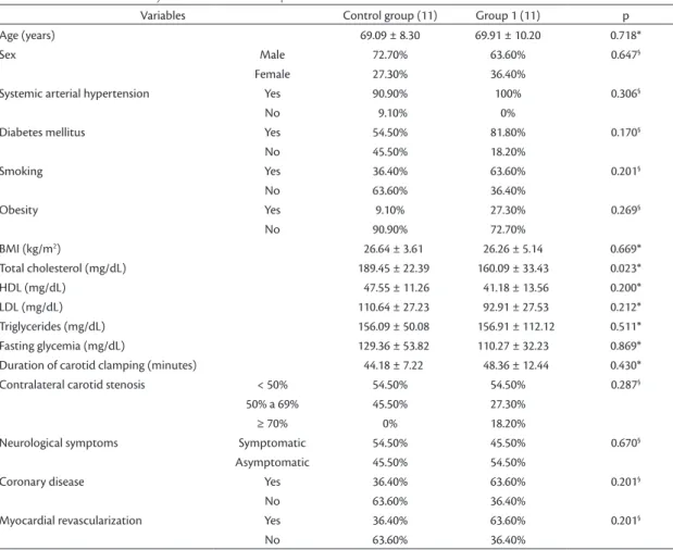
At 1 hour and 6 hours after EAC, serum concentrations of hsCRP were higher in group 1 than in the control group. In both groups, peak hsCRP inlammatory activity occurred at 24 hours after EAC (Figure 1). There were no statistically signiicant differences between the groups in terms of hsCRP levels.
The inlammatory proiles of TNF-alpha concentrations were similar for the two groups, with a reduction in levels 1 hour after EAC and an increase at 6 hours and 24 hours after EAC. However, group 1 exhibited a lower concentration of TNF-alpha than the control group at all postoperative analysis times (Figure 2). The differences in TNF-alpha levels were statistically signiicant at 1 hour (p=0.031), 6 hours (p=0.015) and 24 hours (p=0.017) after EAC.
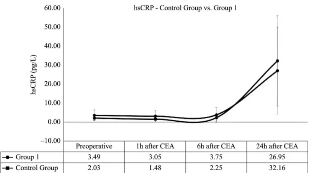
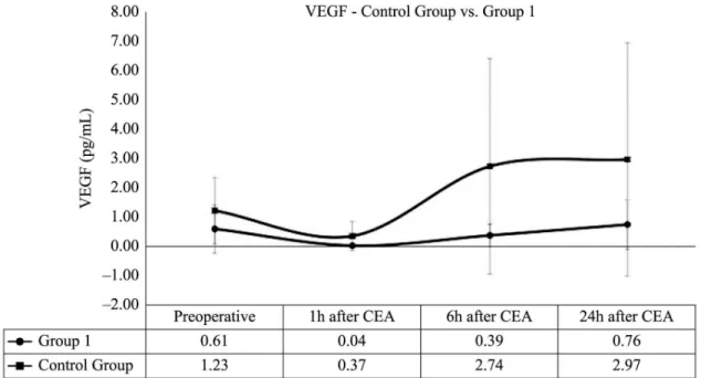
Concentrations of VEGF exhibited similar inflammatory responses in both groups, with a reduction in concentrations 1 hour after EAC and increases at 6 hours and 24 hours after EAC. However, group 1 exhibited a lower concentration of VEGF than the control group at all postoperative analysis times (Figure 3). The differences in VEGF levels were statistically signiicant at 1 hour (p=0.006) and at 6 hours (p=0.005), after EAC.
Table 2 shows the comparison of serum inlammatory activity of hsCRP, of TNF-alpha and of VEGF, for control group against group 1, at the four observation times.
Comparison of serum inflammatory activity of hsCRP, of TNF-alpha and of VEGF between symptomatic patients in control group and symptomatic patients in group 1
The symptomatic patients in the control group (n=6) exhibited lower concentrations of hsCRP 1 hour after EAC, while at 6 hours and 24 hours after EAC,
serum hsCRP concentrations were lower in group 1. The differences between these subsets’ hsCRP levels were not statistically signiicant.
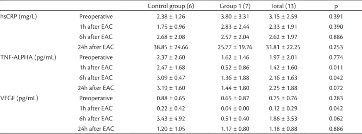
The TNF-alpha inlammatory activity in group 1 was lower than in the control group at all observation times. Differences in TNF-alpha levels attained statistical signiicance at 1 hour (p=0.011) and 6 hours (p=0.042) after EAC.
Symptomatic patients in group 1 exhibited lower concentrations of VEGF than symptomatic patients in control group at all postoperative analysis times. Only the differences in VEGF levels 1 hour after EAC attained statistical signiicance (p=0.011).
Table 3 lists the comparisons of serum inlammatory activity results for hsCRP, for TNF-alpha and for VEGF, for the symptomatic patients in the control
Figure 3. Serum inlammatory activity of VEGF for control group and group 1.
group versus the symptomatic patients in group 1, at the four observation times.
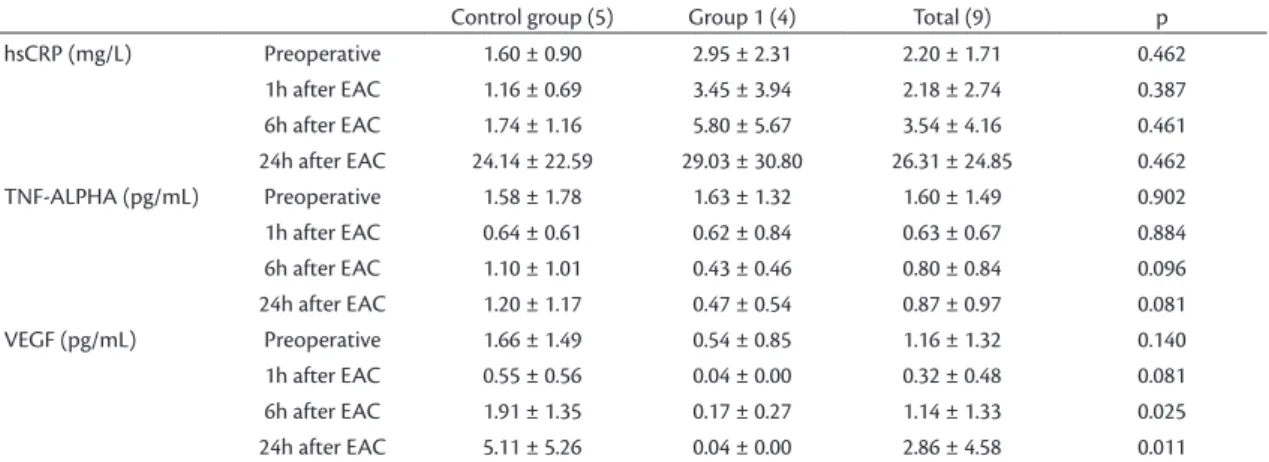
Comparison of serum inflammatory activity of hsCRP, of TNF-alpha and of VEGF between asymptomatic patients in the control group and asymptomatic patients in group 1
The asymptomatic patients in group 1 (n=4) exhibited higher hsCRP concentrations than the asymptomatic patients in the control group at all observation times. The differences between these subsets’ hsCRP levels were not statistically signiicant.
Inlammatory activity of TNF-alpha was lower in group 1 than in the control group at all postoperative observation times. There were no statistically signiicant differences between the TNF-alpha levels of these subsets.
The asymptomatic patients in group 1 exhibited lower concentrations of VEGF at all observation times
than the asymptomatic patients in the control group. The differences between VEGF levels in these subsets were statistically signiicant at 6 hours (p=0.025) and 24 hours (p=0.011) after EAC.
Table 4 lists the comparisons of serum inlammatory activity results for hsCRP, for TNF-alpha and for VEGF, for the asymptomatic patients in the control group versus the asymptomatic patients in group 1, at the four observation times.
Comparison of the inflammatory activity of TNF-alpha and of VEGF in tissue specimens from symptomatic and asymptomatic patients in the control group and in group 1
Symptomatic patients exhibited higher tissue concentrations of VEGF, whereas the asymptomatic patients exhibited higher tissue levels of TNF-alpha. There were no statistically signiicant differences
Table 2. Serum inlammatory activity of hsCRP, of TNF-alpha and of VEGF, for control group and group 1, at four diferent
observation times.
Control group (11) Group 1 (11) Total (22) p hsCRP (mg/L) Preoperative 2.03 ± 1.14 3.49 ± 2.89 2.76 ± 2.27 0.189
1h after EAC 1.48 ± 0.86 3.05 ± 2.88 2.27 ± 2.23 0.177 6h after EAC 2.25 ± 1.72 3.75 ± 3.84 3.00 ± 3.00 0.576 24h after EAC 32.16 ± 23.82 26.95 ± 22.84 29.56 ± 22.93 0.450 TNF-ALPHA (pg/mL) Preoperative 2.01 ± 2.20 1.62 ± 1.34 1.82 ± 1.79 0.869 1h after EAC 1.63 ± 1.57 0.56 ± 0.81 1.10 ± 1.34 0.031 6h after EAC 2.19 ± 1.26 1.02 ± 1.55 1.61 ± 1.50 0.015 24h after EAC 2.28 ± 1.70 1.09 ± 1.51 1.68 ± 1.69 0.017 VEGF (pg/mL) Preoperative 1.23 ± 1.13 0.61 ± 0.82 0.92 ± 1.01 0.070 1h after EAC 0.37 ± 0.49 0.04 ± 0.00 0.20 ± 0.38 0.006 6h after EAC 2.74 ± 3.67 0.39 ± 0.38 1.56 ± 2.81 0.005 24h after EAC 2.97 ± 3.97 0.76 ± 0.84 1.87 ± 3.02 0.069
Table 3. Serum inlammatory activity of hsCRP, of TNF-alpha and of VEGF, for symptomatic patients in the control group and in
group 1, at four diferent observation times.
Control group (6) Group 1 (7) Total (13) p hsCRP (mg/L) Preoperative 2.38 ± 1.26 3.80 ± 3.31 3.15 ± 2.59 0.391
in tissue concentrations of TNF-alpha or of VEGF between symptomatic and asymptomatic patients.
The asymptomatic and symptomatic patients in group 1 exhibited lower tissue concentrations of TNF-alpha and of VEGF than those in the control group. There were no statistically signiicant differences in tissue concentrations of TNF-alpha or of VEGF between patients in the control group and patients in group 1.
Table 5 lists the results for tissue inlammatory activity of TNF-alpha and of VEGF for patients in the control group and in group 1.
DISCUSSION
This study has demonstrated for the irst time that intraoperative administration of hydrocortisone interferes in the inlammatory responses of TNF-alpha and VEGF, reducing their concentrations over a period of 24 hours after EAC. Additionally, we also observed a tendency for hydrocortisone to have an effect not only at the serum level, but also at the tissue level, controlling inlammatory activity of TNF-alpha and of VEGF in the carotid plaque.
Hermus et al.7 state that cellular inlammatory
activity, expressed by serum and tissue production of inlammatory biomarkers such as interleukins, metalloproteinases, hsCRP, TNF-alpha and VEGF, contributes to progression and destabilization of the
carotid plaque, resulting in TIA and CVA.7 Additionally,
the magnitude of the systemic and tissue inlammatory
process, assessed by measurement of these speciic biomarkers may relect the risk of postoperative complications and progression of the contralateral
carotid disease.7
Raja et al.12 claim that the systemic inlammatory
response after cardiovascular surgery is due to ischemia-reperfusion injury to target organs caused by clamping of arteries and restoration of perfusion after release of the hemostatic forceps. Additionally, persistence of an elevated degree of postoperative inlammation can potentially be prejudicial to patients
subjected to cardiovascular surgical interventions.12
However, Rubens et al.13 and Liguori et al.14 have
concluded that the systemic inlammatory response is variable and can be inluenced by patient comorbidities; intraoperative non-pharmacological interventions; type of anesthesia; perioperative hemodynamic conditions; surgical aspects (surgical incision, duration of the procedure, duration of arterial clamping and need for blood transfusion); and postoperative recovery. In order to ensure that the same perioperative conditions prevailed, we standardized general anesthesia and classical endarterectomy. The serum hsCRP, TNF-alpha and VEGF assays were performed on samples taken after removal of carotid clamping, so that the results would relect the maximum production of these biomarkers during carotid clamping. In contrast, the hydrocortisone was administered during induction of anesthesia, in order to assess its immunomodulatory
Table 5. Tissue inlammatory activity of TNF-alpha and of VEGF, for patients in the control group and in group 1.
Control group (11) Group 1 (11) Total (22) p TNF-ALPHA (pg/mL) Symptomatic 1.29 ± 0.33 1.16 ± 0.52 1.22 ± 0.43 0.550
Asymptomatic 1.40 ± 0.46 1.16 ± 0.56 1.30 ± 0.49 0.521 VEGF (pg/mL) Symptomatic 11.02 ± 17.85 5.33 ± 5.76 7.96 ± 12.57 0.886 Asymptomatic 8.15 ± 13.13 1.78 ± 0.99 5.32 ± 9.89 0.327
Table 4. Serum inlammatory activity of hsCRP, of TNF-alpha and of VEGF, for asymptomatic patients in the control group and in
group 1, at four diferent observation times.
Control group (5) Group 1 (4) Total (9) p
effect, which has a short half-life (1.5 to 2 hours), not only on the postoperative inlammatory activity of serum hsCRP, TNF-alpha and VEGF, but also on TNF-alpha and VEGF in the carotid plaque.
Previous publications have already demonstrated that glucocorticoids can interfere in the systemic inlammatory response, reducing concentrations of serum biomarkers and attenuating activation of the complement system and leukocyte and neutrophil
adhesion,13,14 but this effect has not been demonstrated
with respect to hsCRP, TNF-alpha or VEGF, and the action of glucocorticoids on the behavior of biomarkers present in patients treated with EAC has not been assessed. After administering 30 mg/kg of intravenous methylprednisolone before surgery and before unclamping the thoracic aorta in 16 patients treated with revascularization of the myocardium,
Kawamura et al.15 observed reductions in plasma
concentrations of interleukins IL-6 and IL-8 at 1 hour, 2 hours and 3 hours after the aorta was unclamped.
Komori et al.16 administered 1g of intravenous
methylprednisolone to ten patients 2 hours before conventional surgical repair of infrarenal abdominal aortic aneurysms and detected lower concentrations of IL-6 after unclamping the abdominal aorta and on the irst day after the operation and lower CRP levels on the irst day after the operation, compared with patients who were not given the preoperative dose of methylprednisolone.
In our study, the symptomatic patients exhibited higher serum concentrations of hsCRP, TNF-alpha and VEGF than the asymptomatic patients for the preoperative measurement and the postoperative measurements, with the exception of the hsCRP concentration at 6 hours after EAC and the VEGF
concentration at 24 hours after EAC. Koutouzis et al.17
also observed higher preoperative concentrations of hsCRP and TNF-alpha in symptomatic patients; but the preoperative levels of hsCRP and TNF-alpha were lower in their symptomatic and asymptomatic
patients than in our patients. In contrast, Puz et al.18
reported higher preoperative TNF-alpha levels in both symptomatic patients (14.58 pg/mL) and in asymptomatic patients (13.93 pg/mL), when compared with our patients. We believe that these differences were the result of the characteristics of the patients analyzed, the methods used to assay these biomarkers, the methods for classiication of patients according to presence or absence of prior neurological symptoms and to the interval of time that elapsed between symptoms and measurement of the biomarkers.
Of the biomarkers analyzed, hsCRP exhibited the greatest postoperative inlammatory activity, and the
maximum concentration was observed 24 hours after
EAC. Alvarez Garcia et al.19 identiied an association
between elevated hsCRP levels and a history of neurological events and presence of unstable carotid plaques, but did not demonstrate differences between the levels of this biomarker and the severity of the
neurological ischemic event. Heider et al.20 observed
ischemic brain injuries in 22.4% of their patients after EAC, concluding that elevated preoperative levels of hsCRP are associated with a greater risk of cerebral ischemic injuries after EAC. In our study, serum hsCRP levels exhibited a discrete change in inlammatory behavior in response to administration of hydrocortisone, but signiicant reductions in their postoperative concentrations were not observed in
symptomatic or asymptomatic patients. Morrow et al.21
have previously concluded that serum hsCRP levels do not change in response to hormone changes or anti-inlammatory drugs. Possible explanations for the mild effect exerted by the hydrocortisone on serum hsCRP levels in our study are founded on the short half-life (1.5 to 2 hours) of hydrocortisone, its administration during induction of anesthesia and the long half-life of hsCRP (18 hours). Strategies that could be tested in future studies with the aim of reducing the inlammatory response of hsCRP include increasing the dose of hydrocortisone, using a corticoid with a longer half-life or administering the hydrocortisone 24 hours after EAC.
In contrast with hsCRP, we observed a considerable reduction in the postoperative concentrations of TNF-alpha, at all observation times, in response to administration of hydrocortisone. The lower inlammatory activity of TNF-alpha was still identiiable when symptomatic patients were analyzed separately, with a reduction in concentrations at 1 hour and 6 hours after EAC, in response to administration of hydrocortisone. TNF-alpha stimulates thickening of the vascular endothelium, provoking deposition of collagen by ibroblasts and contributes to hypotension, coagulopathy
and renal dysfunction.22 Welsh et al.23 observed a
correlation between TNF-alpha levels and the risk of
recurrent CVA. Additionally, Profumo et al.24 state
that maintenance of elevated TNF-alpha levels after EAC is associated with progression of contralateral carotid disease.
plaques through the process of chemoattraction of monocytes and other inlammatory cells, since it induces expression of metalloproteinases and cellular adhesion molecules, promotes neoangiogenesis and
stimulates expression and synthesis of thromboplastin.25
Szabó et al.26 assessed 82 patients treated with eversion
EAC, observing than carotid restenosis occurred in patients who exhibited elevated serum VEGF levels four days after the operation.
We did not observe statistical differences between symptomatic patients and asymptomatic patients in terms of concentrations of TNF-alpha or VEGF in
carotid plaques. Grufman et al.27 also failed to detect
statistical differences between tissue concentrations of TNF-alpha between symptomatic patients and asymptomatic patients. However, administration of the intraoperative dose of hydrocortisone did exhibit a tendency to reduce the concentrations of TNF-alpha and VEGF in the carotid plaque, suggesting an attempt to control the inlammatory activity in the carotid plaque.
This study suffers from certain limitations. While the hydrocortisone interfered in the inlammatory activity of the serum biomarkers, our results do not provide prognostic information about the patients in terms of progression of contralateral carotid disease and or carotid restenosis. Our results do however provide a basis for conducting further studies to assess the effects of intraoperative administration of hydrocortisone on the prognosis of patients treated with EAC.
In conclusion, the immunomodulatory effect of hydrocortisone reduces postoperative serum concentrations of TNF-alpha and VEGF, particularly in symptomatic patients. Intraoperative administration of hydrocortisone exhibited a tendency to reduce tissue concentrations of TNF-alpha and VEGF.
REFERENCES
1. Teixeira BC, Lopes AL, Macedo RCO, et al. Inflammatory markers, endothelial function and cardiovascular risk. J Vasc Bras. 2014;13(2):108-15. http://dx.doi.org/10.1590/jvb.2014.054.
2. Balanescu S, Calmac L, Constantinescu D, Marinescu M, Onut R, Dorobantu M. Systemic inflammation and early atheroma formation: are they related? Maedica. 2010;5(4):292-301. PMid:21977173.
3. Tuttolomondo A, Di Raimondo D, Pecoraro R, Arnao V, Pinto A, Licata G. Atherosclerosis as an inflammatory disease. Curr Pharm Des. 2012;18(28):4266-88. http://dx.doi.org/10.2174/138161212802481237. PMid:22390643.
4. Moller MJ, Qin Z, Toursarkissian B. Tissue markers in human atherosclerotic carotid artery plaque. Ann Vasc Surg. 2012;26(8):1160-5. http://dx.doi.org/10.1016/j.avsg.2012.06.008. PMid:23068427.
5. Mauriello A, Sangiorgi GM, Virmani R, et al. A pathobiologic link between risk factors profile and morphological markers of carotid
instability. Atherosclerosis. 2010;208(2):572-80. http://dx.doi. org/10.1016/j.atherosclerosis.2009.07.048. PMid:19683236.
6. Wekesa AL, Cross KS, O’Donovan O, et al. Predicting carotid artery disease and plaque instability from cell-derived microparticles. Eur J Vasc Endovasc Surg. 2014;48(5):489-95. http://dx.doi.org/10.1016/j. ejvs.2014.08.007. PMid:25218652.
7. Hermus L, Lefrandt JD, Tio RA, Breek JC, Zeebregts CJ. Carotid plaque formation and serum biomarkers. Atherosclerosis. 2010;213(1):21-9. http://dx.doi.org/10.1016/j.atherosclerosis.2010.05.013. PMid:20627248.
8. Stahn C, Buttgereit F. Genomic and nongenomic effects of glucocorticoids. Nat Clin Pract Rheumatol. 2008;4(10):525-33. http://dx.doi.org/10.1038/ncprheum0898. PMid:18762788.
9. Imamura K, Hayashi F, Suzumura A. [Cytokine production by peripheral blood monocytes/macrophages in the patients with multiple sclerosis and its suppression by methylprednisolone]. Rinsho Shinkeigaku. 1992;32(3):276-80. PMid:1628450.
10. Elenkov IJ. Neurohormonal-cytokine interactions: implications for inflammation, common human diseases and well-being. Neurochem Int. 2008;52(1-2):40-51. http://dx.doi.org/10.1016/j. neuint.2007.06.037. PMid:17716784.
11. NASCET Investigators. Clinical alert: benefit of carotid endarterectomy for patients with high-grade stenosis of the internal carotid artery. National Institute of Neurological Disorders and Stroke Stroke and Trauma Division. North American Symptomatic Carotid Endarterectomy Trial (NASCET) investigators. Stroke. 1991;22(6):816-7. http://dx.doi.org/10.1161/01.STR.22.6.816. PMid:2057984.
12. Raja SG, Dreyfus GD. Modulation of systemic inflammatory response after cardiac surgery. Asian Cardiovasc Thorac Ann. 2005;13(4):382-95. http://dx.doi.org/10.1177/021849230501300422. PMid:16304234.
13. Rubens FD, Mesana T. The inflammatory response to cardiopulmonary bypass: a therapeutic overview. Perfusion. 2004;19(Suppl 1):S5-12. http://dx.doi.org/10.1191/0267659104pf717oa. PMid:15161059.
14. Liguori GR, Kanas AF, Moreira LFP. Managing the inflammatory response after cardiopulmonary bypass: review of the studies in animal models. Rev Bras Cir Cardiovasc. 2014;29(1):93-102. http:// dx.doi.org/10.5935/1678-9741.20140017. PMid:24896169. 15. Kawamura T, Inada K, Okada H, Okada K, Wakusawa R.
Methylprednisolone inhibits increase of interleukin 8 and 6 during open heart surgery. Can J Anaesth. 1995;42(5 Pt 1):399-403. http:// dx.doi.org/10.1007/BF03015485. PMid:7614647.
16. Komori K, Ishida M, Matsumoto T, et al. Cytokine patterns and the effects of a preoperative steroid treatment in the patients with abdominal aortic aneurysms. Int Angiol. 1999;18(3):193-7. PMid:10688417.
17. Koutouzis M, Rallidis LS, Peros G, et al. Serum interleukin-6 is elevated in symptomatic carotid bifurcation disease. Acta Neurol Scand. 2009;119(2):119-25. http://dx.doi.org/10.1111/j.1600-0404.2008.01068.x. PMid:18638042.
18. Puz P, Lasek-Bal A, Ziaja D, Kazibutowska Z, Ziaja K. Inflammatory markers in patients with internal carotid artery stenosis. Arch Med Sci. 2013;9(2):254-60. http://dx.doi.org/10.5114/aoms.2013.34533. PMid:23671435.
19. Alvarez Garcia B, Ruiz C, Chacon P, Sabin JA, Matas M. High-sensitivity C-reactive protein in high-grade carotid stenosis: risk marker for unstable carotid plaque. J Vasc Surg. 2003;38(5):1018-24. http:// dx.doi.org/10.1016/S0741-5214(03)00709-2. PMid:14603210.
2007;46(3):449-54. http://dx.doi.org/10.1016/j.jvs.2007.05.035. PMid:17826232.
21. Morrow DA, Ridker PM. C-reactive protein, inflammation, and coronary risk. Med Clin North Am. 2000;84(1):149-61, ix. http:// dx.doi.org/10.1016/S0025-7125(05)70211-X. PMid:10685132.
22. Baki ED, Sivaci RG, Kokulu S, Ela Y, Aldemir M. Effects of anesthetic choice on inflammatory response in cardiac surgery. Inflammation and Cell Signaling. 2014;1:e75.
23. Welsh P, Woodward M, Rumley A, Lowe G. Associations of circulating TNFalpha and IL-18 with myocardial infarction and cardiovascular risk markers: the Glasgow Myocardial Infarction Study. Cytokine. 2009;47(2):143-7. http://dx.doi.org/10.1016/j. cyto.2009.06.002. PMid:19581111.
24. Profumo E, Esposito C, Buttari B, et al. Intracellular expression of cytokines in peripheral blood from patients with atherosclerosis before and after carotid endarterectomy. Atherosclerosis. 2007;191(2):340-7. http://dx.doi.org/10.1016/j.atherosclerosis.2006.03.030. PMid:16678185.
25. Russell DA, Abbott CR, Gough MJ. Vascular endothelial growth factor is associated with histological instability of carotid plaques. Br J Surg. 2008;95(5):576-81. http://dx.doi.org/10.1002/bjs.6100. PMid:18344184.
26. Szabó A, Laki J, Madsen HO, et al. Early rise in serum VEGF and PDGF levels predisposes patients with a normal MBL2 genotype to restenosis after eversion endarterectomy. Stroke. 2007;38(8):2247-53. http://dx.doi.org/10.1161/STROKEAHA.106.475954. PMid:17626901.
27. Grufman H, Gonçalves I, Edsfeldt A, et al. Plasma levels of high-sensitive C-reactive protein do not correlate with inflammatory activity in carotid atherosclerotic plaques. J Intern Med. 2014;275(2):127-33. http://dx.doi.org/10.1111/joim.12133. PMid:24010553.
*
Correspondence Sthefano Atique Gabriel Rua Dr. Melo Alves, 685 - Cerqueira Cesar CEP 01417-010 - São Paulo (SP), Brazil E-mail: [email protected]
Author information SAG - Vascular and endovascular surgeon, Department of Vascular Surgery, Hospital e Maternidade Celso Pierro; Professor, Pontifícia Universidade Católica de Campinas (PUC-Campinas). LA - Tenured professor, Universidade de São Paulo (USP), Department of Cytology, Laboratório Central, Faculdade de Medicina da USP. VLC - Tenured professor, Universidade de São Paulo (USP), Department of Pathology, Faculdade de Medicina da USP. CBB - Vascular and endovascular surgeon, Department of Vascular Ultrasound, Instituto Dante Pazzanese de Cardiologia. OCJ - MSc in Surgery, Department of Vascular Surgery; Professor, Pontifícia Universidade Católica de Campinas (PUC-Campinas). JLBA - PhD in Surgery, Universidade Estadual de Campinas (Unicamp), Department of Surgery; Professor, Pontifícia Universidade Católica de Campinas (PUC-Campinas). RAC - PhD in Surgery, Faculdade de Ciências Médicas da Santa Casa de Misericórdia de São Paulo, Department of Vascular/Endovascular Surgery; Professor, Faculdade de Ciências Médicas da Santa Casa de Misericórdia de São Paulo.
Author contributions Conception and design: SAG, JLBA, RAC Analysis and interpretation: SAG, LA, VLC, CBB, OCJ, JLBA, RAC Data collection: SAG, CBB, LA, VLC Writing the article: SAG, LA, VLC, CBB, OCJ, JLBA, RAC Critical revision of the article: SAG, LA, VLC, CBB, OCJ, JLBA, RAC Final approval of the article*: SAG, LA, VLC, CBB, OCJ, JLBA, RAC Statistical analysis: SAG, CBB, JLBA, RAC Overall responsibility: SAG, JLBA, RAC