Expression analysis on 14-3-3 proteins in regenerative liver following partial
hepatectomy
Deming Xue
1,2, Yang Xue
3, Zhipeng Niu
1,2, Xueqiang Guo
1,2and Cunshuan Xu
1,21
College of Life Science, Henan Normal University, Xinxiang, Henan, China
2
Key Laboratory for Cell Differentiation Regulation, Xinxiang, Henan, China
3
Academy of Fine Arts, Henan Normal University, Xinxiang, Henan, China
Abstract
14-3-3 proteins play a vital part in the regulation of cell cycle and apoptosis as signaling integration points. During liver regeneration, the quiescent hepatocytes go through hypertrophy and proliferation to restore liver weight. There-fore, we speculated that 14-3-3 proteins regulate the progression of liver regeneration. In this study, we analyzed the expression patterns of 14-3-3 proteins during liver regeneration of rat to provide an insight into the regenerative mechanism using western blotting. Only four isoforms (g,e,sandt/q) of the 14-3-3 proteins were expressed in re-generative liver after partial hepatectomy (PH). The dual effects, the significant down-regulation of 14-3-3eand the significant up-regulation of 14-3-3t/qat 2 h after PH, might play particularly important roles in S-phase entry. The sig-nificant peaks of 14-3-3sat 30 h and ofeandt/qat 24 h might be closely related not only to the G2/M transition but
also to the size of hepatocytes. Possibly, the peak of 14-3-3eexpression seen at 168 h plays critical roles in the termi-nation of liver regeneration by inhibiting cellular proliferation.
Keywords: liver regeneration, 14-3-3 proteins, western blotting.
Received: February 06, 2017; Accepted: July 20, 2017.
Introduction
Liver has a remarkable capability of regenerating fol-lowing different kinds of damage (Cienfuegoset al., 2014). Liver is composed of numerous types of cells including hepatocytes, sinusoidal endothelial cells, stellate cells, Kupffer-Browicz cells, and biliary epithelial cells (Kanget al., 2012). Nevertheless, hepatocytes, which account for approximately 80% of the liver mass and around 70% of liver cells, perform most of the functions of metabolism and synthesis (Si-Tayebet al., 2010). In seriously injured liver with damaged proliferation of hepatocytes, progenitor cells are thought to be helpful for regeneration through prolifera-tion and differentiaprolifera-tion (Alisonet al., 2009). In contrast, re-generation following partial hepatectomy (PH) does not need such a progenitor cell. The remnant liver undergoes compensatory hypertrophy and recovers the initial liver weight within approximately a week in rodents (Michalopoulos, 2007). The multi-lobe structure of the liver permits resecting one or more lobes to obtain various degrees of loss of liver weight. Because resecting the liver lobes does not injure the remnant tissue, PH is thought to be a very good experimental model for research into the
regen-erative mechanisms of this tissue. (Miyaoka and Miyajima, 2013).
The family of 14-3-3 proteins comprises a group of highly homologous acidic proteins expressed in all euka-ryotic organisms. This family is made up of seven isoforms
in human and rodent tissues (b/a,g,z/d,s,e,h,t/q) and plays an important role in the regulation of many cellular processes, including cell cycle, cell differentiation, apop-tosis, DNA repair, motility and adhesion. 14-3-3 proteins function as phosphoserine/phosphothreonine-binding mo-dules that take part in phospho-dependent protein-protein interactions (Gardino and Yaffe, 2011; Wuet al., 2015). Their expression is tissue-specific.
Cell division and apoptosis take place during hepatic regeneration following 2/3 PH (Sakamoto et al., 1999). Therefore, the 14-3-3 protein family may be closely linked to hepatic regeneration. Previously we reported the expres-sion patterns of 14-3-3 mRNAs in regenerative liver fol-lowing 2/3 PH by real-time qRT-PCR (Xueet al., 2015). In the current study, we further analyzed the expression pat-terns of 14-3-3 proteins by western blotting.
Materials and Methods
Animals and PH model
Sprague Dawley male rats (200±10 g) were obtained from the Animal Center of Henan Normal University. The rats were permittedad libitumaccess to food and water. For the PH model, rats were anaesthetized by ether inhalation, and the left lateral and median lobes, which account for two-thirds of the total liver weight, were resected (Stolzet al., 1999). The remaining liver lobes were obtained at 0, 2, 6, 12, 24, 30, 36, 72, 120 and 168 h after PH. All samples of liver were quickly frozen in liquid nitrogen, and then stored at -80 °C. All rats were placed in the facility of Animal Cen-ter of Henan Normal University, and all procedures were performed according to the Animal Protection Law of China.
Protein extraction and western blotting
The regenerating liver tissues stored in liquid nitro-gen were ground into fine powder and then suspended in extraction buffer (7 M urea, 2 M thiourea, 4% CHAPS). Next, the suspension was vortex-mixed for 1 h at 4 °C, and subsequently centrifuged at 20,000 xgfor 1 h at 4 °C. The supernatants were collected and stored at –80 °C for further use. The protein concentration was assessed with the com-mercial RC DCTMProtein Assay Kit according to the manu-facturer’s instructions (BIO-RAD, USA). Protein samples, 50 mg, were separated by electrophoresis on 12% SDS/PAGE gels and subsequently electrophoretically transferred to polyvinylidene difluoride membranes (Milli-pore). The membranes were blocked with 5% non-fat milk, washed, and subsequently probed with antibodies against 14-3-3b/a, g, z/d,s, e, h, t/q (all 1:1000) and GAPDH (1:2000) (Sangon Biotech Co. Ltd., Shanghai, China)over-night at 4 °C. After being washed, the membranes were in-cubated with horseradish peroxidase-conjugated secondary antibodies (Sangon Biotech Co. Ltd., Shanghai, China), de-tected with an enhanced chemiluminescence detection kit (Boster Corporation, China) and then imaged in an ImageQuant LAS 4000 mini (GE Healthcare Bio-Sciences Corporation) system.
Statistical analysis
Analysis of the western blots was carried out by a standard technique. The gray intensities of the bands were quantified by Image J software. The relative gray level value of the target band was normalized against that of the respective internal control GAPDH. Statistical analysis on protein expression was then carried out using SPSS version 16.0 (SPSS, Inc., Chicago, IL, USA) software. All data were reported as means± standard deviation (n=3). Stu-dent’st-tests were used for analyzing the expression differ-ence between 0 h and the other time points after P, with p<0.05 indicating statistical significance.
Results
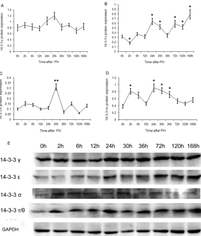
The expression patterns of the seven 14-3-3 protein isoforms in regenerative liver after PH were assessed by western blotting using an enhanced chemiluminescence de-tection kit. The results showed that only four (g,e,sand
t/q) of the 14-3-3 isoforms were expressed (Figure 1). When compared with 0 h, the expression of 14-3-3gat the other time points showed wavy oscillations without signifi-cant difference (t < t0.05(4)=2.776, p > 0.05) (Figure 1A).
14-3-3eexpression, which showed a minimum at 2 h, was significantly down-regulated in comparison with that at 0 h (t=3.105>t0.05(4)=2.776, p<0.05). 14-3-3eexpression
was significantly up-regulated at 24, 30, 72, 120 and 168 h (t=3.871, 2.970, 2.895, 2.970 and 3.899 > t0.05(4) = 2.776,
p<0.05). The transcript level at 168 h was the highest, and that at 30 h and 120 h was the lowest among these (Figure 1B).
14-3-3sexpression, with a peak at 30 h after PH, was significantly up-regulated in comparison with that at 0 h (t = 5.750 > t0.01(4)= 4.601, p<0.01), whereas those at the other
time points showed no significant difference, exhibiting wavy oscillations (t < t0.05(4)= 2.776, p > 0.05) (Figure 1C).
When compared with 0 h after PH, the 14-3-3t/q ex-pression at 2, 24, 30 and 36 h was significantly up-regulated (t = 3.948, 3.660, 3.952 and 2.860 > t0.05(4)= 2.776, p<0.05).
The transcript level at 24 and 36 h were the highest and low-est, respectively (Figure 1D).
Discussion
Liver regeneration has been extensively investigated, but many essential mechanisms are still vague, for instance, the mechanisms of cell hypertrophy, cell division, and reg-ulation of organ size (Miyaoka and Miyajima, 2013). For a long time, it has been thought that after 70% PH all remnant hepatocytes would undergo one or two rounds of cell divi-sion for recovery of the initial cell number and liver weight (Duncanet al., 2009). Recent studies have reported that af-ter 30% PH, the hepatocyte size of the remnant liver in-creases and recovers its initial weight, but the hepatocyte number does not change. In comparison, upon 70% PH, hepatocyte hypertrophy occurs for several hours following PH, before the cells proliferate. Although nearly all hepato-cytes enter S-phase, only approximately half go through the cell division cycle and increase cell number. As a result, af-ter 70% PH the liver recovers its original weight through both hypertrophy and proliferation of hepatocytes (Miyao-kaet al., 2012).
The sophisticated process of regeneration is divided into three different stages: an initial phase, a proliferative phase, and a terminative phase. In rats, within less than 15 min following PH, quiescent hepatocytes withdraw from the G0phase and go into G1phase. The first proliferation
syn-thesis peak was shown to occur between 22 and 24 hours, followed by the karyokinesis peak between 28 and 30 hours (Corlu and Loyer, 2012). The 14-3-3 proteins act on the G1/S and G2/M transitions by combining with cell cycle
regulation proteins and regulating their function (Hermeking and Benzinger, 2006; Gardino and Yaffe,
2011), and therefore, they presumably play critical roles in the regulation of hepatic regeneration.
Successful activation of different cyclin-dependent kinases (CDKs) is required for passing through the cel1cycle. These kinases are regulated by transient binding of regulatory subunits, phosphorylation and
phorylation (Sherr and Roberts, 1995; Harper and Brooks, 2005).
The 14-3-3 proteins participate in the modulation of transition from G1into S through a variety of mechanisms.
They associate with and negatively modulate cell division cycle phosphatases (CDC25), which are also concerned with the regulation of CDK complexes that are extremely important for the transition from G1to S. CDC25A, a key
element for the entry into S phase by activating CDK2 through dephosphorylation, is inhibited by 14-3-3ethrough cytoplasmic sequestration (Chenet al., 2003). In addition, direct binding of 14-3-3sto the kinases CDK2 and CDK4 negatively regulates S-phase entry (Larongaet al., 2000). The 14-3-3 proteins can immediately bind to the CDK in-hibitor p27 as well, which opposes p27-mediated G1arrest.
Following phosphorylation by the serine-threonine kinase AKT at Thr-198 and Thr-157, p27 associates with the 14-3-3b,g,z,e,handtisoforms, and does not function as CDK inhibitor because of its cytoplasmic sequestration (Fujitaet al., 2002; Lianget al., 2002; Shinet al., 2002), as the association hinders the binding of p27 to importina
(Sekimotoet al., 2004). Through this mechanism, the 14-3-3 proteins may contribute to cell cycle progression. Fur-thermore, the p90 ribosomal protein S6 kinase also associ-ates with and directly phosphorylassoci-ates p27KIP1on Thr-198, resulting in the binding to 14-3-3e,h, s, andtisoforms (Fujitaet al., 2003). At 2 h after 2/3 PH, the significant down-regulation of 14-3-3e and the significant up-regu-lation of 14-3-3tmay lead to the release of CDC25A from 14-3-3eand the association of p27KIP1with 14-3-3t, respec-tively. These dual effects might play particularly important roles in S-phase entry.
14-3-3 proteins are also necessary for the transition from G2into M (Fordet al., 1994). In mammalian cells, a
key step for mitotic entry is the activation of CDC2 kinase (Norburyet al., 1991). During S phase, CDC2 activity is in-hibited through phosphorylation by the MYT1/MIK1 and WEE1 kinases at Thr-14 and Tyr-15 (Parker and Piwnica-Worms, 1992; Liu et al., 1997). CDC25C dephos-phorylates CDC2 at Thr-14 and Tyr-15, which leads to CDC2 activation and initiates entry into M phase (Gautier et al., 1991). Activating CDC25C is indispensable for pro-moting cell cycle progress, whereas its restraint is con-nected with the activation of the G2/M checkpoint
(Donzelli and Draetta, 2003). CDC25C activity is sup-pressed following phosphorylation on the residue Ser-216, mediated by CHK1, CHK2 or C-TAK1. This occurs through subsequent association with 14-3-3 isoforms and sequestration in the cytoplasm. This causes the increase in CDC2 phosphorylation, leading to decreased activity and repression of entry into mitosis (Sanchezet al., 1997; Peng et al., 1998; Chaturvediet al., 1999). Seemingly, not all of the 14-3-3 isoforms are able to associate with and modulate CDC25C. CDC25C is able to interact with 14-3-3tandzin
lung adenocarcinoma A549 cells (Qi and Martinez, 2003). Only 14-3-3gandeamong seven different 14-3-3 isoforms specifically associate with CDC25C in U2OS cells and thereby restrain CDC25C from inducing premature chro-matinic condensation. In contrast, 14-3-3sdoes not associ-ate with CDC25C, but obstructs premature chromatinic condensation induced by the CDC25C mutant S216A and wild type CDC25C (Dalalet al., 2004). This shows that 14-3-3sregulates mitotic entry downstream of CDC25C.
CDC25B activity is also inhibited by binding to 14-3-3 isoforms, which blocks substrate access of CDK1-cyclinB complexes to the CDC25B catalytic site (Astuti and Gabrielli, 2011). CDC25B associates with different isoforms of the 14-3-3 family, and it was shown to interact with 14-3-3b,z,s,eandhin vivoand with 14-3-3b,zand
hin a yeast two-hybrid test (Milset al., 2000; Uchidaet al., 2004).
According to the experimental results and the above discussion, we propose the following hypotheses. The sig-nificant peaks of 14-3-3eexpression,tat 24 h and ofsat 30 h after PH are related to the regulation of not only the G2/M
transition, but also the size of hepatocytes. They may play critical roles in preventing the remnant hepatocytes from prematurely entering into mitosis after PH. As a result, the size of the hepatocytes increases. It is reported that the terminative process in liver regeneration has not been ade-quately investigated, compared with the initiation (Miyao-ka and Miyajima, 2013). The significant up-regulation of 14-3-3efrom 72 to 168 h after PH might be one of the im-portant factors inhibiting the G1/S-transition by a direct
as-sociation with CDC25A, and thus, would be key for the termination of hepatic regeneration.
Acknowledgments
This work was funded by grants from the Interna-tional Collaborative Project on Science and Technology of Henan Province (No. 152102410038), the Base and Fron-tier Technology Research Project of Henan Province (No. 132300410135) and the National Natural Science Founda-tion of China (No. 31572270).
References
Alison MR, Islam S and Lim S (2009) Stem cells in liver regenera-tion, fibrosis and cancer: The good, the bad and the ugly. J Pathol 217:282-298.
Astuti P and Gabrielli B (2011) Phosphorylation of Cdc25B3 Ser169 regulates 14-3-3 binding to Ser151 and Cdc25B ac-tivity. Cell Cycle 10:1960-1967.
Chen MS, Ryan CE and Piwnica-Worms H (2003) Chk1 kinase negatively regulates mitotic function of Cdc25A phospha-tase through 14-3-3 binding. Mol Cell Biol 23:7488-7497. Cienfuegos JA, Rotellar F, Baixauli J, Martínez-Regueira F,
Pardo F and Hernández-Lizoáin JL (2014) Liver regenera-tion - The best kept secret. A model of tissue injury response. Rev Esp Enferm Dig 106:171-194.
Corlu A and Loyer P (2012) Regulation of the G1/S transition in hepatocytes: Involvement of the cyclin-dependent kinase Cdk1 in the DNA replication. Int J Hepatol 2012:689324. Dalal SN, Yaffe MB and DeCaprio JA (2004) 14-3-3 family
members act coordinately to regulate mitotic progression. Cell Cycle 3:672-677.
Donzelli M and Draetta GF (2003) Regulating mammalian check-points through Cdc25 inactivation. EMBO Rep 4:671-677. Duncan AW, Dorrell C and Grompe M (2009) Stem cells and liver
regeneration. Gastroenterology 137:466-481.
Ford JC, al-Khodairy F, Fotou E, Sheldrick KS, Griffiths DJ and Carr AM (1994) 14-3-3 protein homologs required for the DNA damage checkpoint in fission yeast. Science 265:533-535.
Fujita N, Sato S, Katayama K and Tsuruo T (2002) Akt-dependent phosphorylation of p27Kip1 promotes binding to 14-3-3 and cytoplasmic localization. J Biol Chem 277:28706-28713. Fujita N, Sato S and Tsuruo T (2003) Phosphorylation of p27Kip1
at threonine 198 by p90 ribosomal protein S6 kinases pro-motes its binding to 14-3-3 and cytoplasmic localization. J Biol Chem 278:49254-49260.
Gardino AK and Yaffe MB (2011) 14-3-3 proteins as signaling in-tegration points for cell cycle control and apoptosis. Semin Cell Dev Biol 22:688-695.
Gautier J, Solomon MJ, Booher RN, Bazan JF and Kirschner MW (1991) CDC25 is a specific tyrosine phosphatase that di-rectly activates p34cdc2. Cell 67:197-211.
Harper JV and Brooks G (2005) The mammalian cell cycle: An overview. Methods Mol Biol 296:113-153.
Hermeking H and Benzinger A (2006) 14-3-3 proteins in cell cy-cle regulation. Semin Cancer Biol 16:183-192.
Kang LI, Mars WM and Michalopoulos GK (2012) Signals and cells involved in regulating liver regeneration. Cells 1:1261-1292.
Laronga C, Yang HY, Neal C and Lee MH (2000) Association of the cyclin-dependent kinases and 14-3-3sigma negatively regulates cell cycle progression. J Biol Chem 275:23106-23112.
Liang J, Zubovitz J, Petrocelli T, Kotchetkov R, Connor MK, Han K, Lee JH, Ciarallo S, Catzavelos C, Beniston R, et al.
(2002) PKB/Akt phosphorylates p27, impairs nuclear im-port of p27 and opposes p27-mediated G1arrest. Nat Med
8:1153-1160.
Liu F, Stanton JJ, Wu Z and Piwnica-Worms H (1997) The human Myt1 kinase preferentially phosphorylates Cdc2 on threo-nine 14 and localizes to the endoplasmic reticulum and Golgi complex. Mol Cell Biol 17:571-583.
Michalopoulos GK (2007) Liver regeneration. J Cell Physiol 213:286-300.
Mils V, Baldin V, Goubin F, Pinta I, Papin C, Waye M, Eychene A and Ducommun B (2000) Specific interaction between 14-3-3 isoforms and the human CDC25B phosphatase. Oncogene 19:1257-1265.
Miyaoka Y and Miyajima A (2013) To divide or not to divide: Re-visiting liver regeneration. Cell Div 8:8.
Miyaoka Y, Ebato K, Kato H, Arakawa S, Shimizu S and Miyajima A (2012) Hypertrophy and unconventional cell di-vision of hepatocytes underlie liver regeneration. Curr Biol 22:1166-1175.
Norbury C, Blow J and Nurse P (1991) Regulatory phosphoryl-ation of the p34cdc2 protein kinase in vertebrates. EMBO J 10:3321-3329.
Parker LL and Piwnica-Worms H (1992) Inactivation of the p34cdc2-cyclin B complex by the human WEE1 tyrosine kinase. Science 257:1955-1957.
Peng CY, Graves PR, Ogg S, Thoma RS, Byrnes 3rd MJ, Wu Z, Stephenson MT and Piwnica-Worms H (1998) C-TAK1 protein kinase phosphorylates human Cdc25C on serine 216 and promotes 14-3-3 protein binding. Cell Growth Differ 9:197-208.
Qi W and Martinez JD (2003) Reduction of 14-3-3 proteins corre-lates with increased sensitivity to killing of human lung can-cer cells by ionizing radiation. Radiat Res 160:217-223. Sakamoto T, Liu Z, Murase N, Ezure T, Yokomuro S, Poli V and
Demetris AJ (1999) Mitosis and apoptosis in the liver of interleukin-6-deficient mice after partial hepatectomy. Hepatology 29:403-411.
Sanchez Y, Wong C, Thoma RS, Richman R, Wu Z, Piwnica-Worms H and Elledge SJ (1997) Conservation of the Chk1 checkpoint pathway in mammals: Linkage of DNA damage to Cdk regulation through Cdc25. Science 277:1497-1501. Sekimoto T, Fukumoto M and Yoneda Y (2004) 14-3-3
sup-presses the nuclear localization of threonine 157-phos-phorylated p27 (Kip1). EMBO J 23:1934-1942.
Sherr CJ and Roberts JM (1995) Inhibitors of mammalian G1
cyclin-dependent kinases. Genes Dev 9:1149-1163. Shin I, Yakes FM, Rojo F, Shin NY, Bakin AV, Baselga J and
Arteaga CL (2002) PKB/Akt mediates cell-cycle progres-sion by phosphorylation of p27 (Kip1) at threonine 157 and modulation of its cellular localization. Nat Med 8:1145-1152.
Si-Tayeb K, Lemaigre FP and Duncan SA (2010) Organogenesis and development of the liver. Dev Cell 18:175-189. Stolz DB, Mars WM, Petersen BE, Kim TH and Michalopoulos
GK (1999) Growth factor signal transduction immediately after two-thirds partial hepatectomy in the rat. Cancer Res 59:3954-3960.
Uchida S, Kuma A, Ohtsubo M, Shimura M, Hirata M, Nakagama H, Matsunaga T, Ishizaka Y and Yamashita K (2004) Bind-ing of 14-3-3beta but not 14-3-3 sigma controls the cytoplas-mic localization of CDC25B: Binding site preferences of 14-3-3 subtypes and the subcellular localization of CDC25B. J Cell Sci 117:3011-3020.
Wu YJ, Jan YJ, Ko BS, Liang SM and Liou JY (2015) Involve-ment of 14-3-3 proteins in regulating tumor progression of hepatocellular carcinoma. Cancers 7:1022-1036.
Xue DM, Guo XQ, Chen R, Niu ZP and Xu CS (2015) 14-3-3 gene expression in regenerating rat liver after 2/3 partial hepatectomy. Genet Mol Res 14:2023-2030.
Associate Editor: Jeremy A. Squire