Endothelial Cells
Jeffrey Kroon, Simon Tol, Sven van Amstel, Judith A. Elias, Mar Fernandez-Borja*
Department of Molecular Cell Biology, Sanquin Research and Landsteiner Laboratory, University of Amsterdam, Amsterdam, The Netherlands
Abstract
The inflammatory response of endothelial cells triggered by cytokines such as TNFaand IL1bplays a pivotal role in innate immunity. Upon pro-inflammatory cytokine stimulation, endothelial cells produce chemokines and cytokines that attract and activate leukocytes, and express high levels of leukocyte adhesion molecules. This process is mediated by intracellular signaling cascades triggered by activation of e.g. the TNFa receptor (TNFR) that lead to the activation of the NFkB transcription factor and of MAP kinases, which in turn activate inflammatory gene transcription. We found that the small GTPase RhoB was strongly and rapidly upregulated in primary human endothelial cells by TNFa, IL1b and LPS. We subsequently investigated the role of RhoB in the regulation of TNFR signaling in endothelial cells by silencing RhoB expression with siRNA. We provide evidence that the TNFa-induced activation of p38 MAP kinase is strongly dependent on RhoB, but not on RhoA, while JNK activation is regulated by both RhoB and RhoA. Consistent with the important role of p38 MAP kinase in inflammation, we demonstrate that loss of RhoB impairs TNFa-induced ICAM-1 expression and reduces cell production of IL6 and IL8. In addition, we show that RhoB silencing alters the intracellular traffic of TNFaafter endocytosis. Since RhoB is a known regulator of the intracellular traffic of membrane receptors, our data suggest that RhoB controls TNFasignaling through the regulation of the TNFR traffic.
Citation:Kroon J, Tol S, van Amstel S, Elias JA, Fernandez-Borja M (2013) The Small GTPase RhoB Regulates TNFaSignaling in Endothelial Cells. PLoS ONE 8(9): e75031. doi:10.1371/journal.pone.0075031
Editor:Venuprasad K. Poojary, Baylor Institute for Immunology Research, United States of America
ReceivedJune 29, 2010;AcceptedJuly 26, 2013;PublishedSeptember 26, 2013
Copyright:ß2013 Kroon et al. This is an open-access article distributed under the terms of the Creative Commons Attribution License, which permits unrestricted use, distribution, and reproduction in any medium, provided the original author and source are credited.
Funding:M. Fernandez-Borja was funded by project 112 from the Landsteiner Foundation for Blood Transfusion. The funders had no role in study design, data collection and analysis, decision to publish, or preparation of the manuscript.
Competing Interests:The authors have declared that no competing interests exist. * E-mail: [email protected]
Introduction
Tumor necrosis factora(TNFa) is a pleiotropic pro-inflamma-tory cytokine that plays a pivotal role in the innate immune response to infection and tissue injury. Vascular endothelial cells respond to TNFaby upregulating the expression of cytokines and chemokines, such as IL-6 and IL-8, and of endothelial leukocyte adhesion molecules, such as VCAM-1, ICAM-1 and E-selectin [1]. These molecules enable TNFa-activated endothelial cells to attract, activate and recruit circulating leukocytes, which subse-quently extravasate to reach the site of infection or injury. The inflammatory program induced by TNFais a result of intracellular signaling triggered by the TNFa-receptor (TNFR) [2,3]. Upon ligand binding, TNFR trimerizes and recruits TRAF-2 (TNFR-associated factor 2) and RIP1 (receptor interacting protein 1) to its cytoplasmic death domain. The formation of this signaling complex leads to the activation of the transcription factor NFkB and of the MAP kinases JNK and p38. Subsequently, the TNFR is rapidly endocytosed and eventually degraded in the lysosomes [4,5]. However, TNFR internalization is clearly not only a mechanism of receptor downregulation but also of signaling compartmentalization, providing temporal and spatial regulation of the diverse signaling cascades triggered by the activated receptor [6]. While signaling from the TNFR leading to NFkB activation takes place at the plasma membrane, there is com-pelling evidence that TNFR pro-apoptotic signaling occurs on endosomes [4,7]. In addition, several molecules involved in TNFR signaling are found on the surface of endosomal and lysosomal
compartments [8,9]. Finally, one study has demonstrated that internalization of the TNFR from the plasma membrane is a required step for the activation of p38 and JNK MAP kinases [10]. RhoB is a short-lived Rho GTPase whose expression is inducible by a variety of stimuli including growth factors, such as EGF and PDGF [11] and stress stimuli such as DNA-damaging drugs, UV irradiation and reactive oxygen species [12,13]. RhoB is 83% identical to RhoA, a constitutively expressed GTPase and a well-established regulator of actomyosin-based contractility and of serum-induced transcription. Although these two GTPases bind to a similar set of proteins in solution, their non-overlapping intracellular distribution provides specificity to their respective actions [14]. Whereas RhoA is cytosolic and translocates to the plasma membrane upon activation, RhoB localizes to endosomes/multive-sicular bodies [15]. Multiveendosomes/multive-sicular bodies are primarily involved in the sorting of membrane proteins for their delivery to lysosomes for degradation. Consistently, RhoB regulates the sorting and degrada-tion of growth factor and cytokine receptors [16–21]. In agreement with the role of Rho GTPases as critical regulators of actin dynamics, RhoB appears to control vesicle traffic through the regulation of actin polymerization on endosomes [22,23], possibly through the recruitment and activation of Diaphanous proteins [23,24].
variety of stimuli including growth factors, UV radiation and oxidative stress [11–13]. Here we report that RhoB protein is rapidly upregulated in primary human endothelial cells by TNFa, IL1band bacterial lipopolysaccharide (LPS). We have addressed the role of RhoB in TNFR signaling by using siRNA-mediated knockdown of RhoB. We present evidence that RhoB is essential for the activation of p38 and JNK MAP kinases, but not NFkB, by TNFa. Finally, we show that RhoB silenced cells accumulate endocytosed TNFa pointing to a defect in traffic kinetics and/or receptor sorting. In summary, our data suggest that RhoB has as role in TNFasignaling through the regulation of TNFR intracellular traffic.
Materials and Methods
Cell culture
Pooled primary human umbilical vein endothelial cells (HUVEC, Lonza) were seeded on fibronectin-coated culture flasks
and maintained in EGM-2 medium (Lonza) in a humidified atmosphere of 95% air and 5% CO2 at 37uC.
Reagents and antibodies
Recombinant human TNFa, IL1b, IFNc, VEGF and TGFb were from R&D Systems. Bacterial lipopolysaccharide (LPS) was from Sigma.The pharmacological inhibitors cycloheximide, MG132 and N-acetyl-cysteine (NAC) were from Sigma. SC-514, SP600125 and SB230580 were from Calbiochem. Rabbit and mouse anti-RhoB, mouse anti-RhoA, mouse anti-phospho-ERK, mouse phospho-JNK, goat VCAM-1 and mouse anti-NFkB p50 were obtained from Santa Cruz Biotechnology. Rabbit anti-phospho-p38 and mouse anti-IkBawere obtained from Cell Signaling. Mouse anti-a-tubulin (DM1A) was from Sigma. Mouse anti-EEA1 and mouse anti-RhoGDI were from BD Biosciences. Secondary antibodies labeled with Alexa488 or Alexa568 for
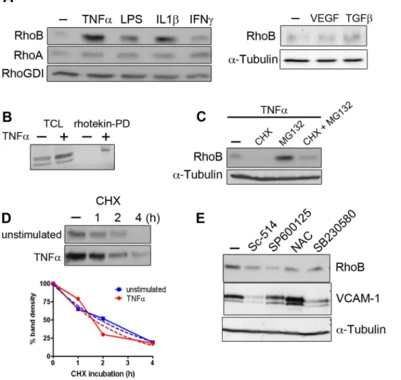
Figure 1. Pro-inflammatory mediators induce ‘de novo’ RhoB synthesis and RhoB activation in human umbilical vein endothelial cells.(A) Lysates of HUVEC incubated for 16 h with the indicated stimuli were analyzed for the expression of RhoB and RhoA by western blot. RhoGDI and tubulin were detected to control for equal loading; (B) Pull-down of GTP-Rho from HUVEC stimulated or not with TNFafor 4 h. Precipitates were analyzed for the presence of RhoB; (C) RhoB expression was induced by a 4 h TNFastimulation, and subsequently proteasome inhibitor MG132 and/ or the protein synthesis inhibitor cycloheximide was added to the cells for an additional 4 hours incubation; (D) RhoB detection in lysates of HUVEC stimulated with TNFafor 4 hours before the addition of cycloheximide (CHX) for 1, 2 or 4 hours. A digital scan of the film was made and the intensity of the RhoB bands was measured using ImageJ software. Data are shown as percentage of the RhoB present in the absence of cycloheximide. Unstimulated (solid blue line) and TNFa-stimulated cells (solid red line). Fitted regression lines obtained by linear regression analysis are shown as dotted lines; (E) Endothelial cells were incubated with TNFain combination with the NFkB inhibitor sc-514, the JNK inhibitor SP600125, the p38 inhibitor SB230580 or the anti-oxidant N-acetyl cysteine (NAC). RhoB and VCAM-1 were detected in cell lysates.a-Tubulin was detected to control for equal loading.
doi:10.1371/journal.pone.0075031.g001
immunofluorescence were purchased from Invitrogen. Secondary antibodies labeled with horseradish peroxidase for immunoblot-ting were from Amersham.
Immunoblotting and phosho-MAP kinase arrays
Cells were lysed in cold NP-40 buffer (1% NP40, 100 mM NaCl, 100 mM MgCl2, 10% glycerol, 50 mM Tris pH 7.4)
containing a cocktail of phosphatase and protease inhibitors
(Thermo Scientific). Protein content of lysates was quantified with the Precision Red Advanced Protein Assay Reagent (Cytoskele-ton). Equal protein concentrations were loaded in SDS-PAGE gels and analyzed by Western blotting. Equal loading was additionally controlled by detection of Rho-GDI and atubulin. The pixel density of each band was determined with ImageJ and values corrected by the correspondingatubulin intensities. These values Figure 2. RhoB does not regulate TNFa-induced NFkB
activa-tion.(A) Efficiency of RhoB silencing with either a pool of 3 siRNAs (RhoB pool) or with two single siRNAs from the pool individually (#1 and#2) was analyzed in cells stimulated with TNFafor 16 hours; (B) TNFawas added for 30 min to cells transfected with RhoB siRNAs as in (A) or with an siRNA control. Cell lysates were analyzed for the presence of IkBa. a-Tubulin was detected to control for equal loading. Graphs represent band densities corrected for protein loading and are normalized to control transfected cells. No significant differences were found between control and RhoB-deficient cells; (C) Endothelial cells transfected with siRNA control or with a pool of RhoB siRNAs were stimulated with TNFafor 0 and 30 min, fixed and permeabilized and then incubated with an antibody to the p50 NFkB subunit.
doi:10.1371/journal.pone.0075031.g002
Figure 3. RhoB regulates MAP kinase activation by TNFa.(A)
Cells transfected with siRNA control or with a pool of RhoB siRNAs were stimulated or not with TNFafor 30 min. Lysates were prepared and incubated with an anti-phospho-MAP kinase antibody array. Pixel intensity of spots in the array was determined, corrected for background and represented as percentage of the positive controls included in the array; (B) Western blot analysis of phospho-ERK1/2, phospho-p38 and phospho-hsp27 in HUVEC transfected with siRNA control, with a pool of RhoB siRNAs (RhoB pool) or with single RhoB siRNAs (RhoB#1 and#2).
were normalized to those of siRNA control-transfected cells. Non-parametric one-way ANOVA Tukey test was used to evaluate statistical significance of at least 3 independent experiments. A two-way ANOVA with Bonferroni post-test was used to evaluate statistical significance when the effects of several siRNA effects were compared at different time points on TNF stimulation. Results are expressed as mean 6 SEM (*p,0.05; **p,0.01; ***p,0.001).
Human phospho-mitogen activated kinase protein antibody arrays were purchased from R&D Systems and used according to manufacturer instructions. Briefly, cell lysates were incubated with the antibody arrays overnight at 4uC. After washing, arrays were incubated with a mixture of phospho-site specific biotinylated antibodies for 2 hours at room temperature. Bound biotinylated antibodies were detected with HRP-streptavidin. Arrays were developed in ECL and exposed to X-Omat films. Digital scans of the films were analyzed for pixel density with ImageJ. Averaged background values corresponding to the negative controls were subtracted from the values of each spot.
Values for duplicates on the array were averaged and represented as a percentage of the pixel density of the positive controls included in the array.
RhoB activity assay
Rho-GTP pulldown assays were performed as previously described [25]. Cells were lysed on ice in lysis buffer containing 50 mM Tris pH 7.6, 500 mM NaCl, 1% Triton X-100, 0.1% SDS, 0.5% deoxycholate, 10 mM MgCl2, 100 and a cocktail of
protease inhibitors (Sigma). Lysates were clarified by centrifuga-tion at 14,0006g 5 min and incubated with Glutathion S-transferase (GST)-Rho binding domain beads. After washing four times in 50 mM Tris pH 7.6, 150 mM NaCl, 1% Triton X-100, 0.5 mM MgCl2, 100mM orthovanadate, with protease
inhibitors, bound Rho proteins were solubilized with SDS-sample buffer and analyzed by SDS-PAGE with specific antibodies for RhoB.
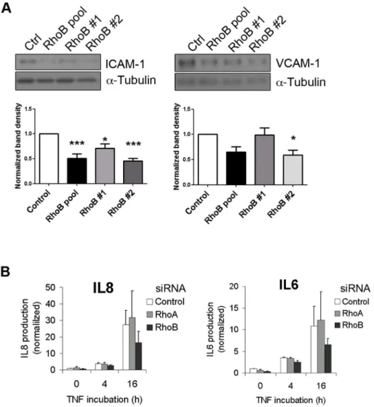
Figure 4. RhoB silencing impairs TNFa-induced pro-inflammatory molecule expression.(A) Lysates of cells transfected with siRNAs as in
Figure 3 were stimulated with TNFafor 4 h and analyzed for total ICAM-1 and VCAM-1 expression by western blotting; (B) ELISA analysis of IL6 and IL8 present in conditioned media of cells transfected with a pool of RhoB siRNAs, with RhoA siRNA or a control siRNA were stimulated with TNFafor 4 or 16 h. Graph shows normalized values after dividing by the IL concentration in the medium of unstimulated siRNA control-transfected cells (12356592 pg/mL IL8 and 18767.5 pg/mL IL6) (mean6SEM, n = 3; *p,0,05).
doi:10.1371/journal.pone.0075031.g004
RhoB and RhoA knock-down with siRNA
Cells were transfected with a pool of 3 siRNA duplexes for RhoB (sc-29472, Santa Cruz Biotechnology, 59-39 CCCUUG-UCUGUAACAUAGAAs(siRhoB#1); CCACACUUGUACGC-UGUAA(siRhoB#2); CCAGUGGUACUUCUACUAA(siRhoB#3) or with either siRNA#1 or #2. siRNA duplex for RhoA (sc-29471, Santa Cruz Biotechnology, 59-39 GGCAGAGAUAUG-GCAAACA). As control we used a non targeting 20–25 nucleo-tide siRNA designed as a negative control (Control siRNA-A, sc-37007, Santa Cruz Biotechnology). Transfection of HUVEC was performed using the siRNA transfection reagent and medium from Santa Cruz Biotechnology according to the manufacturer’s protocol. Briefly, HUVEC were seeded the day previous to transfection in EGM-2 without antibiotics. For transfection, cells were washed in transfection medium and incubated with a mixture of siRNA and transfection reagent for 5 hours at 37uC and 5% CO2. Cells were then rinsed and incubated for a period of 48 to
72 hours before stimulation and analysis.
TNFaendocytosis
To analyse TNFa endocytosis and intracellular traffic, cells were incubated with biotin-labelled human TNFa(R&D Systems) for 1 hour at 4uC followed by 30 minutes incubation with FITC-avidin (R&D Systems) also at 4uC. After washing, cells were either immediately fixed in 3.7% formaldehyde or transferred to 37uC to allow TNFa internalization and fixed after different incubation times for immunofluorescence analysis. Flow cytometry analysis was performed as indicated above but cells were detached from the dish with trypsin-EDTA (Lonza) after each incubation time. After addition of Trypsin Neutralizer Solution (Lonza), cells were kept on ice. Flow cytometry analysis of FITC-positive cells was performed in a FACS Canto (BD Biosciences).
Immunofluorescence
Cells seeded on fibronectin-coated glass coverslips were fixed in formaldehyde 3.7%, permeabilized with 0.1% Triton X-100 and
blocked with PBS containing 0.5% bovine serum albumin (PBS-BSA). Primary antibodies were incubated for 1 hour at room temperature followed by 30 minutes incubation with secondary antibodies. All antibodies were diluted in PBS-BSA. Images of stained cells were collected with a LSM510 confocal microscope (Zeiss). Images were analyzed for quantitative co-localization using Zen 2009 software (Zeiss).
Cytokine ELISA
Cytokine levels of IL6 and IL8 were measured in the supernatants of HUVEC transfected with Control, RhoB or RhoA siRNAs and stimulated with TNFafor 4 and 16 hours using commercially available enzyme-linked immunosorbent assay (ELISA) kits (PeliKine CompactTMhuman ELISA kits, Sanquin, Amsterdam, The Netherlands) as previously described [26]. The plates were read in an ELISA reader (Labsystems Multiskan Multisoft, Helsinki, Finland) at 450 nm, with 540 nm as a reference.
Statistical analyses
Statistically significant differences between data means were determined by two-tailed paired Student’s t test using Excel software (Microsoft); P,0.05 was considered statistically signifi-cant.
Results
We found that the pro-inflammatory mediators TNFa, IL1b and bacterial LPS (lipopolysaccharide) potently stimulated RhoB expression in primary human endothelial cells, while RhoA expression was unchanged (Figure 1A). In contrast, other endothelial stimuli such as interferon c (INFc, transforming growth factor b (TGFb and vascular endothelial growth factor (VEGF) had little effect on RhoB expression (Figure 1A).
To assess whether, in addition to RhoB protein levels, TNFa also increases the levels of activated RhoB, we precipitated GTP-Rho with the GTP-Rho-binding domain of rhotekin coupled to Figure 5. TNFais internalized into RhoB-positive vesicles.(A) Cells were stained for RhoB before and after stimulation with TNFafor 4 hours
(upper panels) and before and after proteasome inhibition with MG132 for 1 hour (lower panels); (B) Cells were stimulated with TNFafor 4 h and stained for RhoB (green) and EEA1 (red). Arrows point to vesicles positive for both proteins; (C) Cells were incubated at 4uC with biotin-TNFaand FITC-streptavidin (green) and transferred to 37uC for 30 min to allow internalization. Following fixation/permeabilization, cells were stained for RhoB (red). A magnification of the area within the box is shown on the right. Arrows point to vesicles where TNFacolocalises with RhoB. Bars: 10mm.
Sepharose beads [25] and detected RhoB by western blotting. GTP-RhoB could be detected in lysates of cells stimulated with TNFa(Figure 1B), which indicates that TNFaincreases the level of activated RhoB in endothelial cells.
Next, we sought to determine the mechanism by which TNFa induced RhoB up-regulation. Two mechanisms regulate RhoB levels in cells: increased synthesis and protein stabilization [12,27– 29]. To discriminate between these two possibilities, we used inhibitors of protein synthesis (cycloheximide) and of proteasomal
degradation (MG132). We incubated HUVEC with TNFaalone for 4 hours, in order to accumulate RhoB, and subsequently added cycloheximide and/or MG132 for an additional 2 hours. Cycloheximide incubation caused the complete depletion of RhoB whereas MG132 increased RhoB levels when compared to cells stimulated with TNFa alone (Figure 1C). These results indicate that TNFa upregulates RhoB protein synthesis and that newly synthesized RhoB is rapidly degraded by the proteasome. Consistently, inhibition of both synthesis and degradation after TNFastimulation resulted in RhoB levels comparable to those in unstimulated cells (Figure 1C). To estimate the half-life of RhoB in resting and in TNFa-stimulated cells, the kinetics of RhoB degradation was examined. Endothelial cells were first stimulated with TNFa for 4 hours to accumulate RhoB in cells. Then, protein synthesis was inhibited by the addition of cycloheximide for 1, 2 and 4 hours and RhoB levels were analyzed by western blotting. A progressive loss of RhoB was observed correlating with the duration of cycloheximide treatment in both control and TNFa-treated cells (Figure 1D). We plotted the band intensity as a percentage of the RhoB present in the absence of cycloheximide and fitted the data points using a one-phase exponential decay function (Figure 1D). The estimated values for the half-life of RhoB was 2.3 hours in unstimulated cells and 1.7 hours in TNFa -stimulated cells. Thus, TNFa does not significantly change the half-life of RhoB, suggesting that TNFadoes not enhance RhoB protein stability.
TNFa activates gene transcription through the activation of both NFkB and MAP kinase pathways [3]. To assess the involvement of these pathways in the upregulation of RhoB by TNFa, we tested different pharmacological inhibitors of NFkB and p38 and JNK MAP kinases [30]. Although the inhibitor of NFkB, sc-514 and the ROS scavenger N-acetyl-cysteine (NAC) impaired RhoB induction by TNFa, the largest effect was observed with the JNK inhibitor SP600125 (Figure 1E).
To explore the role of RhoB in TNFa-induced inflammation we examined the two main signaling pathways triggered by TNFa,; NFkB and MAPK, after silencing RhoB expression using siRNA-mediated knock-down with a pool of 3 different siRNAs or with single siRNAs (#1 and#2) from this pool individually (Figure 2A). Activation of NFkB by TNFaresults in the phosphorylation and subsequent degradation of inhibitory IkB proteins [3]. Neither RhoB, nor RhoA downregulation affected the breakdown of the IkBa chain following TNFa stimulation (Figure 2B and Figur-e S1A). ConsistFigur-ent with thFigur-esFigur-e rFigur-esults, RhoB silFigur-encing did not prevent NFkB nuclear translocation, as determined by immuno-fluorescent staining for the p65 NFkB subunit (Figure 2C). Thus, RhoB does not regulate NFkB activation by TNFa.
We next used anti-phospho MAP kinase antibody arrays to analyze changes in phosphorylation of various cellular serine/ threonine kinases in endothelial cells transfected with RhoB siRNA or control siRNA before and after TNFastimulation. The results of this analysis showed that TNFa-induced phosphorylation of JNK (JNK1, JNK2), p38aand heat shock protein 27 (hsp27, a substrate of the p38 MAP kinase pathway) is abrogated in RhoB-deficient cells (Figure 3A). In support of the validity of the MAP kinase array analysis, RhoB silencing also reduced the levels of phosphorylated Akt and GSK3b, as previously described in endothelial cells [31] and in keratinocytes [32]. We further confirmed the effects of RhoB silencing on the levels of phosphorylated MAP kinases by traditional western blotting. Similar to the results with phospho-MAP kinase arrays, RhoB knock-down resulted in decreased levels of phospho-JNK, phospho-p38 and phospho-hsp27 upon TNFa stimulation (Figure 3B and Figure S1B). In contrast, RhoB silencing had no Figure 6. RhoB-silencing causes accumulation of internalized
TNFain cells.(A) Cells transfected with siRNA control or a pool of 3
RhoB siRNAs were incubated with biotin-TNFaand FITC streptavidin at 4uC and then transferred to 37uC for 10, 30 or 90 min. Cells were fixed/ permeabilized and stained for RhoB. Images were obtained with a confocal microscope (lower panels). (B) Cells transfected and incubated with biotin-TNFaand FITC streptavidin as in (A) were detached with trypsin and FITC fluorescence were analyzed by flow cytometry. Bar: 20mm.
doi:10.1371/journal.pone.0075031.g006
effect on ERK activation, indicating that RhoB only regulates stress-activated MAPKs. To test for the specificity of RhoB in p38 activation, we examined p38 activation in RhoA-silenced cells (Figure S1B). These data suggest that both RhoB and RhoA are required for TNFa-induced JNK activation while RhoB specifi-cally regulates p38 activation. Interestingly, we found that blocking RhoA expression upregulates the cellular levels of RhoB by approximately 6-fold in unstimulated and 3-fold in TNFa -stimulated cells. RhoB knock-down only moderately increases RhoA levels by less than 2-fold.
MAPKs p38 and JNK regulate TNFa-induced pro-inflamma-tory gene expression [33,34]. Since our data show that RhoB regulates MAPK activation by TNFa, we next tested whether RhoB is required for the pro-inflammatory response of endothelial cells. To this end, we analyzed the expression of VCAM-1 and ICAM-1, as well as of the production of IL8 and IL6 (Figure 4). RhoB silencing significantly reduced TNFa-induced ICAM-1 expression. Although VCAM-1 levels appeared slightly reduced, the changes were not statistically significant (Figure 4A). In addition, RhoB silencing diminished the endothelial production of IL8 and IL6 (Figure 4B). Thus, RhoB appears to be required for optimal expression of pro-inflammatory molecules by endothelial cells upon TNFastimulation.
RhoB modulates a variety of signal transduction pathways through the regulation of receptor traffic [16–21]., therefore we addressed the question whether RhoB also controls intracellular traffic of the TNFR. RhoB localized to vesicles in endothelial cells stimulated with TNFa(Figure 5A, upper panels). This distribution is similar to that found in unstimulated cells treated with proteasome inhibitor in order to accumulate RhoB to detectable levels (Figure 5A, lower panels). Thus, TNFadoes not appear to change the subcellular distribution of RhoB, which localizes to EEA1-positive early endosomes (Figure 5B) [23,35].
Upon TNFabinding, the TNFR is internalized into endosomes, which subsequently fuse with trans-Golgi network-derived vesicles, and is finally transported to lysosomes [4]. To address the question whether RhoB regulates TNFR traffic we used biotin-TNFa/FITC-streptavidin as previously described [4]. Cells were incubated at 4uC with biotin-TNFafollowed by incubation with FITC-streptavidin. Subsequently, internalization of biotin-TNFa/FITC-streptavidin complexes was allowed by transfer of the cells to 37uC for various periods of time. Detection of RhoB shows that internalized TNFacolocalizes with RhoB (Figure 5C), suggesting that the TNFR traffics through RhoB-positive endo-somes.
We then studied TNFainternalization upon RhoB silencing by immunofluorescence (Figure 6A) and flow cytometry (Figure 6B) in a time-course experiment. No apparent differences were observed in the amount of biotin-labelled TNFabound to the cell surface between control and RhoB-deficient cells, which indicates that RhoB does not control surface expression of the TNFR. Also after 10 and 30 minutes of internalization, similar intracellular amounts of TNFa were found in control and RhoB-negative cells (Figure 6A). However, after 90 minutes, RhoB-deficient cells contained a larger amount of TNFathan control cells (Figure 6A), suggesting that RhoB plays a role in TNFR traffic. We performed the same experiment and quantitatively analyzed cell-associated fluorescence by flow cytometry (Figure 6B). These results recapitulate those found by confocal microscopic analysis. The absence of TNFa-positive cells at time point 0 can be explained by the fact that cells are suspended by trypsinization, which is likely to cause the loss of membrane-bound TNFa.
Discussion
Rho GTPases are key signaling components controlling the inflammatory response elicited by pro-inflammatory cytokines [36]. In our study, we examined the role of RhoB in the inflammatory response elicited by TNFa in primary human endothelial cells.
We show here that pro-inflammatory mediators such as TNFa, IL1b and LPS upregulate RhoB expression, whereas RhoA appears to be constitutively expressed. Our data suggest that TNFaincreases RhoB protein synthesis without affecting protein stability. The half-life of RhoB in unstimulated endothelial cells was of,2 hours, as previously shown in other cell types, and of
1.7 hours in TNFa-stimulated cells, indicating that TNFadoes not promote RhoB protein stabilization. Our data suggest that TNFa induces the transcriptional activation of the immediate-early gene encoding RhoB mainly through a JNK-dependent pathway, previously involved in the transcriptional upregulation of RhoB [37,38].
Following our initial observation that TNFapotently stimulates RhoB protein synthesis and activity, we argued that RhoB might have a role in TNFa-dependent signaling. First, we show that TNFa increases active GTP-RhoB in endothelial cells. This increase may be a consequence of the increase in RhoB protein rather than of enhanced RhoB activation by TNFa-regulated guanine nucleotide-exchange factors (GEFs). Although the ex-change factor that activates RhoB in endothelial cells is currently unknown, recent work showed that GEF-H1 mediates LPS-induced RhoB activation in dendritic cells [39]. Future studies will determine if GEF-H1 is also involved in RhoB activation by TNFa in endothelial cells.
The enhanced activity of RhoB in TNFa-stimulated cells suggested a role for RhoB in TNFR signaling. We studied the activation of the two main signaling cascades triggered by TNFa; the NFkB and MAPK pathways, after RhoB silencing with siRNA. To control for specificity of RhoB action, we silenced RhoA, a close member of the Rho GTPase subfamily of Ras GTPases. Our studies revealed that RhoB does not regulate NFkB activation by TNFa. However, activation of p38 MAP kinase by TNFa is critically dependent on RhoB, while both RhoB and RhoA are required for JNK activation. To the best of our knowledge, this is the first study on the specific role of endogenous RhoA or RhoB in the activation of p38 and JNK by TNFa in primary human endothelial cells. Our findings are supported by previous studies using Rho-targeting toxins or Rho mutants [40,41]. Inhibition of all Rho isoforms (RhoA, B and C) with C3 toxin was shown to impair TNFa-induced p38 activation in endothelial cells [40]. In addition, expression of active mutants of RhoA and RhoB was shown to activate JNK in 293T cells [41]. Thus, RhoB regulates TNFa-dependent activation of stress-activated MAPKs in endothelial cells.
the pro-inflammatory response of endothelial cells to TNFa through the regulation of p38 activation.
RhoB is an endosomal GTPase that regulates endosome dynamics through the recruitment of actin-polymerizing proteins of the formin family [23,24]. Accordingly, RhoB controls the endocytic traffic and signaling of growth factor and chemokine receptors [16–21]. We assessed whether RhoB also regulates TNFR traffic by analyzing TNFa internalization in RhoB-deficient cells. Although we could not detect the TNFR by immunofluorescence due to high background and low specific signal of anti-TNFR antibodies, we are confident the intracellular traffic of TNFareflects that of TNFa/TNFR complexes [4]. First, we show that RhoB localizes to endosomes and that internalized TNFatraffics through RhoB-positive endosomes. RhoB silencing causes the intracellular accumulation of endocytosed TNFa whereas, in control cells TNFa disappears in time. These data show that RhoB is involved in the regulation of the intracellular trafficking of TNFa and suggest that RhoB is required for the sorting of the TNFR to the degradative pathway, in a similar manner as previously described for the CXCR2 receptor [19].
Even though we do not yet have direct proof, we speculate that RhoB participates in the activation of MAPKs by TNFathrough the regulation of TNFR traffic. In support of our hypothesis, EEA-1 positive endosomes carry activated MAP kinases [49,50] and inhibition of receptor endocytosis hampers downstream activation of these kinases, suggesting that kinase activation takes place in an intracellular endocytic compartment [51]. Specifically, TNFR internalization is required for the activation of MAPK and Akt but not for IkBa degradation [10], suggesting that TNFR activates MAP kinases from an intracellular compartment whereas NFkB activation occurs at the plasma membrane. Similarly, TNFR-induced caspase activation takes place on endosomes [4]. Finally,
endocytic compartments have been involved in the TNFa -dependent expression of cytokines and adhesion molecules [8,52]. In summary, our study shows that RhoB is critically required for the inflammatory response of endothelial cells to TNFa, likely through MAP kinase activation downstream of the TNFR. In addition, our data suggest that RhoB may regulate TNFR signaling through its regulation of TNFR endocytic traffic kinetics and/or of receptor sorting.
Supporting Information
Figure S1 (A) Cells transfected with a pool of 3 RhoB siRNAs, with a RhoA siRNA or with siRNA control were stimulated or not with TNFa for 30 minutes and IkBa was detected by western blotting of cell lysates.a-Tubulin was detected as control for equal protein loading; (B) Cells transfected with siRNAs mentioned above were stimulated with TNFafor 30 minutes. Subsequently, phospho-p38 and phospho-JNK were detected by western blotting of cell lysates.a-Tubulin was detected as control for equal protein loading.
(TIF)
Acknowledgments
We thank Else de Groot for performing the ELISA assays and Tomasz Poplonski for proofreading our manuscript. We are grateful to Dr Jaap van Buul for critically reading our manuscript.
Author Contributions
Conceived and designed the experiments: MFB. Performed the experi-ments: JK ST SvA JAE. Analyzed the data: JK SvA MFB. Wrote the paper: MFB.
References
1. Bradley JR (2008) TNF-mediated inflammatory disease. J Pathol 214: 149–160. 2. Hehlgans T, Pfeffer K (2005) The intriguing biology of the tumour necrosis factor/tumour necrosis factor receptor superfamily: players, rules and the games. Immunology 115: 1–20.
3. Karin M, Gallagher E (2009) TNFR signaling: ubiquitin-conjugated TRAFfic signals control stop-and-go for MAPK signaling complexes. Immunol Rev 228: 225–240.
4. Schneider-Brachert W, Tchikov V, Neumeyer J, Jakob M, Winoto-Morbach S, et al. (2004) Compartmentalization of TNF receptor 1 signaling: internalized TNF receptosomes as death signaling vesicles. Immunity 21: 415–428. 5. Tsujimoto M, Yip YK, Vilcek J (1985) Tumor necrosis factor: specific binding
and internalization in sensitive and resistant cells. Proc Natl Acad Sci U S A 82: 7626–7630.
6. Schutze S, Tchikov V, Schneider-Brachert W (2008) Regulation of TNFR1 and CD95 signalling by receptor compartmentalization. Nat Rev Mol Cell Biol 9: 655–662.
7. Mahul-Mellier AL, Strappazzon F, Petiot A, Chatellard-Causse C, Torch S, et al. (2008) Alix and ALG-2 are involved in tumor necrosis factor receptor 1-induced cell death. J Biol Chem 283: 34954–34965.
8. Dodeller F, Gottar M, Huesken D, Iourgenko V, Cenni B (2008) The lysosomal transmembrane protein 9B regulates the activity of inflammatory signaling pathways. J Biol Chem 283: 21487–21494.
9. Liao W, Xiao Q, Tchikov V, Fujita K, Yang W, et al. (2008) CARP-2 is an endosome-associated ubiquitin ligase for RIP and regulates Tinduced NF-kappaB activation. Curr Biol 18: 641–649.
10. Woo CH, Kim TH, Choi JA, Ryu HC, Lee JE, et al. (2006) Inhibition of receptor internalization attenuates the TNFalpha-induced ROS generation in non-phagocytic cells. Biochem Biophys Res Commun 351: 972–978. 11. Jahner D, Hunter T (1991) The ras-related gene rhoB is an immediate-early
gene inducible by v-Fps, epidermal growth factor, and platelet-derived growth factor in rat fibroblasts. Mol Cell Biol 11: 3682–3690.
12. Fritz G, Kaina B, Aktories K (1995) The ras-related small GTP-binding protein RhoB is immediate-early inducible by DNA damaging treatments. J Biol Chem 270: 25172–25177.
13. Kajimoto H, Hashimoto K, Bonnet SN, Haromy A, Harry G, et al. (2007) Oxygen activates the Rho/Rho-kinase pathway and induces RhoB and ROCK-1 expression in human and rabbit ductus arteriosus by increasing mitochondria-derived reactive oxygen species: a newly recognized mechanism for sustaining ductal constriction. Circulation 115: 1777–1788.
14. Wheeler AP, Ridley AJ (2004) Why three Rho proteins? RhoA, RhoB, RhoC, and cell motility. Exp Cell Res 301: 43–49.
15. Adamson P, Paterson HF, Hall A (1992) Intracellular localization of the P21rho proteins. J Cell Biol 119: 617–627.
16. Gampel A, Parker PJ, Mellor H (1999) Regulation of epidermal growth factor receptor traffic by the small GTPase rhoB. Curr Biol 9: 955–958.
17. Huang M, Duhadaway JB, Prendergast GC, Laury-Kleintop LD (2007) RhoB regulates PDGFR-beta trafficking and signaling in vascular smooth muscle cells. Arterioscler Thromb Vasc Biol 27: 2597–2605.
18. Lajoie-Mazenc I, Tovar D, Penary M, Lortal B, Allart S, et al. (2008) MAP1A light chain-2 interacts with GTP-RhoB to control epidermal growth factor (EGF)-dependent EGF receptor signaling. J Biol Chem 283: 4155–4164. 19. Neel NF, Lapierre LA, Goldenring JR, Richmond A (2007) RhoB plays an
essential role in CXCR2 sorting decisions. J Cell Sci 120: 1559–1571. 20. Rondanino C, Rojas R, Ruiz WG, Wang E, Hughey RP, et al. (2007)
RhoB-dependent modulation of postendocytic traffic in polarized Madin-Darby canine kidney cells. Traffic 8: 932–949.
21. Sandilands E, Akbarzadeh S, Vecchione A, McEwan DG, Frame MC, et al. (2007) Src kinase modulates the activation, transport and signalling dynamics of fibroblast growth factor receptors. EMBO Rep 8: 1162–1169.
22. Sandilands E, Cans C, Fincham VJ, Brunton VG, Mellor H, et al. (2004) RhoB and actin polymerization coordinate Src activation with endosome-mediated delivery to the membrane. Dev Cell 7: 855–869.
23. Fernandez-Borja M, Janssen L, Verwoerd D, Hordijk P, Neefjes J (2005) RhoB regulates endosome transport by promoting actin assembly on endosomal membranes through Dia1. J Cell Sci 118: 2661–2670.
24. Wallar BJ, Deward AD, Resau JH, Alberts AS (2007) RhoB and the mammalian Diaphanous-related formin mDia2 in endosome trafficking. Exp Cell Res 313: 560–571.
25. Garcia-Mata R, Wennerberg K, Arthur WT, Noren NK, Ellerbroek SM, et al. (2006) Analysis of activated GAPs and GEFs in cell lysates. Methods Enzymol 406: 425–437.
26. van der Pouw Kraan TC, Boeije LC, de Groot ER, Stapel SO, Snijders A, et al. (1997) Reduced production of IL-12 and IL-12-dependent IFN-gamma release in patients with allergic asthma. J Immunol 158: 5560–5565.
27. Engel ME, Datta PK, Moses HL (1998) RhoB is stabilized by transforming growth factor beta and antagonizes transcriptional activation. J Biol Chem 273: 9921–9926.
28. Fritz G, Kaina B (2001) Transcriptional activation of the small GTPase gene rhoB by genotoxic stress is regulated via a CCAAT element. Nucleic Acids Res 29: 792–798.
29. Westmark CJ, Bartleson VB, Malter JS (2005) RhoB mRNA is stabilized by HuR after UV light. Oncogene 24: 502–511.
30. Kishore N, Sommers C, Mathialagan S, Guzova J, Yao M, et al. (2003) A selective IKK-2 inhibitor blocks NF-kappa B-dependent gene expression in interleukin-1 beta-stimulated synovial fibroblasts. J Biol Chem 278: 32861– 32871.
31. Adini I, Rabinovitz I, Sun JF, Prendergast GC, Benjamin LE (2003) RhoB controls Akt trafficking and stage-specific survival of endothelial cells during vascular development. Genes Dev 17: 2721–2732.
32. Canguilhem B, Pradines A, Baudouin C, Boby C, Lajoie-Mazenc I, et al. (2005) RhoB protects human keratinocytes from UVB-induced apoptosis through epidermal growth factor receptor signaling. J Biol Chem 280: 43257–43263. 33. Mannam P, Zhang X, Shan P, Zhang Y, Shinn AS, et al. (2013) Endothelial
MKK3 is a critical mediator of lethal murine endotoxemia and acute lung injury. J Immunol 190: 1264–1275.
34. Su X, Ao L, Zou N, Song Y, Yang X, et al. (2008) Post-transcriptional regulation of TNF-induced expression of ICAM-1 and IL-8 in human lung microvascular endothelial cells: an obligatory role for the p38 MAPK-MK2 pathway dissociated with HSP27. Biochim Biophys Acta 1783: 1623–1631.
35. Llado A, Timpson P, Vila de MS, Moreto J, Pol A, et al. (2008) Protein kinase Cdelta and calmodulin regulate epidermal growth factor receptor recycling from early endosomes through Arp2/3 complex and cortactin. Mol Biol Cell 19: 17– 29.
36. Rolfe BE, Worth NF, World CJ, Campbell JH, Campbell GR (2005) Rho and vascular disease. Atherosclerosis 183: 1–16.
37. Kim BK, Kim HM, Chung KS, Kim DM, Park SK, et al. (2011) Upregulation of RhoB via c-Jun N-terminal kinase signaling induces apoptosis of the human gastric carcinoma NUGC-3 cells treated with NSC12618. Carcinogenesis 32: 254–261.
38. Kim DM, Won M, Chung CS, Kim S, Yim HJ, et al. (2010) JNK-mediated transcriptional upregulation of RhoB is critical for apoptosis of HCT-116 colon cancer cells by a novel diarylsulfonylurea derivative. Apoptosis 15: 1540–1548. 39. Kamon H, Kawabe T, Kitamura H, Lee J, Kamimura D, et al. (2006) TRIF-GEFH1-RhoB pathway is involved in MHCII expression on dendritic cells that is critical for CD4 T-cell activation. EMBO J 25: 4108–4119.
40. Nwariaku FE, Rothenbach P, Liu Z, Zhu X, Turnage RH, et al. (2003) Rho inhibition decreases TNF-induced endothelial MAPK activation and monolayer permeability. J Appl Physiol 95: 1889–1895.
41. Teramoto H, Crespo P, Coso OA, Igishi T, Xu N, et al. (1996) The small GTP-binding protein rho activates c-Jun N-terminal kinases/stress-activated protein kinases in human kidney 293T cells. Evidence for a Pak-independent signaling pathway. J Biol Chem 271: 25731–25734.
42. Kotlyarov A, Neininger A, Schubert C, Eckert R, Birchmeier C, et al. (1999) MAPKAP kinase 2 is essential for LPS-induced TNF-alpha biosynthesis. Nat Cell Biol 1: 94–97.
43. Kyriakis JM, Avruch J (2001) Mammalian mitogen-activated protein kinase signal transduction pathways activated by stress and inflammation. Physiol Rev 81: 807–869.
44. Lasa M, Mahtani KR, Finch A, Brewer G, Saklatvala J, et al. (2000) Regulation of cyclooxygenase 2 mRNA stability by the mitogen-activated protein kinase p38 signaling cascade. Mol Cell Biol 20: 4265–4274.
45. Neininger A, Kontoyiannis D, Kotlyarov A, Winzen R, Eckert R, et al. (2002) MK2 targets AU-rich elements and regulates biosynthesis of tumor necrosis factor and interleukin-6 independently at different post-transcriptional levels. J Biol Chem 277: 3065–3068.
46. Saklatvala J, Dean J, Clark A (2003) Control of the expression of inflammatory response genes. Biochem Soc Symp 95–106.
47. Winzen R, Kracht M, Ritter B, Wilhelm A, Chen CY, et al. (1999) The p38 MAP kinase pathway signals for cytokine-induced mRNA stabilization via MAP kinase-activated protein kinase 2 and an AU-rich region-targeted mechanism. EMBO J 18: 4969–4980.
48. Wysk M, Yang DD, Lu HT, Flavell RA, Davis RJ (1999) Requirement of mitogen-activated protein kinase kinase 3 (MKK3) for tumor necrosis factor-induced cytokine expression. Proc Natl Acad Sci U S A 96: 3763–3768. 49. Delcroix JD, Valletta JS, Wu C, Hunt SJ, Kowal AS, et al. (2003) NGF signaling
in sensory neurons: evidence that early endosomes carry NGF retrograde signals. Neuron 39: 69–84.
50. Pelkmans L, Zerial M (2005) Kinase-regulated quantal assemblies and kiss-and-run recycling of caveolae. Nature 436: 128–133.
51. Miaczynska M, Pelkmans L, Zerial M (2004) Not just a sink: endosomes in control of signal transduction. Curr Opin Cell Biol 16: 400–406.