Association between HPV infection and prostate cancer in a Mexican
population
Olivia Medel-Flores
1, Vania Alejandra Valenzuela-Rodríguez
1, Rodolfo Ocadiz-Delgado
3, Leonardo Josué
Castro-Muñoz
1, Sandra Hernández-Leyva
2, Gabriel Lara-Hernández
2, Jesús-Gabriel Silva-Escobedo
2,
Patricio Gariglio Vidal
3and Virginia Sánchez-Monroy
11
Laboratorio de Biomedicina Molecular I, Escuela Nacional de Medicina y Homeopatía, Instituto
Politécnico Nacional, Ciudad de México, Mexico.
2
Laboratorio Multidisciplinario de Investigación, Escuela Militar de Graduados de Sanidad, Secretaría de
la Defensa Nacional, Ciudad de México, Mexico.
3
Departamento de Genética y Biología Molecular, Centro de Investigación y de Estudios Avanzados del
Instituto Politécnico Nacional, Ciudad de México, Mexico.
Abstract
The aim of this study was to evaluate the association between prostate cancer (PCa) and Human papillomavirus (HPV) infection in the Mexican population. We studied 356 paraffin-embedded tissues from unrelated Mexican men with PCa or benign prostatic hyperplasia (BPH), with the latter serving as control. HPV detection was performed by polymerase chain reaction (PCR) using universal primers, and viral genotypes were detected using sequencing or multiplex PCR. Light microscopy analyses enabled the identification of koilocytes in samples subsequently analyzed for HPV detection byin situ PCR and for p16-INK4A expression by immunohistochemistry. The results showed that high risk- (HR) HPVs were detected in 37/189 (19.6%) PCa specimens compared to 16/167 (9.6%) of BHP speci-mens (odds ratio 2.3; 95% CI= 1.2 to 4.3;p=0.01). These data suggest HR-HPV may play a role in PCa. HPV 52 and 58 were the most frequent genotypes (33 and 17%, respectively) detected in the population studied. Koilocytes were detected in allin situ PCR-HPV-positive samples, representing a pathognomonic feature of infection, and we ob-served the overexpression of p16-INK4A in HPV-positive samples compared to HPV-negative samples, indirectly suggesting the presence of HR-HPV E7 oncoprotein. These results suggest that HPV infection plays an important role in prostate cancer development.
Keywords: HPV, prostate, cancer, koilocytes.
Received: October 26, 2017; Accepted: March 29, 2018.
Introduction
Prostate cancer (PCa) is the second most common cancer and the fifth leading cause of death from cancer in men (Ferlay et al., 2012). Infectious agents represent a risk factor in cancer pathogenesis (Chen et al., 2014). Clinical and epidemiological evidence has demonstrated that infections may lead to chronic inflammation, which induces an inflammatory microenvironment that pro-motes the proliferation and survival of malignant cells, angiogenesis and metastasis, subverts adaptive immune responses, and alters responses to hormones and chemo-therapeutic agents (Coussens and Werb, 2002; Mantovani et al., 2008).
Human papillomavirus (HPV) infection is one of the most common sexually transmitted infections (STIs) worldwide (Heideggeret al., 2015). Based on the findings of epidemiological and mechanistic studies, HPV types 16, 18, 31, 33, 35, 39, 45, 51, 52, 56, 58, and 59 have been classified by the International Agency for Research on Cancer (IARC) as human carcinogens (Chenet al., 2014). High-risk (HR) genotypes of HPV cause cancer, particu-larly cervical, anal, vulvar/vaginal, penile, and oropharyngeal (Gillison et al., 2015; Gao and Smith, 2016; Stratton and Culkin, 2016; Nelson and Benson, 2017).
HPV infection is also one of the causes of intra-prostatic inflammation, and there is evidence showing that chronic inflammation is involved in the regulation of cel-lular events in prostate carcinogenesis (Jianget al., 2013; Sfanos et al., 2013; Caini et al., 2014; Taverna et al., 2015).
DOI: http://dx.doi.org/10.1590/1678-4685-GMB-2017-0331
Send correspondence to Virginia Sánchez-Monroy. Laboratorio de Biomedicina Molecular I, Escuela Nacional de Medicina y Homeopatía, Instituto Politécnico Nacional, Ciudad de Mexico, C.P. 07320, México. E-mail: vickysm17@hotmail.com.
A recent meta-analysis of 26 tissue-based case-con-trol studies showed a significantly increased risk of PCa in the presence of HPV infection (Yang et al., 2015). In México, the association between the detection of HPV DNA in prostatic tissue and the frequency of viral geno-types has been poorly investigated (Martinez-Fierroet al., 2010; Dávila-Rodríguezet al., 2016), and mainly cervical tissues have been studied as is summarized in Table 1. The present study examined the association of HPV detection
and viral genotypes in prostate carcinomas in Mexican men.
Materials and Methods
Study population
The present study was conducted at the Central Mili-tary Hospital of the National Defense Ministry, Mexico City. We studied 356 paraffin-embedded tissue samples
Table 1- HPV detection in prostate and cervical tissue samples from Mexican population.
Sample type Global prevalence Genotypes detected Reference Benign prostatic hyperplasia and
adenocarcinoma
15% 18, 51, 52, 66 Dávila-Rodríguezet al., 2016
Prostatitis, normal hyperplasic and carcinoma prostate tissues
13% 33, 45, 52, 58, 66, 68, 83, 44, 81, CP6108 Martínez-Fierroet al., 2010
NC, SIL 8% SI: 59, 51, 45 Jácome-Galarzaet al.,2017 MI:
52-53, 51-59, 61-67, 66-11, 16-62, 53-62, 59-CP6 108, 45-66, 45-51
NC 36% SI: 51, 52, 16, 33 Gallegos-Bolañoset al., 2017
MI: 16-51, 16-52
NC, SIL, CT 71% SI: 16 Romero-Moreloset al., 2017 MI:16-52, 16-45
NC 21% 58 Conde-Ferráezet al., 2017
NC, D, CC 20% 59, 52, 16, 56 Fajardo-Ramírezet al., 2017 AC 42% 16, 18, 45, 58 González-Losaet al., 2017 SIL, CC 91% SI: 16 58 31 18 70 Ortega-Cervanteset al., 2016
MI: 16-18, 16-51, 16-52, 16-59, 16-66, 16-70
NC, ASCUS, SIL, CC 68% 33, 16, 18, 51 DelaRosa-Martínezet al., 2016 NC, CIN1, CIN3, CC 53% 16, 18, 45, 52, 58, 39, 62, 51, 84, 53, CP6108 Aguilar-Lemarroyet al., 2015 NC, AC, CC 18% 16, 58, 52 Magaña-Contreraset al., 2015
NC, SIL, CC 67% SI: 16, 18, 31, 59, 58, 33, 45, 52, 58 Salcedoet al., 2014 MI: 16-31, 16-33 16-45, 16-52, 16-58
CC 99% 16, 18, 45, 31 Guardado-Estradaet al., 2014
NC, SIL, CC 57% 16, 18, 58, 31, 33, 45 Peralta-Rodríguezet al., 2012
NC 21% 6 11 Cancheet al., 2011
NC, SIL 44% 16, 18, 58, 11, 53, 35, 45 Orozco-Colínet al., 2010
NC, SIL, CC 80% 16, 33 Illades-Aguiaret al., 2010 NC, SIL 31% 16, 18, 31, 6, 11 Velázquez-Márquezet al., 2010 NC, SIL, CC 25% 6 11, 16, 18, 31 Velázquez-Márquezet al., 2009
from unrelated men over 40 years old, who had undergone radical prostatectomy.
The samples were divided into 2 groups designated controls and cases. The control group comprised 167 be-nign prostatic hyperplasia (BPH) tissue samples, and the case group comprised 189 tissue samples from men diag-nosed with PCa, which was confirmed by histological anal-ysis. The Institutional Human Research Ethical Committee approved the protocol.
DNA extraction and molecular assays
DNA was extracted from paraffin-embedded tissue samples using the DNeasy Blood and Tissue Kit (QIAGEN Ltd., Crawley, U.K.) according to the manufacturer’s pro-tocol. DNA concentrations were spectrophotometrically determined at 260 nm. The integrity of the DNA samples was assessed by electrophoresis in 1% agarose gels, with the human beta-globin gene being amplified by polymerase chain reaction (PCR) as internal control. HPV detection was performed using consensus primers to amplify part of the L1 gene HPV region, following the previously demon-strated efficacy of PCR amplification from a variety of gen-ital HPV types (Manos et al., 1989). All samples were amplified using three pairs of degenerate primers MY09/MY11, GP5+/6+, and L1C1. The sizes of the ampli-fication products were approximately 450, 150, and 250 bp, respectively. Following PCR for the detection of HPV ge-notypes, all amplicons were purified using ExoSAP-IT (USB) and sequenced in an ABI PRISM 3130 automated DNA sequencer (Applied Biosystems) using the ABI PRISM BigDye Terminator v3.1 Cycle Sequencing Kit (Applied Biosystems). As Sanger sequencing is not reliable in cases of multiple infection, all HPV-positive samples were also analyzed for the detection of multiple genotypes by means of the MPCR Kit for Human Papilloma Virus Set 2 (Maxim Biotech, Inc). The kit is based on multiplex PCR, which simultaneously amplifies HPV genotypes 6, 11, 16, 18, 31, 33, 52, and 58.
Histopathological analyses and light microscopy detection of koilocytes in HPV-positive samples
All HPV-positive tissue samples were stained with hematoxylin and eosin (HE). Briefly, the tissues were incu-bated with Harris’ hematoxylin for 15 min and subse-quently washed with distilled water, followed by acid alcohol, running water, and 2% sodium bicarbonate. Subse-quently, the samples were fixed in 80% ethanol for 2 min, placed in alcoholic eosin solution for 10 min, and then the samples were decolorized with 90% ethanol to remove ex-cess dye. Finally, the samples were analyzed using a light field optical microscope at 20´magnification.
In situ
HPV detectionTo identify the high-risk HPV (HR-HPV) (HPV-16, -18, -31, -33, -52b and -58) E6/E7 viral genes,in situPCR
was performed using E6/E7 specific primers, as previously described (Fujinagaet al., 1991; Manjarrezet al., 2006). Forin situanalysis of E6/E7 gene amplification, directin situ PCR was performed as previously described, with some modifications (Nuovo, 2001; Ocadiz-Delgadoet al., 2012, 2013). Briefly, dried dewaxed sections on DNase/RNase-free electrocharged slides were incubated with Proteinase K. After thoroughly washing with ultrapure water, PCR optimal solution (master mix) containing digo-xigenin-11-(2’-deoxy-uridine-5’)-triphosphate (DIG-11-dUTP; Roche, USA) was added (Nuovo, 2001). Negative controls were generated without a forward primer. In situ PCR was performed using a Perkin Elmer system (USA). The amplification of DNA was accomplished using a hot start method with two consensus sequence primer pairs within E6 and E7 of high-risk HPV (pU-1M and pU-2R primers) (Fujinagaet al., 1991) and 5 U of recombinantTaq DNA polymerase (Thermo Fisher Scientific, USA). The cycling conditions were 2 min at 94 ºC and 18 cycles of 94 ºC for 1 min, 60 ºC for 1 min and 72 ºC for 1 min. Clips and AmpliCover discs were removed and the slides were washed in PBS, followed by 5 min in 100% EtOH prior to air drying.
Detection of
in situ
PCR productsWe used an indirect immunolabeling method with a primary anti-digoxigenin antibody (Fab fragments; Roche) conjugated to alkaline phosphatase to detect the PCR prod-uct. Briefly, blocking was performed in 5% BSA (Sigma, USA) in PBS for 30 min. The slides were subsequently drained and an anti-DIG antibody (diluted 1:200 in 100 mM Tris-HCl, pH 7.4, and 150 mM NaCl) was applied (100 mL per sample) for 2 h at room temperature. The de-tection of alkaline phosphatase was performed for 10 min using an NBT/BCIP kit (Roche). After detection, the slides were rinsed in distilled water for 5 min and counterstained with Fast Green. The slides were air-dried and subse-quently mounted in Permount histological mounting me-dium (Fisher Scientific, USA).
Digital image capture and analysis
Images were obtained using a DFC290 HD digital camera (Leica Microsystems, USA). The image files were opened with PhotoImpact software (Ulead PhotoImpact SE ver. 3.02; Ulead Systems, U.S.A.) and digitally processed to obtain a more homogeneous signal.
Immunohistochemistry
blocked with 10% bovine serum albumin (Sigma) in PBS for 30 min. Incubation with a monoclonal p16-INK4A anti-body (Santa Cruz Biotechnology, Santa Cruz, CA) was per-formed overnight at 4 °C. Protein detection was perper-formed using the Mouse/Rabbit PolyDetector HRP/DAB Detec-tion System (Bio SB, USA) (Ocadiz-Delgadoet al., 2012; Cortés-Malagónet al., 2013). Brown precipitates were ob-served, indicating the presence of the p16-INKA4 protein.
Statistical analysis
The chi-squared test was performed, and the odds ra-tio was determined with 95% confidence intervals using SPSS statistical software, version 17.0 (SPSS Inc., Chi-cago, IL), as a measure of the association between HPV in-fection and the risk of PCa.
Results
In this study, we examined the presence of HPV in paraffin-embedded tissue samples from unrelated Mexican men with PCa or BPH. Using three pairs of degenerate primers targeting the L1 late gene, HPV was detected in 14.9% (53/356) of all tissues analyzed, showing higher lev-els in tissues with PCa (37/189 or 19.6%) than in tissues with BPH (16/167 or 9.6%), suggesting that HPV infection could be a risk factor for PCa (odds ratio 2.3, 95% CI 1.23–4.3,p=0.01).
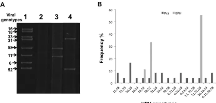
Genotype detection was evaluated by sequencing from L1 gene amplicons or PCR multiplex. The detection of HR HPV genotypes was 81.4% (83% in the BPH group and 79% in the PCa group), which is much higher than the occurrence of the low-risk (LR) HPV genotypes at 19% (17% in the BPH group and 21% in the PCa group). The vi-ral genotypes observed in the samples in order of decreas-ing prevalence were HPV 52 (33.3%), HPV 58 (17.17%), HPV 11 (12.7%), HPV 18 (10.8%), HPV 16 (7.8%), HPV 33 (6.9%), HPV 6 (5.9%), and HPV 31 (4.0%) (Table 2). The detection of multiple HPV genotypes in the same sam-ple (2-4 types) was 62.3% (33/53), which was higher than the detection of a single HPV genotype at 37.7% (20/53). Detection of multiple HPV genotypes was not dominant in
any of the groups, and both groups showed a high fre-quency of multiple infection (PCa 65% vs BPH 56.3%). Of the 33 men detected with multiple infection, 19 (57.6%) were co-infected by two types, 12 (36.3%) were co-infec-ted by three types, and 2 (6.1%) were co-infecco-infec-ted by four types (Figure 1).
With respect to HPV genotype distribution in the study groups, in HPV infections involving a single geno-type, HPV 52 was the most common genotype found in both groups, while in HPV infections with multiple geno-types, the most common genotypes were HPV 58 and 52 for the PCa group, and HPV 52, followed by HPV 11 and 58, for the control group (Table 1). The most frequent combi-nations of genotypes detected were 16/18 for the PCa group and 11/52/58 for the BPH group (Figure 1).
Additionally, we identified koilocytes, cells contain-ing an acentric, hyperchromatic nucleus displaced by a large perinuclear vacuole. Although the genesis of the cyto-plasmic vacuole remained unclear, particularly because both HPV DNA replication and virion assembly exclu-sively occur in the nucleus, in clinical biopsies from cervi-cal cells, koilocytosis is observed in both LR and HR HPV infections (Krawczyket al., 2008). Therefore, in this study, we demonstrated that all HPV-positive samples showed koilocytosis. (Figure 2). Moreover, by using immunohis-tochemical assays, p16-INK4A protein overexpression was demonstrated in HPV-positive PCa tissue, indirectly sug-gesting the presence of HR-HPV E7 oncoprotein (Figure 3). As expected, HPV-negative tissue showed low levels p16-INK4A protein expression (Figure 3).
Discussion
In this study, we examined the presence of HPV in two study groups: a control group comprising BPH tissues and a case group comprising cancerous tissues. We found HPV in 53 (14.9%) of the 356 tissues analyzed, which is similar to the findings in other reports of tissue analysis from Latin America (18.63%) (Yanget al., 2015) and Mex-ico (11.5%-14.9%) (Martínez-Fierroet al., 2010; Dávila-Rodríguezet al., 2016). Consistent with the meta-analysis
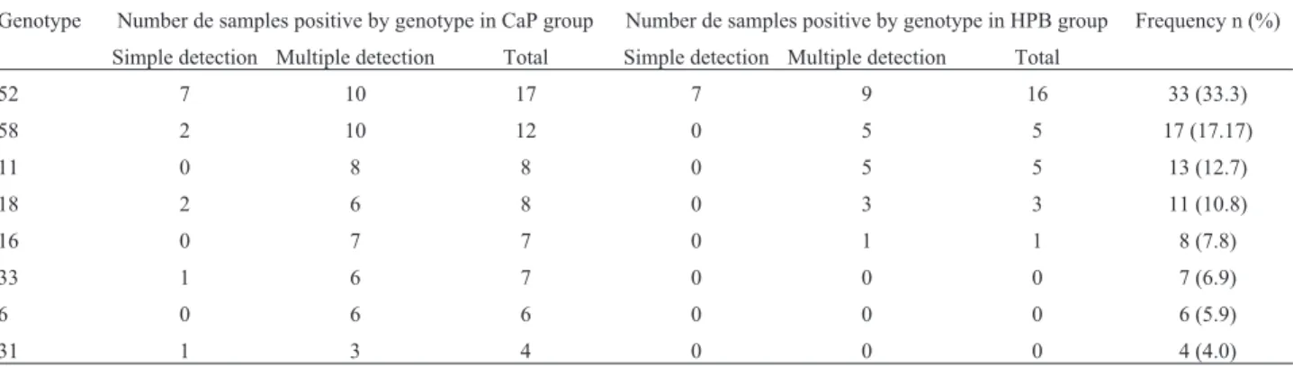
Table 2- Frequency of HPV genotypes detected from study samples.
Genotype Number de samples positive by genotype in CaP group Number de samples positive by genotype in HPB group Frequency n (%) Simple detection Multiple detection Total Simple detection Multiple detection Total
52 7 10 17 7 9 16 33 (33.3)
58 2 10 12 0 5 5 17 (17.17)
11 0 8 8 0 5 5 13 (12.7)
18 2 6 8 0 3 3 11 (10.8)
16 0 7 7 0 1 1 8 (7.8)
33 1 6 7 0 0 0 7 (6.9)
6 0 6 6 0 0 0 6 (5.9)
Figure 1- HPV genotypes detected in this study. (A) Electrophoresis of PCR products. Lane 1: positive control (HPV genotypes detected), lane 2: nega-tive control (no added DNA), lanes 3 and 4: representanega-tive samples. (B) HPV genotypes frequency in biological samples analyzed.
Figure 2- Histopathological and molecular analysis of prostate cancer tissues. (A) Koilocytes were observed in several HPV-infected prostate cancer
tis-sues. Arrows indicate the koilocytes in a representative image. (B)In situHR-HPV detection. HR-HPV E6/E7 DNA was detected in prostate cancer sec-tions employingin situPCR as indicated in the Materials and Methods. The signal was mainly nuclear (indicated by empty arrows). Magnification: 10´
and 40´. (C) Solid arrows indicate a positive signal of HPV DNA amplification in koilocytes (K). Magnification: 63´. The numbers indicate the control
and other reports from Mexico, in the present study, we de-tected an association between HPV and PCa, with different frequencies of HPV in the two study groups. It is important to consider that because it is difficult to obtain normal pros-tate tissues, in this study, we used BPH tissues as controls; however, the use of normal prostate tissues as controls may show a higher association between HPV and PCa.
Some reports have described a role for HPV in PCa, suggesting that infection triggers chronic recurrent inflam-mation, and the prostate gland could be infected owing to its anatomic proximity to the anogenital and urinary sites, thus being in support of the association of cancer with HPV (Rabkin et al., 1992; Guma et al., 2016; Tolstov et al., 2014). The detection of HR genotypes was much higher than that of LR genotypes, which confirms the frequent identification of HR HPVs in PCa seen in many studies (Bae, 2015; Yanget al., 2015).
In contrast to other reports showing that the HPV types 16 and 18 are the most prevalent, in this work, the most prevalent HPV types were 52 and 58 in both study groups. These differences may be related to the fact that the distribution of HPV varies among different populations, as has been well recognized in previous epidemiological stud-ies (Bae, 2015; Yanget al., 2015). Moreover, the genotypes detected in our prostate tissue samples are consistent with previous reports from Mexican populations (Martínez-Fierro et al., 2010; Dávila-Rodríguez et al., 2016), and
these genotypes are also prevalent in cervical cancer samples from different areas of Mexico, as summarized in Table 1, demonstrating the importance of sexual transmis-sion as a route for dissemination of the virus.
Some researchers have reported that co-infections in cervical cells may be associated with higher persistence rates of certain HR-HPV types compared with those of LR-HPV types or single infection (Trottieret al., 2006; De Brotet al., 2017). In this study, the detection of HR-HPV genotypes was much higher than that of LR-HPV geno-types in both groups; however, the most frequent combina-tions of genotypes detected were 16/18 and 11/52/58 for PCa and BPH, respectively. Therefore, evidence suggests that BPH with HR HPV co-infections could be a precursor of PCa, consistent with chronic recurrent inflammation as a known cause of PCa (De Marzoet al., 2007; Elkahwaji, 2012; Sfanoset al., 2014).
A long-recognized, pathognomonic feature of HPV infection is the appearance of halo or koilocytotic cells. Here, we detected koilocytes, which have been identified in prostate tissues in other studies (Whitaker et al., 2013). Koilocytes were detected in 100% of the samples with HPV. Moreover, the in situ PCR detection of HR-HPVE6E7 genes and p16-INK4A overexpression in PCa tissues similar to that in human prostate epithelial cell lines (Koet al., 2003; Theodoreet al., 2010) and a male case of urothelial carcinoma with squamous differentiation
associ-Figure 3- Immunohistochemical detection of p16INK4A in prostate cancer. A strong positive (empty arrows) signal of p16INK4A protein was detected
ated withHPV in another report (Gumaet al., 2016) are suggestive of early and late ongoing oncogenic processes in BHP and PCa, respectively. This hypothesis is supported by a recent meta-analysis that demonstrated that BPH was associated with an increased incidence of PCa (Daiet al., 2016) and the fact that HR-HPVs have been identified in both benign and malignant prostate tissues (Linet al., 2011; Bae, 2015; Yanget al., 2015). Moreover, recent evidence has shown that in Australian men, HR-HPVs are present in benign prostate tissues before the development of HPV-associated PCa (Glennet al., 2017).
The results presented here are important for the fol-lowing reasons: (i) development of diagnostic assays; (ii) evaluation of the impact of vaccination in cancer preven-tion strategies, especially since many HPV genotypes are not covered by the current quadrivalent HPV vaccine used in Mexico; and (iii) description of highly prevalent genito-urinary tract HPV infections in sexually active men in México (Lajouset al., 2005; Giulianoet al., 2008; Mén-dez-Martínezet al., 2014) that may be important reservoirs of persistent HPV and play an important role in the patho-genesis and progression of BPH and PCa (Gandagliaet al., 2017).
In conclusion, the high frequency of detection of HPV in PCa, combinations of genotypes with oncogenic potential that dominated in the PCa group, identification of HPV associated koilocytes, and overexpression of p16INK4A in prostate cancer specimens constitute evi-dence suggesting the association of HPV with PCa and a potential role for the virus in the etiology of PCa.
Acknowledgments
This work was supported by the Instituto Politécnico Nacional (Secretaria de Investigación y Posgrado proyec-tos: 20171979, 20180484), part of work was developed in Escuela Militar de Graduados de Sanidad.
References
Aguilar-Lemarroy A, Vallejo-Ruiz V, Cortés-Gutiérrez EI, Sal-gado-Bernabé ME, Ramos-González NP, Ortega-Cervantes L, Arias-Flores R, Medina-Díaz IM, Hernández-Garza Fet al. (2015) Human papillomavirus infections in Mexican women with normal cytology, precancerous lesions, and cervical cancer: type-specific prevalence and HPV coin-fections. J Med Virol. 87:871-884.
Bae JM (2015) Human papillomavirus 16 infection as a potential risk factor for prostate cancer: An adaptive meta-analysis. Epidemiol Health. 37:e2015005.
Caini S, Gandini S, Dudas M, Bremer V and Severi E (2014) Sex-ually transmitted infections and prostate cancer risk: a sys-tematic review and meta-analysis. Cancer Epidemiol 38:329-338.
Canche JR, Canul J, Suárez R, de Anda R and González MR (2011) Infection by human papilloma virus amongst female inmates in a social re-adaptation centre in South-West Mex-ico. Rev Esp Sanid Penit 13:84-90.
Chen CJ, Hsu WL, Yang HI, Lee MH, Chen HC, Chien YC and You SL (2014) Epidemiology of virus infection and human cancer. Recent Results Cancer Res 193:11-32.
Cortés-Malagón EM, Bonilla-Delgado J, Díaz-Chavez J, Hidal-go-Miranda A, Romero-Cordoba S, Uren A, Celik H, McCormick M, Munguía-Moreno JA, Ibarra-Sierra Eet al. (2013) Gene expression profile regulated by the HPV16E7 oncoprotein and estradiol in cervical tissue. Virology 447:155-165.
Conde-Ferráez L, Martíez JR, Ayora-Talavera G and Losa MD (2017) Human papillomavirus andChlamydia trachomatis infection in gyneco-obstetric outpatients from a Mexican hospital. Indian J Med Microbiol 35:74-79.
Coussens LM and Werb Z (2002) Inflammation and cancer. Na-ture 420:860-867.
Dai X, Fang X, Ma Y and Xianyu J (2016) Benign prostatic hyper-plasia and the risk of prostate cancer and bladder cancer: A meta-analysis of observational studies. Medicine (Balti-more) 95:e3493.
Dávila-Rodríguez MI, Ignacio MCV, Aragón TAR, Olache JD, Castelan ME, Lara MS and Cortés GEI (2016) Human papilloma virus detection by INNOLipa HPV in prostate tis-sue from men of Northeast Mexico. Asian Pac J Cancer Prev 17:4863-4865.
De Brot L, Pellegrini B, Moretti ST, Carraro DM and Soares FA (2017) Infections with multiple high-risk HPV types are as-sociated with high-grade and persistent low-grade intra-ephitelial lesions of the cervix. Cancer 125:138-143. DelaRosa-Martínez R, Sánchez-Garza M and López-Revilla R
(2016) HPV genotype distribution and anomalous associa-tion of HPV33 to cervical neoplastic lesions in San Luis Potosí, Mexico. Infect Agent Cancer 11:16
De Marzo AM, Platz EA, Sutcliffe S, Xu J, Grönberg H, Drake CG, Nakai Y, Isaacs WB and Nelson WG (2007) Inflamma-tion in prostate carcinogenesis. Nat Rev Cancer 7:256-69. Elkahwaji JE (2012) The role of inflammatory mediators in the
development of prostatic hyperplasia and prostate cancer. Res Rep Urol 5:1-10.
Fajardo-Ramírez OR, Barboza-Cerda MC, Ortíz-López R, Rojas-Martínez A, Garza-Rodríguez ML, Sepulveda-Flores A, Gonzalez-Guerrero JF, Bernal-Silva S, Cerda-Flores RM, Calleja-Macías IE, Rodríguez-Flores S, Sandoval-Guzmán E, Plascencia-Solis T, Pérez-Reyes Pet al.(2017) Prevalence and 3-year persistence of human papillomavirus serotypes in asymptomatic patients in Northern Mexico. Int J Gynaecol Obstet 136:40-46.
Ferlay J, Soerimataram I, Dikshit R, Eser S, Mathers C, Rebelo M, Parkin DM, Forman D and Bray F (2012) Cancer incidence and mortality: sources, methods and major patterns in GLO-BOCAN 2012. Int J Cancer 136:E359-386.
Fujinaga Y, Shimada M, Okazawa K, Fukushima M, Kato I and Fujinaga K (1991) Simultaneous detection and typing of genital human papillomavirus DNA using the polymerase chain reaction. J Gen Virol 72:1039-1044.
Gallegos-Bolaños J, Rivera-Domínguez JA, Presno-Bernal JM and Cervantes-Villagrana RD (2017) High prevalalence of oc-infection between human papillomavirus (HP) 51 and 52 in Mexican population. BMC Cancer 17:531.
in-flammation in the development and progression of benign and malignant diseases. Curr Opin Urol 27:99-106. Gao G and Smith DI (2016) Human papillomavirus and the
devel-opment of different cancers. Cytogenet Genome Res 150:185-193.
Gillison ML, Chaturvedi AK, Anderson WF and Fakhry C (2015) Epidemiology of human papillomavirus-positive head and neck squamous cell carcinoma. J Clin Oncol 33:3235-3242. Giuliano AR, Lazcano-Ponce E, Villa LL, Fores R, Salmeron J,
Lee JH, Papenfuss MR, Abrahamsen M, Jolles E, Nielson CM et al.(2008) The human papillomavirus infection in men study: Human papillomavirus prevalence and type dis-tribution among men residing in Brazil, Mexico, and the United States. Cancer Epidemiol Biomarkers Prev 17:2036-2043.
Glenn WK, Ngan CC, Amos TG, Edwards RJ, Swift J, Lutze-Mann L, Shang F, Whitaker NJ and Lawson JS (2017) High risk human papilloma viruses (HPVs) are present in benign prostate tissues before development of HPV associated pros-tate cancer. Infect Agent Cancer 12:46.
González-Losa M del R, Rosado-Lopez I, Valdez-González N and Puerto-Solís M (2004) High prevalence of human papil-lomavirus type 58 in Mexican colposcopy patients. J Clin Virol 29:202-205.
González-Losa MD, Puerto-Solís M, Ayora-Talavera G, Gómez-Carvallo J, Euán-López A, Cisneros-Cutz JI, Rosado-López A, Echeverría Salazar J and Conde-Ferráez L (2017) Preva-lence of anal infection due to high-risk human papillo-mavirus and analysis of E2 gene integrity among women with cervical abnormalities. Enferm Infecc Microbiol Clin pii:S0213-005X(16)30390-1.
Guardado-Estrada M, Juárez-Torres E, Román-Bassaure E, Me-dina-Martinez I, Alfaro A, Benuto RE, Dean M, Villegas-Sepulveda N and Berumen J. (2014) The distribution of high-risk human papillomaviruses is different in young and old patients with cervical cancer. PLoS One 9:e109406. Guma S, Malglantay R, Lau R, Wieczorek R, Melamed J, Deng
FM, Zhou M, Makarov D, Lee P, Pincus MR and Pei ZH (2016) Papillary urothelial carcinoma with squamous differ-entiation in association with human papiloma virus: case re-port and literature review. Am J Clin Exp Urol 4:12-16. Heidegger I, Borena W and Pichler R (2015) The role of human
papilloma virus in urological malignancies. Anticancer Res 35:2513-2519.
Illades-Aguiar B, Alarcón-Romero L del C, Antonio-Véjar V, Zamudio-López N, Sales-Linares N, Flores-Alfaro E, Fer-nández-Tilapa G, Vences-Velázquez A, Muñoz-Valle JF and Leyva-Vázquez MA (2010) Prevalence and distribution of human papillomavirus types in cervical cancer, squamous intraepithelial lesions, and with no intraepithelial lesions in women from Southern Mexico. Gynecol Oncol 117:291-296.
Jácome-Galarza I, Ito-Nakashimada MA, Figueroa-Aguilar G, García-Latorre E, Salazar MI, López-Orduña E, Camacho AD, Valdez-Alarcón JJ, Hernández JM and León-Ávila G (2017) Prevalence of human papillomavirus in women from the State of Michoacan, Mexico, showed high frequency of unusual virus genotypes. Rev Invest Clin 69:262-269. Jiang J, Li J, Yunxia Z, Zhu H, Liu J and Pumill C (2013) The role
of prostatitis in prostate cancer: Meta-analysis. PLoS One 8:e85179.
Ko D, Gu Y, Yasunaga Y, Nakamura K, Srivastava S, Moul JW, Sesterhenn IA, McLeod DG, Arnstein P, Taylor DOet al. (2003) A novel neoplastic primary tumor-derived human prostate epitelial cell line. Int J Oncol 22:1311-1317. Krawczyk E, Siprynowicz FA, Liu X, Dai Y, Hartmann DP,
Hanover J and Schlegel R (2008) Koilocytosis: A coopera-tive interaction between the humanpapillomavirus E5 and E6 oncoproteins. Am J Pathol 173: 682-688.
Lajous M, Mueller N, Cruz-Valdéz A, Aguilar LV, Franceschi S, Hernadez-Avila M and Lazcano-Ponce E (2005) Determi-nants of prevalence, acquisition, and persistence of human papillomavirus in healthy Mexican military men. Cancer Epidemiol Biomarkers Prev 14:1710-1716.
Lazcano-Ponce E, Herrero R, Muñoz N, Cruz A, Shah KV, Alon-so P, Hernández P, Salmerón J and Hernández M (2001) Ep-idemiology of HPV infection among Mexican women with normal cervical cytology. Int J Cancer 91:412-420. Lin Y, Mao Q, Zheng X, Yang K, Chen H, Zhou C and Xie L
(2011) Human papillomavirus 16 or 18 infection and pros-tate cancer risk: A meta-analysis. Ir J Med Sci. 180:497-503. López-Revilla R, Martínez-Contreras LA and Sánchez-Garza M
(2008) Prevalence of high-risk human papillomavirus types in Mexican women with cervical intraepithelial neoplasia and invasive carcinoma. Infect Agent Cancer 3:3.
Magaña-Contreras M, Contreras-Paredes A, Chavez-Blanco A, Lizano M, De la Cruz-Hernandez Y and De la Cruz-Hern-andez E (2015) Prevalence of sexually transmitted patho-gens associated with HPV infection in cervical samples in a Mexican population. J Med Virol 87:2098-2105.
Manjarrez ME, Ocadiz R, Valle L, Pacheco C, Marroquin A, De la Torre C, Selman M and Gariglio P (2006) Detection of hu-man papillomavirus and relevant tumor suppressors and oncoproteins in laryngeal tumors. Clin Cancer Res. 12:6946-6951.
Manos MM, Ting Y, Wright DK, Lewis AJ, Broker TR and Wolinsky SM (1989) Use of polymerase chain reaction am-plification for the detection of genital human papilloma-viruses. Cancer Cells 7:209-214.
Mantovani A, Allavena P, Sica A and Balkwill F (2008) Can-cer-related inflammation. Nature 454:436-444.
Martinez-Fierro ML, Leach RJ, Gomez-Guerra LS, Garza-Gua-jardo R, Johnson-Pais T, Beuten J, Morales-Rodriguez IB, Hernandez-Ordoñez MA, Calderón Cardenas G, Ortiz-Lo-pez Ret al.(2010) Identification of viral infections in the prostate and evaluation of their association with cancer. BMC Cancer 10:326.
Méndez-Martínez R, Rivera-Martínez NE, Crabtree-Ramírez B, Sierra-Madero JG, Caro-Vega Y, Galván SC, de León DC and García Carrancá A (2014) Multiple human papillo-mavirus infections are highly prevalent in the anal canal of human immunodeficiency virus-positive men who have sex with men. BMC Infect Dis 14:671.
Montoya-Fuentes H, Suárez RAE, Ramírez-Muñoz MP, Aré-valo-Lagunas I, Morán MMC, Gallegos AMP, Flores-Mar-tínez SE, Rosales QS and Sánchez CJ (2001) The detection of human papillomavirus 16, 18, 35 and 58 in cervical-ute-rine cancer and advanced degree of squamous intraepithelial lesions in WesternMexico: Clinical-molecular correlation. Ginecol Obstet Mex 69:137-142.
Nuovo GJ (2001) Co-labeling usingin situPCR: A review. J Histochem Cytochem 49:1329-1339.
Ocadiz-Delgado R, Castañeda-Saucedo E, Indra AK, Hernan-dez-Pando R, Flores-Guizar P Cruz-Colin JL, Recillas-Targa F, Perez-Ishiwara G, Covarrubias L and Gariglio P (2012) RXRadeletion and E6E7 oncogene expression are sufficient to induce cervical malignant lesionsin vivo. Can-cer Lett 317:226-236.
Ocadiz-Delgado R, Albino-Sanchez ME, Garcia-Villa E, Aguilar-Gonzalez MG, Cabello C, Rosete D, Mejia F, Man-jarrez-Zavala ME, Ondarza-Aguilera C, Rivera-Rosales RM et al.(2013)In situmolecular identification of the influenza A (H1N1) 2009 Neuraminidase in patients with severe and fatal infections during a pandemic in Mexico City. BMC In-fect Dis 13:20.
Orozco-Colín A, Carrillo-García A, Méndez-Tenorio A, Ponce-de-León S, Mohar A, Maldonado-Rodríguez R, Guerra-Arias R, Flores-Gil O, Sotelo-Regil R and Lizano M (2010) Geographical variation in human papillomavirus prevalence in Mexican women with normal cytology. Int J Infect Dis 14:e1082-1087.
Ortega-Cervantes L, Aguilar-Lemarroy A, Rojas-García AE, Bar-rón-Vivanco BS, Vallejo-Ruiz V, León DC, Hernández YY, Jáuregui-Martínez A and Medina-Díaz IM (2016) Human papilloma virus genotypes in women from Nayarit,Mexico, with squamous intraepithelial lesions and cervical cancer. Int J Health Sci 10:327-338.
Peralta-Rodríguez R, Romero-Morelos P, Villegas-Ruíz V, Men-doza-Rodríguez M, Taniguchi-Ponciano K, González-Ye-bra B, Marrero-Rodríguez D and Salcedo M. (2012) Preva-lence of human papillomavirus in the cervical epithelium of Mexican women: Meta-analysis. Infect Agent Cancer 7:34. Piña-Sánchez P, Hernández-Hernández DM, López-Romero R, Vázquez-Ortíz G, Pérez-Plasencia C, Lizano-Soberón M, González-Sánchez JL, Cruz-Talonia F and Salcedo M (2006) Human papillomavirus-specific viral types are com-mon in Mexican women affected by cervical lesions. Int J Gynecol Cancer. 16:1041-1047.
Rabkin CS, Biggar RJ, Melbye M and Curtis RE (1992) Second primary cancers following anal and cervical carcinoma: Evi-dence of shared etiologic factors. Am J Epidemiol. 136:54-58.
Romero-Morelos P, Uribe-Jiménez A, Bandala C, Poot-Vélez A, Ornelas-Corral N, Rodríguez-Esquivel M, Valdespino-Za-vala M, Taniguchi K, Marrero-Rodríguez D, López-Romero R and Salcedo M (2017) Genotyping of the human papiloma virus in a group of Mexican women treated in a highy spe-cialist hospital: Multiple infections and their potential tran-scendence in the current vaccination programme. Med Clin (Bar) 149:287-292.
Salcedo M, Pina-Sanchez P, Vallejo-Ruiz V, Monroy-García A, Aguilar-Lemarroy A, Cortes-Gutierrez EI, Santos-Lopez G, Montoya-Fuentes H, Grijalva R, Madrid-Marina V et al. (2014) Human papillomavirus genotypes among females in Mexico: A study from the Mexican institute for social secu-rity. Asian Pac J Cancer Prev 15:10061-10066.
Sambrook J, Fritsch T and Maniatis T (1989) Molecular Cloning Cold Spring Harbor Press, Nova York.
Sánchez-Anguiano LF, Alvarado-Esquivel C, Reyes-Romero MA and Carrera-Rodríguez M (2006) Human papillomavirus in-fections in women seeking cervical Papanicolaou cytology of Durango, Mexico: Prevalence and genotypes. BMC In-fect Dis 20:27.
Sfanos KS, Isaacs WB and De Marzo AM (2013) Infections and inflammation in prostate cancer. Am J Clin Exp Urol 1:3-11. Sfanos KS, Hempel HA and De Marzo AM (2014) The role of in-flammation in prostate cancer. Adv Exp Med Biol 816:153-181.
Stratton KL and Culkin DJ (2016) A contemporary review of HPV and penile cancer. Oncology 30:245-249
Taverna G, Pedretti E, Di Caro G, Borroni EM, Marchesi F and Grizzi F (2015) Inflammation and prostate cancer: Friends or foe? Inflamm Res 64:275-286.
Theodore S, Sharp S, Zhou J, Turner T, Li H, Miki J, Ji Y, Patel V, Yates C and Rhim JS (2010) Establishment and character-ization of a pair of non-malignant and malignant tumor de-rived cell lines from an African American prostate cancer patient. Int J Oncol 37:1477-1482.
Tolstov Y, Hadaschik B, Pahernik S, Hohenfellner M and Duen-sing S (2014) Human papillomaviruses in urological malig-nancies: A critical assessment. Urol Oncol 32:46.e19-27. Torroella-Kouri M, Morsberger S, Carrillo A, Mohar A, Meneses
A, Ibarra M, Daniel RW, Ghaffari AM, Solorza G and Shah KV (1998) HPV prevalence among Mexican women with neoplastic and normal cervixes. Gynecol Oncol 70:115-120. Trottier H, Mahmud S, Costa MC, Sobrinho JP, Duarte Franco E,
Rohan TE, Ferenczy A, Villa LL and Franco EL (2006) Hu-man papillomavirus infections with multiple types and risk of cervical neoplasia. Cancer Epidemiol Biomarkers. 15:1274-1280.
Velázquez-Márquez N, Paredes-Tello MA, Pérez-Terrón H, San-tos-López G, Reyes-Leyva J and Vallejo-Ruiz V (2009) Prevalence of human papillomavirus genotypes in women from a rural region of Puebla, Mexico. Int J Infect Dis 13:690-695.
Velázquez-Márquez N, Jimenéz-Aranda L, Sánchez-Alonso P, Santos-López G, Reyes-Leyva J and Vallejo-Ruiz V (2010) Human papillomavirus infection in women from Tlaxcala, Mexico. Braz J Microbiol 41:749-756.
Yang L, Xie S, Feng X, Chen Y, Zheng T, Dai M, Zhou CK, Hu Z, Li N and Hang D (2015) Worldwide prevalence of human papillomavirus and relative risk of prostate cancer: A meta-analysis. Sci Rep 5:14667.
Whitaker NJ, Glenn WK, Sahrudin A, Orde MM, Delprado W and Lawson JS (2013) Human papillomavirus and Epstein Barr virus in prostate cancer: Koilocytes indicate potential onco-genic influences of human papillomavirus in prostate can-cer. Prostate 73:236-241.
Associate Editor: Anamaria Aranha Camargo

![Figure 3 - Immunohistochemical detection of p16INK4A in prostate cancer. A strong positive (empty arrows) signal of p16INK4A protein was detected in HPV-positive tissues compared with HPV-negative [HPV(-)] tissues](https://thumb-eu.123doks.com/thumbv2/123dok_br/16056399.696325/6.918.99.823.99.536/immunohistochemical-detection-prostate-positive-detected-positive-compared-negative.webp)
