José Marcus Raso EulálioI,Manoel Luiz FerreiraI, Paulo César SilvaI, José Eduardo Ferreira MansoII, Andrei Ferreira Costa NicolauIII, Thales Penna de CarvalhoIII, Julia Radicetti de Siqueira Paiva e SilvaIV, Adrielle Rodas FernandesIV, Alberto SchanaiderV
A technical note on low cost rat laparoscopy an initial
experience
1Acta Cir Bras. 2018;33(9):853-861
Abstract
Purpose: To evaluate a novel and adapted low-cost set model for laparoscopic surgery in rats.
Methods: Nine Wistar rats underwent two different laparoscopic procedures, splenectomy
(n=3) and distal pancreatectomy with splenectomy (n = 6), after assembling a low-cost set replacing the conventional one (monitor, micro camera, image processor, light source, laparoscope and insufflator). The new set included an Android Tablet 10.5 ‘’, a 5mm USB Endoscope and semiautomatic sphygmomanometer monitor.
Results: The same surgeon performed the laparoscopic procedures. Total surgical time
ranged from 36 to 60 minutes with a mean of 45.8 minutes. Three rats died during the distal pancreatic and splenectomy procedure (33.3%), due to respiratory failure (n = 1), uncontrolled abdominal hemorrhage (n=1) and iatrogenic gastric perforation (n = 1). We followed the other six rats (66.6%) for seven days with no further evidence of complications.
Conclusions: The laparoscopic partial pancreatectomy and splenectomy can be performed
with the novel low-cost set assembled in the present experimental study. Both specific
training and skills development are required to validate more advanced laparoscopic
procedures and achieve a desirable outcome.
Key words: Laparoscopy. Pancreatectomy. Splenectomy. Models, Animal. Rats.
DOI: http://dx.doi.org/10.1590/s0102-865020180090000014
IPhD, Associate Professor, Postgraduate Program in Surgical Sciences, Department of Surgery, School of Medicine, Universidade Federal do Rio de Janeiro (UFRJ), Brazil. Technical procedures; acquisition, analysis and interpretation of data, manuscript preparation.
IIPhD, Associate Professor, Postgraduate Program in Surgical Sciences, Department of Surgery, School of Medicine, UFRJ, Rio de Janeiro-RJ, Brazil. Acquisition, analysis and interpretation of data.
IIIFellow Master degree, Postgraduate Program, Department of Surgery, School of Medicine, UFRJ, Rio de Janeiro-RJ, Brazil. Technical procedures; acquisition, analysis and interpretation of data.
IVGraduate student, Department of Surgery, School of Medicine, UFRJ, Rio de Janeiro-RJ, Brazil. Acquisition of data,
technical procedures.
Nine adult isogenic Wistar rats (Rattus norvegicus albinus), weighing from 250 to 412g, were obtained from the Centre of Experimental Surgery, School of Medicine of UFRJ. They were housed in appropriate
environmental conditions, on a circadian cycle
in a temperature-controlled room (24oC), fed with standard industrial rat chow and water ad libitum.
The study developed a complete laparoscopy set, using low cost surgical instruments, adapted for surgery in rats. The usual set to perform the method presume the availability of (01) monitor, (2) micro camera, (3) image processor, (4) light source,
(5) laparoscope and (6) insufflator. The set of
instruments proposed in the present study
replaced all six items aforementioned and
was composed by (1) Android 10.5” Tablet, (2)
5mm USB Endoscope and (3) semiautomatic
sphygmomanometer monitor (Figure 1).
Tablet android 10.5
For the surgical procedures, the authors used an Android 10.5” Samsung
SM-T800 tablet with a 6.0.1 software version. This
replaces the monitor and the image processor of the micro camera. The USB Camera program,
available free in the Android platform, allows
the capture and recording of images (Figure 1).
USB 5mm endoscope
The experimental model usedaDigital Supereyes Y003 5.4mm Endoscopy and the Lensoul 5.5mm OTG Endoscope. These two models are composed by rigid (Y003) or semi-rigid (Lensoul) rods, up to 5.5mm in diameter,
with an USB proximal connection that links to
the tablet and another distal end with six led spotlights, and a 2.0 megapixel micro camera.
This setting replaces the micro camera, the
light source and the laparoscope. These accessories were connected to the 10.5” tablet
■
Introduction
During the past 20 years, the development of new surgical techniques such as laparoscopy has improved the treatment
of surgical patients by minimizing surgical trauma, accelerating postoperative recovery,
and reducing the length of hospital stay1. Several complex open surgical techniques can now be performed by minimally invasive
surgery. In most clinical trials, higher operating theatre cost is offset by shorter hospital stays, less medication requirements, shorter periods of convalescence, and faster return to work and to normal activity2,3.
One of the most commonly used animals in the experimental surgical research is the Wistar rat. This specie is easy to handle and has a low maintenance cost. However, the high-cost of laparoscopic sets and the need for
specific techniques for this specimen size are the main limiting points for the development
of laparoscopic experimental procedures in rats4,5.
Several articles have shown the
feasibility of laparoscopic surgery techniques in rats in the last 25 years6-8. As a rule, the
surgical manipulations and tissue resections are performed using conventional material for human patients adapted to the rat size.
A novel low-cost set adapted for laparoscopic surgery in rats may be useful not
only for training this technique, particularly in counties with few financial resources, but also
to allow surgical researches that mimic the
operative field in humans beings.
■
Methods
The study was in conformity to
Brazilian law for scientific use of animals and international guiding principles for animal care and was approved by the Ethics Committee for
and displayed appropriate images for the procedures (Figure 1).
Semi-automatic sphygmomanometer monitor
We used a G-Tech BP3ABOH upper arm
semi-automatic pressure device with a battery powered monitor and an insufflation system
(Figure 1). It was connected in a Y-shaped rubber tube with one extremity coupled to a
5mm trocar and the other to the pressure cuff. At the distal end of the insufflation bulb a CO2 source was connected (Figure 1 - item 3).
Figure 1 - Set of instruments adapted for laparoscopic surgery in rats. 1. Micro camera with rigid rod 5mm. 2. Micro camera with flexible rod 5.5mm. 3. Adaptation of CO2 conductive tube. 4. Detail of the CO
2 tube connected to the 5mm
trocar. 5. Semi-automatic manometer gauged. 6. Tablet Android 10.5”.
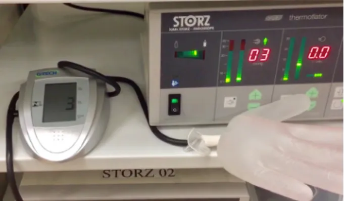
A pressure validation test was made
to evaluate the G-Tech BP3ABOH manometer with this device. This test consisted of the
simultaneous adaptation of a latex glove, which
has a quite similar volume to the rat’s abdomen,
with a Storz insufflator used in medical
laparoscopy and the sphygmomanometer
monitor. The carbon dioxide insufflation
generated equivalent measurements in both
insufflator and sphygmomanometer monitors
(Figure 2).
Figure 2 - Validation of semi-automatic manometer monitor for experimental use in rats. Latex
glove insufflation after simultaneous adaptation to professional insufflator, that brought forth equivalent markings in pressure variations between
1 and 10mmHg. Equivalent measurements can be
seen in both insufflator and sphygmomanometer
monitors.
Anesthetic and surgical technique
After 24h preoperative fasting,
rats underwent general anesthesia by
intraperitoneal injection of ketamine
hydrochloride solution (25mg/kg) and xylazine hydrochloride (3mg/kg)9. Hydration, when
necessary, was given by instillation of 0.9% sodium chloride solution in the peritoneal
cavity under laparoscopic vision. Immediately
after anaesthetic administration, animals were immobilized in the dorsal decubitus and the skin over the surgical site was shaved with 10%
polyvinylpyrrolidone-iodine. Upper legs were
fixed in minimal extension.
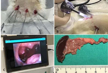
The procedure started with three 1.5
cm incisions in both left and right iliac fossae
and in the middle line over the suprapubic
site. Dissection was continued with muscle
exposure followed by a single purse-string
suture with polyglactin 910 (Vicryl®) 4-0, around each incision. Then, the anterior wall of the abdominal cavity was rised by the purse-string
sutures in order to allow insertion of the three trocars of 5mm. The first trocar inserted in the
the USB micro camera port and to insufflate the
abdominal cavity. The two remaining trocars were placed under direct vision by the micro
camera, previously adapted to the tablet. A
careful manual inflation was done until the
pressure reached 4.0mmHg10,11 (Figure 3).
Figure 3 - General aspects of laparoscopic technique. A. The three purse-string suture done. B. Placement of trocars and the micro camera. C. Intra-abdominal image on the tablet screen. D. Resected spleen and distal pancreas.
In addition to the three trocars, the
surgical procedures also demanded the
following tools: tweezers, 5mm scissors and
a 5mm bipolar forceps. Two surgeons and
one anesthesiologist assistant attended all
procedures.
The pneumoperitoneum was relieved
whenever there was no manipulation to preserve the ventilatory capacity of the
specimens. The rats were monitored with oximetry and heart rate monitors.
Postoperative analgesia was provided
with dypirone diluted in water at a dose of
30mg/kg/day. Behavior, feeding and surgical wounds were evaluated daily. After seven days of follow-up, the animals were killed without pain by isoflurane saturation in a closed camera and laparotomized with evaluation of the
cavity, regarding the presence or absence of
surgical complications (bruise, fistula, ascites, abscess, hematoma, peritonitis).
■
Results
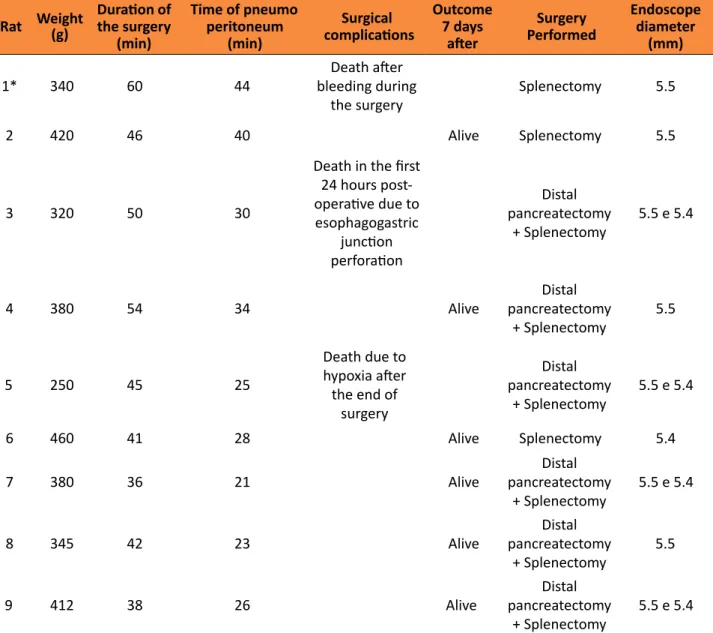
Total surgical time necessary to complete the organ resections ranged from 36
to 60 minutes with a mean of 45.8 minutes.
The pneumoperitoneum time ranged from 23
to 44 minutes with a mean of 30.1 minutes. There were no accidents in the placement of
the trocars. No wound infection was found. Three animals died. The first one
due to uncontrolled abdominal hemorrhage
immediately after the section of the vascular
pedicle of the splenic lobule of the pancreas.
The second animal died in the first 24 hours postoperatively and necropsy displayed a gastric perforation at the esophagogastric junction. The third one, died at the end of operation,
when abdominal wall was already closed, in consequence of a respiratory failure. The other rats were monitored for seven days and showed
Table 1 - Results of 9 laparoscopic procedures in rats.
Rat Weight
(g)
Duration of
the surgery (min)
Time of pneumo peritoneum
(min)
Surgical
complications
Outcome 7 days
after
Surgery
Performed
Endoscope
diameter (mm)
1* 340 60 44
Death after
bleeding during the surgery
Splenectomy 5.5
2 420 46 40 Alive Splenectomy 5.5
3 320 50 30
Death in the first
24 hours post-
operative due to
esophagogastric
junction perforation
Distal pancreatectomy
+ Splenectomy
5.5 e 5.4
4 380 54 34 Alive
Distal pancreatectomy
+ Splenectomy
5.5
5 250 45 25
Death due to
hypoxia after
the end of surgery
Distal pancreatectomy
+ Splenectomy
5.5 e 5.4
6 460 41 28 Alive Splenectomy 5.4
7 380 36 21 Alive
Distal pancreatectomy
+ Splenectomy
5.5 e 5.4
8 345 42 23 Alive
Distal pancreatectomy
+ Splenectomy
5.5
9 412 38 26 Alive
Distal pancreatectomy
+ Splenectomy
5.5 e 5.4
*Female rat.
■
Discussion
Laparoscopy is now the gold standard
method for various operations of the digestive, urinary, endocrine and reproductive tract.
Their acceptance and consistency of results are accompanied by the development of instruments that enable and facilitate an increasing number of procedures1-3. However, the cost of this technology12 creates several
limitations that hamper the development
of experimental models, especially in small
animals.
Experimental laparoscopy is a fundamental modality for both training4,5 and development of surgical techniques. It is also
useful for scientific researches when a minimal
invasive surgical procedure is required7,8. The laparoscopy causes less trauma13, postoperative pain14, dehydration due to exposure of loops15, and often with fewer postoperative
The cost of acquiring the basic set of devices to perform a laparoscopy varies widely according to the country, manufacturer
and model. We found offerings from a semi-system on sale sites (Amazon, EBay) composed
by (01) monitor, (2) micro camera, (3) image processor, (4) light source, (5) laparoscope
and (6) insufflator, ranging from US$ 7500 to US$ 25000. In Brazil, a new laparoscopic
system, with a good quality camera, can easily
exceeds US$ 50000. It becomes even greater in the case of small animals applications that require adaptations and more maintenance
expenses. It is almost impossible to cover these high costs if there are no consistent grants for researchers. Moreover, most of the third word
and even some developing countries like Brazil cannot afford such expenses in an experimental
scenario.
Indeed, the instruments used in this study have very low cost and total sum is up to
US$ 300. A Tablet 10” costs on average US$ 200 (ranging from US$90 to US$700). The semi-automatic sphygmomanometer costs from US$40 to US$60 and the micro camera from US$20 to US$40. Lower accessible prices make it easier to replace any malfunctioning item.
In the present study, these devices were reused without any failure or defect reported. Cleaning for reuse should be immediate and
antisepsis with alcoholic chlorhexidine solution did not impair the function of the appliances.
Peritoneal anesthesia with ketamine and xylazine has the advantage of being simple, effective and easily replicable if the animal
begins to react9. The anesthesiologist assistant
had an essential role in the low-cost technique,
monitoring the vital parameters and the
intra-abdominal pressure of the rats and making delicate insufflation when necessary.
It was observed that the fixation of the upper legs in partial extension helps the spontaneous ventilation. Nevertheless, one
animal died during the procedure due to the
probable effect of the sum of the anesthesia
with the pneumoperitoneum. This specimen had the lowest weight (250g) and the smallest
dimensions. Anesthesia with intubation and mechanical ventilation was not used,
but it surely can be performed to favor the
implementation of more complex and
time-consuming procedures with the low-cost instruments.
The Veress needle was tested previously
and in such circumstances after making a purse-string suture. The pressure cuff was removed
in order to adapt this needle and connect it to the CO2 conducting tube. A Y-shaped system allowed the sphygmomanometer
monitorization. In the Veress needle closed
method of entry, the abdominal pressure may be increased immediately prior to
insertion of the first trocar. We verified that
pneumoperitoneum has to be installed only
after placement of the three trocars, aiming to decrease the time of overload to the ventilatory function of the animals. Furthermore, the open entry technique with direct insertion without prior pneumoperitoneum was utilized by the authors because it is a safe alternative,
faster than the Veress needle technique and
probably associated with less insufflation-related complications such as hypotension and
hypoxia. Rising the purse-string sutures, the
trocars insertion is facilitated. It is necessary
to place the trocars adequately to guarantee
the fixation and absence of gas leakage.
The escape of gas through the trocars holes
makes not only the pneumoperitoneum ineffective but also the procedure unfeasible. The pneumoperitoneum often decreases with manipulation of the tweezers and needs to be
gently corrected by the assistant. Moreover, considering the small holes of the trocars, the
instrumental manipulation must be gentle, regarding the tweezers weight and movements
and also the instrumental mobility. If this care
the surgery. Thus, properly maintenance of the pneumoperitoneum pressure is the cornerstone of a successful procedure. Pressures smaller
than 3 mmHg do not provide good operative field and those above 6 mmHg generate
respiratory failure. Thus it is fundamental to maintain the abdominal pressure between 4 and 6 mmHg4,5,10,11. The relief of the pneumoperitoneum in the presence of hypoxia may improve the animal recover, decreasing the mortality rate. It was demonstrated the accuracy of the sphygmomanometer monitor in
measuring pressures similarly to the traditional insufflator (pressure variations between 1 and 10 mmHg) validating the use of such
adapted device and the pressure controlled by sphygmomanometer was suitable during all procedures.
The adapted set and the technique used were suitable for viewing and
manipulating the intra-abdominal structures by laparoscopy. The left iliac fossa is an
adequate site to enable the trocar view for splenectomy and distal pancreatectomy. A
common difficulty is the presence of the
distended cecum, which must be avoided by
the camera movements. The miniaturization
and proximity of the instruments used were
facilitating factors. Although the definition of 2.0 megapixels is relatively low, visualization of
the rat abdominal cavity was possible due to its small dimensions. The quality of the image
generated by the camera was sufficient to distinguish dissected structures. Likewise, the illumination generated by the LED spotlights
was suitable for the procedure, without any
image artifacts. The 5.4mm and the 5.5mm
diameter of the camera allow for reasonable mobility in the cavity and passes through the 5mm trocar without damaging the trocar seal mechanism.
The vision of a USB camera processed by a Tablet is inferior to a professional laparoscopic micro camera with 3CCDs. However, the
possibility of recognizing the organs, vascular structures and main ducts make this low-cost
technology valid. It was used the two cameras. The Supereyes Y003 (5.4mm) endoscope,
former created for otological evaluation, has the advantage of manually adapting the focus generating very clear images, but in a restricted field. The Lensoul Endoscope (5.5mm) is a
waterproof (IP67) auto-focus cap that allows
better view of larger surgical fields than the other endoscopes. Focus automatism may take longer to compensate the optic movements and generate blurry images soon after moving the optics. Nevertheless, usually this did not
disturb any procedure in our study.
Recording is possible but generates a discrete delay in image processing. When phase recording of the procedure was not
essential, we preferred to perform it with this function disabled. Tablets with more powerful processors are likely to overcome this limitation.
We have shown that both distal pancreatectomy with splenectomy or splenectomy alone can be safely performed
and such resection procedures require the development of surgical skills for dealing with
rats17,18. The two rat’s death because of gastric
perforation and intra-abdominal bleeding occurred early, during or on the day after the
laparoscopic procedures, but the mortality
rate was significantly reduced as soon as the
learning curve ended.
Among the limitations of the low-cost set, we highlight the visualization has a small
delay regarding to the actual movement,
which is almost unnoticeable if there is no
simultaneous recording of the video. The
screen size is smaller than a regular laparoscopy monitor. Since the insufflator is not automatic,
it is necessary that a member of the team
exclusively dedicated to handle the insufflation
as a 30º one.
The adaptations of instruments that make up a novel and basic set of laparoscopic
surgery in rats were validated in the current study. We believe that within rigid parameters,
considering the limitations and potentialities of
the equipment, low-cost laparoscopy in rats can improve experimental research and facilitate the development of new experimental models.
■
Conclusions
Low-cost laparoscopy in rats is a
promising new model of elective laparoscopy. It has a possible translational applicability in complex intra-abdominal operations, mimicking surgical procedures in humans.
■
References
1. Sørensen SM, Savran MM, Konge L, Bjerrum F. Three-dimensional versus two-dimensional vision in laparoscopy:
a systematic review. Surg Endosc. 2016 Jan;30(1):11-23. doi:
10.1007/s00464-015-4189-7. PMID: 25840896.
2. Pang L, Kong J, Wang Y, Zhang Y. Laparoscopic versus open pylorus-preserving
pancreatoduodenectomy. The first meta-analyse of retrospective matched cases.
Acta Cir Bras. 2018 Jan;33(1):40-48. PMID: 29412232.
3. Kasai M, Cipriani F, Gayet B, Aldrighetti L, Ratti F, Sarmiento JM, Scatton O, Kim KH, Dagher I, Topal B, Primrose J, Nomi T, Fuks
D, Abu Hilal M. Laparoscopic versus open
major hepatectomy: a systematic review and meta-analysis of individual patient
data. Surgery. 2018 May;163(5):985-95.
doi:10.1016/j.surg.2018.01.020. PMID:
29555197.
4. van Velthoven RF, Hoffmann P. Methods for
laparoscopic training using animal models. Curr Urol Rep. 2006 Mar;7(2):114-9. PMID: 16526995.
5. Martinez AM, Kalach AC, Espinoza DL.
Millimetric laparoscopic surgery training on a physical trainer using rats. Surg Endosc. 2008 Jan;22(1):246-9. PMID: 18030523.
6. Ypsilantis P, Simopoulos C. A laparoscopic technique of partial hepatectomy in the rat.
J Surg Res. 2016 Oct;205(2):286-91. PMID: 27664874.
7. Meyer F, Ioshii SO, Chin EW, Esser DM, Marcondes RT, Patriani AH, Pimpão Bde F.
Laparoscopic partial nephrectomy in rats.
Acta Cir Bras. 2007 Mar-Apr;22(2):152-6. PMID: 17375224.
8. Targarona EM, Espert JJ, Bombuy E, Trias M. Laparoscopic splenectomy in a rat model: developing an easy technique. J Laparoendosc Adv Surg Tech A. 1999 Dec;9(6):503-6. PMID: 10632512.
9. Wellington D, Mikaelian I, Singer L. Comparison of ketamine-xylazine and
ketamine-dexmedetomidine anesthesia
and intraperitoneal tolerance in rats. J Am Assoc Lab Anim Sci. 2013 Jul;52(4):481-7. PMID: 23849447.
10. Berguer R, Cornelius T, Dalton M. The
optimum pneumoperitoneum pressure
for laparoscopic surgery in the rat model. A detailed cardiorespiratory study. Surg Endosc. 1997 Sep;11(9):915-8. PMID: 9294272.
11. Gutt CN, Riemer V, Brier C, Berguer R, Paolucci V. Standardized technique of
laparoscopic surgery in the rat. Dig Surg. 1998;15(2):135-9. PMID: 9845575.
12. Pędziwiatr M, Wierdak M, Nowakowski M, Pisarska M, Stanek M, Kisielewski M, Matłok M, Major P, Kłęk S, Budzyński A. Cost minimization analysis of laparoscopic
surgery for colorectal cancer within the
enhanced recovery after surgery (ERAS)
protocol: a single-centre, case-matched
study. Wideochir Inne Tech Maloinwazyjne. 2016;11(1):14-21. doi: 10.5114/wiitm.
2016.58617. PMID: 28133495.
13. Yiannakopoulou E, Nikiteas N, Perrea D,
Tsigris C. Minimally invasive surgery and
oxidative stress response: what have we
learned from animal studies? Surg Laparosc Endosc Percutan Tech. 2013 Feb;23(1):25-8. PMID: 23386145.
14. Préfontaine L, Hélie P, Vachon P.
Postoperative pain in Sprague Dawley rats after liver biopsy by laparotomy
versus laparoscopy. Lab Anim (NY). 2015
May;44(5):174-8. doi: 10.1038/laban.731.
PMID: 25897938.
Kuroda Y. Comparison of intestinal transit
recovery between laparoscopic and open surgery using a rat model. Surg Endosc. 2003 Aug;17(8):1237-40. PMID: 12799882. 16. Eulálio JM, Bon-Habib AC, Soares DO,
Corrêa PG, Pineschi GP, Diniz VS, Manso JE, Schanaider A. Critical analysis and systematization of rat pancreatectomy
terminology. Acta Cir Bras. 2016 Oct;31(10):698-704. PMID: 27828605.
17. Targarona EM, Espert JJ, Bombuy E, Trias M. Laparoscopic splenectomy in a rat model: developing an easy technique. J Laparoendosc Adv Surg Tech A. 1999 Dec;9(6):503-6. PMID: 10632512.
18. Silva JJ, Silva AL, Paulo DN. Subtotal laparoscopic splenectomy in rats with
preservation of the inferior pole. Acta
Cir Bras. 2011 Feb;26(1):44-50. PMID: 21271203.
Correspondence:
José Marcus Raso Eulálio
Centro de Ciências da Saúde, UFRJ Centro de Cirurgia Experimental
Avenida Carlos Chagas Filho, 373, Bloco J/2º
andar
21941-90 Rio de Janeiro – RJ Brasil [email protected]
Received: May 08, 2018 Review: July 10, 2018 Accepted: Aug 07, 2018
Conflict of interest: none
Financial sources: CNPq, FAPERJ
1Research performed at Department of Sur-gery, School of Medicine, Universidade