Rebeca Lopes FigueiraI, Karina Miura da CostaII, Ana Laura MarsicoIII, Thamires Melchiades da Silva
MilaniIV, Walusa Assad GonçalvesV, Marcos de Carvalho BorgesVI, Orlando Castro e SilvaVII, Lourenço
SbragiaVIII
Vascular and ventilatory mechanical responses in three
different stages of pulmonary development in the rabbit
model of congenital diaphragmatic hernia
1Acta Cir Bras. 2018;33(10):879-888
Abstract
Purpose: To evaluate the vascular ventilatory response in different stages of lung development and
to compare them to the neonates with congenital diaphragmatic hernia (CDH) in a rabbit model.
Methods: New Zealand rabbits were divided into 8 groups (n=5): E25, E27, E30, and CDH. All
groups were ventilated on a FlexiVent (Scireq, Montreal, QC, Canada), compounding the other 4 groups. The CDH surgery was performed at E25 and the harvest at E30. Dynamic compliance (CRS), dynamic elastance (ERS) and dynamic resistance (RRS) were measured every 4 min/24 min. Median wall thickness (MWT) and airspace were measured. ANOVA Bonferroni tests were used to perform statistical analysis. Significance was considered when p<0.05.
Results: CRS was higher in E30 compared to all other groups (p<0.05). CRS and RRS of CDH
and E27 were similar and were higher in E25 (p<0.05). MWT was decreased according to the gestational age, was increased in E27V and E30V (p<0.05) and decreased in CDHV (p<0.05), airspace was decreased in E25 and increased in all ventilated groups (p<0.05).
Conclusions: The ventilation response of congenital diaphragmatic hernia is like the
pseudoglandular stage of the lung development. These findings add information about the physiology of pulmonary ventilation in CDH.
Key words: Hernias, Diaphragmatic, Congenital. Lung. Rabbits.
DOI: http://dx.doi.org/10.1590/s0102-865020180100000002
IPost Doctoral degree, Postgraduate Laboratory of Experimental Fetal Surgery, Division of Pediatric Surgery, Department of Surgery and Anatomy, Ribeirao Preto Medical School, Universidade de São Paulo (USP), Ribeirao Preto-SP, Brazil.
Scientific and intellectual content of the study; acquisition, analysis and interpretation of data; technical procedures; histopathological examinations; statistical analysis; manuscript preparation and writing.
IIFellow PhD degree, Postgraduate Laboratory of Experimental Fetal Surgery, Division of Pediatric Surgery, Department of Surgery and Anatomy, Ribeirao Preto Medical School, USP, Ribeirao Preto-SP, Brazil. Technical procedures, manuscript
writing, critical revision.
IIIFellow PhD degree, Postgraduate Laboratory of Experimental Fetal Surgery, Division of Pediatric Surgery, Department of Surgery and Anatomy, Ribeirao Preto Medical School, USP, Ribeirao Preto-SP, Brazil. Technical procedures,
histopathological examinations.
IVFellow Master degree, Postgraduate Laboratory of Experimental Pulmonary Physiopathology, Department of Internal
Medicine, Ribeirao Preto Medical School, USP, Ribeirao Preto-SP, Brazil. Technical procedures, collect and organization of the ventilatory paramethers.
VFull Professor, Department of Pediatrics, Ribeirao Preto Medical School, USP, Ribeirao Preto-SP, Brazil. Scientific,
intellectual and design of the study; technical procedures.
VIFull Professor, Laboratory of Experimental Pulmonary Physiopathology, Department of Internal Medicine, Ribeirao Preto
Medical School, USP, Ribeirao Preto-SP, Brazil. Scientific and intellectual content of the study, critical revision, final approval.
VIIFull Professor, Laboratory of Liver Transplantation, Department of Surgery and Anatomy, Ribeirao Preto Medical School,
USP, Ribeirao Preto-SP, Brazil. Scientific and intellectual content of the study.
VIIIAssociate Professor, Head, Department of Surgery and Anatomy, Laboratory of Experimental Fetal Surgery, Department
with less severity of the side contralateral to the defect3. Pulmonary hypertension (PH)
results of a reduction of blood vessels and
hyperplasia of the muscular layer of the peripheral vessels associated to an increase in
the vasoconstricting response, stimulated by both chemical mediators and by autonomic sympathetic innervation5.
In 85% of cases, CDH is located in the left posterolateral region (Bochdalek hernia)6.
The incidence ranges from 0.8-5:10.000 births, with a slight predominance of males, and high mortality rates, requiring multidisciplinary support, extended hospitalization and post-discharge follow-up(5,6.
The care within the first hours of life is critical for newborns with CDH. It may involve strategies such as immediate intubation, nasogastric tube, hemodynamic stabilization, sedation, anesthesia and mechanical ventilation, associated or not with inhaled nitric oxide (NO) and extracorporeal membrane oxygenation (ECMO) [9]. Due to the respiratory particularities of the newborn with CDH, mechanical ventilation is considered a challenge, with damages associated with its prolonged use, oxygen toxicity, bronchopulmonary dysplasia and excessive pulmonary distension, which are called ventilation-induced lung injury10. According
to experimental studies, its pathophysiology associates with residual volumes and larger PEEP, i.e., excessive enlargement of the alveoli and their repetitive reopening11.
Experimental models of CDH are essential to study the etiology, physiopathology, and to develop new strategies of treatment in
this field of research12. The most commonly
used animal models for CDH are rats, mouses, sheep and rabbits13. In rats and mice, the
primary models are the toxicological or genetic knockout, contributing to the investigation of CDH’s etiology, especially in pathways of the
diaphragm development3,14,13. The surgical
model is usually performed in bigger animals
■
Introduction
The pulmonary development in primates
has five phases and begins around the fourth week of gestation with the formation of two pulmonary buds derived from the endoderm (embryogenic phase). The primary bronchi and segments are formed by ramifications, which are completed around week 16 (pseudoglandular phase). The lumen of the bronchi and the terminal bronchioles become large, and the vascularization is more prominent between weeks 17-26th (canalicular phase). Between 24 / 26-36th weeks of gestation until the term, an additional subdivision of bronchioles and formation of primitive alveoli occurs (saccular phase). After 36 weeks (alveolar phase), a gas exchange surface increases due to the decrease of the epithelial layer, resulting in thin-walled alveoli1. In parallel to the lung
development occurs the pulmonary vascular
growing, playing an essential role by acting as
a limiting factor in branching morphogenesis2,
also at pseudoglandular pulmonary stage, the fusion of the four components originates the diaphragm: transverse septum (anterior), muscular wall (posterior), pleuroperitoneal membranes (dorsolateral) and esophageal
mesentery (dorsal)3. When the fusion between
the components is not complete, there is a persistence of the pericardial-peritoneal canal, which communicates the chest and the abdomen, resulting in the herniation of the abdomen content to the thoracic cavity defining
congenital diaphragmatic hernia (CDH)4.
The pulmonary and vascular
development are decreased in CDH, resulting in an immature lung and vessels, which characterizes the two main challenges in the disease: the pulmonary hypoplasia
and pulmonary hypertension5. Pulmonary
hypoplasia results from less ramification, both bronchiolar and vascular, causing a decrease in the number of terminal bronchioles and
to study the mechanical effect of the viscera
on the airways, as hypoplasia and pulmonary hypertension. In rabbits, the creation of the defect is on day 25 (term = 30 days), at the end of pseudoglandular and the beginning of canalicular phases on the rabbit pulmonary development stage, corresponding to the 20th week of gestation in humans (canalicular
phase)14.
Considering the importance of the
pulmonary vasculature response in the study
of CDH and its alterations after ventilation,
this study aimed to verify the parameters of respiratory dynamics and the median wall
thickness changes in the CDH/ventilatory model in rabbits in three different stages of
pulmonary development.
■
Methods
The study was approved by the ethics committee of studies in animal experimentation (CEUA) of Ribeirão Preto Medical School, protocol # 100/2017.
The animals were kept at the experimental surgery vivarium at the
appropriate temperature and light for two
weeks and received ration for rabbits, and water ad libitum until the harvest.
Experimental groups
New Zealand rabbits were divided into eight experimental groups (n=5 rabbits per group):
E25: control harvested at embryonic day 25th;
E25V: ventilated control harvested at embryonic day 25th;
E27: control harvested at embryonic day 27th;
E27V: ventilated control harvested at embryonic day 27th;
E30: control harvested at embryonic day 30th (term);
E30V: ventilated control harvested at embryonic day 30th (term);
CDH: fetus submitted to CDH induction on embryonic day 25th, harvested on embryonic day 30th (n=5);
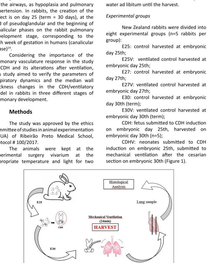
CDHV: neonates submitted to CDH induction on embryonic 25th, submitted to mechanical ventilation after the cesarian section on embryonic 30th (Figure 1).
Figure 1 - Study design of CDHV group. E25: embryonic day 25th. CDH: congenital diaphragmatic hernia. E30:
Surgical procedure
All New Zealand rabbits were duly
weighed and cataloged in their chart. The surgical procedure to create the CDH occurred on day 25 (E25) (term = 30 days), with the pregnant rabbits weighing between 3.7 and 4.5 kg. Induction and anesthetic maintenance were performed by intramuscular injection of ketamine sulfate (ketamin®) 25 to 50 mg/
kg associated with xylazine hydrochloride (Rompun®-Bayer) 5 to 10 mg/kg in the right
posterior thigh. This dose allowed proper anesthesia for 30 to 40 minutes. If necessary, an additional dose was given when the surgical procedure exceeded this period, with half the initial volume. The pregnant rabbit was placed on a surgical thermal table at a constant
temperature of 38°C (Harvard Apparatus®), with
100% oxygen (mask) 1-2 L/min which remained throughout the surgical procedure until total anesthetic recovery. After that, the abdominal region was prepared with an alcoholic solution
of chlorhexidine 2% (Riohex 2%®) and was
operated under aseptic conditions by two surgeons. Before incision, four ml of Lidocaine
1% without vasoconstrictor (Xylestesin®) was
injected in the hypodermic and muscular planes. A median infra-umbilical laparotomy was performed. The uterus was identified (rabbits have uterus bicorne), and one of the horns was gently exposed outside the abdominal cavity. The fetuses were identified from the ovary towards the cervix. The fetus
closest to the ovary was denominated fetus
1, and the next fetus (fetus 2) was the control fetus. The surgical procedure was performed in each uterine horn, in a total of 2-4 fetuses with HDC per rabbit.
Surgical creation of diaphragmatic hernia
The procedure was performed
according to Fauza et al.16, on E25. After
identification of the fetal position, a circumferential suture was performed with Prolene 5-0® at the level of the fetal thorax,
which was seen by transparency through the uterus. After opening the uterine wall, the paw of the left anterior limb was gently exposed. A small dose (0.2 ml) of Lidocaine
1% without vasoconstrictor (Xylestesin®) was
injected into the hypodermic/muscular planes in the posterolateral region of the left thorax, followed by a 3 mm thoracotomy, with exposure of the diaphragm which was incised. The chest
was closed with Prolene® 6-0, and 2 ml of warm
saline (NaCl 0.9%) was administered into the amniotic cavity before closure of the uterine wall to replenish the lost amniotic fluid (Figure 2). Throughout the surgical procedure, the uterine horn was irrigated with a warm saline solution (NaCl 0.9%). After completion of the fetal CDH, the uterine horn was re-inserted into the abdominal cavity, and the same
procedure was performed on other uterus
horn. Finally, the uterine cavity was closed with Vicryl® 2-0, and the skin with Nylon® 4-0
(continuous intradermal suture). At the end of the procedure, the animals received 25 mg/kg Cefazolin and 2.5 mg/kg Medroxyprogesterone
Acetate (Depo Provera®) intramuscularly. After
recovery, the pregnant rabbit was returned to the vivarium, where it remained for another five days until the final harvest, which occurred on E30. All the anesthetic procedure was the same as the maternal surgery, and the doses were adjusted to the offspring according to the fetal and neonatal weight. After the abdominal and uterine opening of the rabbit, the fetuses submitted to CDH and controls were removed from the uterus, cleaned and weighed on an analytical balance (model Ohaus APX-200). After harvesting the required material, the anesthetized rabbit was sacrificed with a lethal
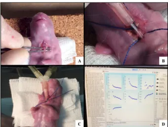
Figure 2 – Surgically creation of CDH. A) Opened uterine wall with exposure of the left anterior limb
of the fetus. B) Left posterolateral thoracotomy. C)
Section of the diaphragm. D) Closure of the uterine
wall.
Mechanical ventilation and in vivo lung ventilatory parameters
The rabbits were anesthetized on the specific gestational day to complete the groups (E25V, E27V, E30V or CDHV) by intramuscular injection of ketamine (ketamin®) 25 to 50
mg/kg associated with xylazine (Rompun®
-Bayer) 5 to 10 mg/kg intramuscularly in the right posterior thigh. A median laparotomy was performed in the same location as the previous surgery. After identification of the fetus with CDH, fetal removal was conducted through a uterine incision. The neonate was weighed and received 1 ml of intraperitoneal ketamine. It was placed in a supine position on a heated table. After cervicotomy, the trachea was isolated, and tracheostomy was performed with a vascular 18G catheter (BD), which was connected to FlexiVent (Scireq, Montreal, QC, Canada). The parameters used for ventilation were: the respiratory rate of 150 breaths/minute, PEEP (positive end-expiratory pressure) of 4 cm H2O, inspiratory time of 0.1s and expiratory time of 0.3 s. Pulmonary mechanics were measured every 4 min for 16
min, and, whenever possible, for 24 min17. The
animals had their temperature checked every 10 minutes. For this purpose, they were kept on a thermal mattress (adult rabbits) / table (newborn rabbits) and the neonates were wrapped in plastic for thermal maintenance (Figure 3).
Figure 3 – Mechanical ventilation. A) Cervicotomy and tracheal identification. B) Tracheostomy with the vascular catheter. C) Connection to the FlexiVent. D) FlexiVent software data.
Harvest and sample processing
After ventilation, the neonates were anesthetized by intramuscular injection of
ketamine (ketamin®) 25 to 50 mg/kg plus
xylazine (Rompun®-Bayer) 5 to 10 mg/kg
intramuscularly in the right posterior thigh. The lung harvest was performing according to the American Society of Thoracic Studies, which suggests the lung collect after the pressure of 20-25 mmHg with formaldehyde through the airway, followed by immediate placement in
formaldehyde.
sections were performed on a Leica microtome (Model RM 2145), with 5μm thickness. They were stained with Masson Trichrome (MT) and
mounted on Entellan®.
Lung and vascular morphometry
For this study, we used left lung samples. The lung sections stained with MT were photographed on a Nikon photomicroscope (Eclipse 80i - Nikon Instruments Inc. Melville, NY, USA) with magnification of 200x and the images were analyzed using Image J2 (National Institutes of Health, Bethesda, MD, USA), a total of 60 sections of lung per group were analyzed. First, image scaling was defined with the Analyze-Set Scale command. The images were analyzed using the color threshold function using Image - Adjust - Color Threshold - Analyze - Measure. Airspace value was measured by subtracting the total parenchyma area minus the total area. Two fields were analyzed per neonate (n = 6 per group). The results were expressed in pixel/μm.
For the pulmonary vasculature analyze, was used a total of 40 arterial histological sections per group were photographed with a magnification of 400X and analyzed using Image-Pro Plus 6.0 software (Media Cybernetics Inc., Rockville, MD-USA). Pre-acinar arterioles between 0 and 30 μm were included. The external diameter (ED) and the internal diameter (ID) were measured, and the media wall thickness (MWT) was calculated
using the formula: MWT = (ED-ID) / ED18.
Statistical analysis
Statistical comparisons were made using one-way analysis of variance (ANOVA), and the Bonferroni method was established as a post hoc test. Student’s t-test was also used when appropriate. Significance values were considered when P<0.05. The program used for statistical analysis was GraphPad Instat, 1997, version 3.0 (GraphPad Prism Software, San Diego, CA, USA).
■
Results
Mechanical ventilation and in vivo lung ventilatory parameters
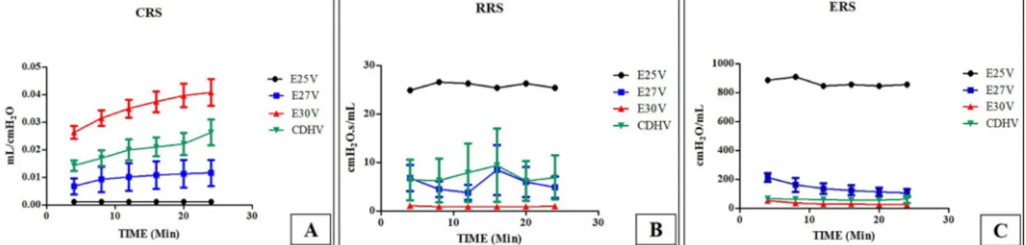
The ventilatory parameters obtained were: dynamic compliance (CRS), dynamic elastance (ERS) and dynamic resistance (RRS). Results showed increased CRS, from the higher to the lower, E30V, CDHV, E27V and E25V, respectively (P<0.05). RRS was increased in E25V group compared to all other groups (P<0.05), and in the group E30V RRS was lower compared to all other groups (P<0.05), with no difference between the groups E27V and CDHV (NS). ERS was increased in the group E25V compared to all groups (P<0.05), and decreased in E27V, E30V, and CDHV with no difference in the after 15min of ventilation. Figure 4 shows the graphs of the respective analyzes.
Figure 4 – Ventilatory parameters analyzed. A) CRS:dynamic compliance. B) RRS: dynamic resistance. C) ERS:
Lung and vascular morphometry
The morphometric analysis of the lung
parenchyma showed increased area of airspace
in the groups ventilated compared to the non-ventilated ones, E27 vs. E27V (P<0.005), E30 vs. E30V (P<0.05), CDH vs. CDHV (P<0.05), except for the E25 vs. E25V groups (NS) that did not present difference (Figures 5 and 6). The group CDH and group E30 were not different in
airspace (NS).
MWT decreased gradually according to the gestational age (E25, E27, E30) and it was influenced by mechanical ventilation, except in the E25V group that did not change compared to E25 group (NS). E27V presented increased MTW compared to E27 (P<0.005), E30V presented increased MTW compared to E30 (P<0.005), and CDHV presented decreased MWT compared to CDH (P<0.005) (Figure 7).
Figure 5 - Airspace and MWT analyzes. A) Graphic of Air Space Area. Unit: μm2. B) Graphic of MWT. Unit: μm.
*P<0.05, **P<0.005.
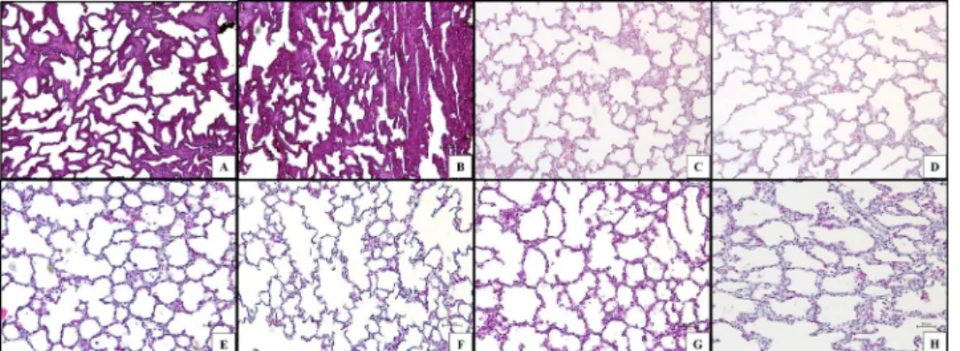
Figure 6 – Masson Trichrome staining of lung tissue sections. A) Group E25. B) Group E25V. C) Group E27. D) Group E27V. E) Group E30. F) Group E30V. G) Group CDH. H) Group CDHV Magnification: x20. Bar: 50μm.
■
Discussion
In this study, we verified the pulmonary
arteriolar response in three different stages of lung development (pseudoglandular,
canalicular and alveolar) and compared the
results with the ventilatory parameters in the rabbit model of CDH.
The ventilatory model in rabbits with CDH it is not new. However, there are
few studies in the literature on the vascular
response in different stages of pulmonary
development in this model. Ohi et al.19
compared the pulmonary growth in the 30th day of gestation (term) with the immature lung in the 25th day of pregnancy, the comparison of Ohi et al.19 was focused on hypoplasia signals
on parenchyma evaluation, the conclusion proved the point of a smaller lung, however, with no definite signs of hypoplasia, indicating a lung with probable normal function. We
did not evaluate the pulmonary hypoplasia
in our study; we measured the airspace as an
indirect morphometrical analysis of pulmonary
immaturity. Our results showed no difference between the groups CDH and E30, which may be explained by the pressure fixation protocol of the lungs, a technical issue in the rabbit
model of CDH already published by Roubliova et
al.20 that compared the littermates tissue with
the non-littermates tissues, showing peculiar differences as in the LMW (wall thickness)
measurements.
Currently, Flemmer et al.13 studied the
neonatal pulmonary impedance in the rabbit model of CDH related to the severity of lung hypoplasia using a ventilatory parameter called forced oscillation technique (FOT), which determine lung tissue forces in small animals, the results showed FOT as good parameter indicator of hypoplastic lungs. In our study, we may say that the ventilatory parameters pointed CDH with similar responses (NS) of E27 (pseudoglandular phase), especially by
reveling equal response in two parameters (RRS and ERS) and decreased CRS. An ERS is numerically equivalent to the inverse of the CRS, being consistent with the values obtained from CRS. RRS, in turn, corresponding to the ratio of resistive pressure by the respiratory
system to air in the alveoli and air13,21.
However, the arteriolar response after ventilation was different between these two groups (CDH and E27), with MWT decreased in the group CDH and increased in the group E27. MWT response after ventilation pointed the acute use of oxygen as a good vasodilator for CDH, but not for the other groups, which presented vasoconstriction after 24min of ventilation. In the groups non-ventilated the CDH group presented higher WMT than E30
(term control) as showed in the model of
CDH in rats, the CDH vascular responses after ventilation presented vasodilatation, while
Control group presented vasoconstriction15.
Other studies in literature also show the increased arteriolar thickness in the rabbit and the rat model of CDH, as Roubliova et al.22 and Taira et al.23, respectively.
In this study it was possible to identify that mechanical ventilation modulates
the response of the parenchyma and the pulmonary arteriolar vasculature. Also the
ventilation response of CDH is similar to the pseudoglandular stage of the lung development (E27), concerning compliance, elastance, and resistance. The median wall thickness response on the immature lungs, E25 and E27, were similar to the E30 after ventilation, showing, when not submitted to ventilation, a gradual decrease of the muscle layer according to the gestational age.
■
Conclusion
and accomplish new information about the CDH model in rabbits.
■
References
1 Mayer S, Metzger R, Kluth D. The embryology of the diaphragm. Semin Pediatr Surg. 2011;20:161–9. doi: 10.1053/j. sempedsurg.2011.03.006.
2 Mous DS, Kool HM, Wijnen R, Tibboel D, Rottier RJ. Pulmonary vascular development in congenital diaphragmatic hernia. Eur Respir Rev. 2018;26:147. doi: 10.1183/16000617.0104-2017.
3 Gallindo RM, Gonçalves FL, Figueira RL, Sbragia L. Prenatal management of congenital diaphragmatic hernia: present, past and future. Rev Bras Ginecol Obstet.
2015;37:140–7. doi:
10.1590/S0100-720320150005203.
4 van Loenhout RB, Tibboel D, Post M, Keijzer R. Congenital diaphragmatic hernia:
comparison of animal models and relevance
to the human situation. Neonatology. 2009;96:137–49. doi: 10.1159/000209850. 5 Yamataka T, Puri P. Pulmonary artery
structural changes in pulmonary
hypertension complicating congenital diaphragmatic hernia. J Pediatr Surg. 1997;32:387–90. doi: 10.1016/S0022-3468(97)90587-X.
6 Kotecha S, Barbato A, Bush A, Claus F, Davenport M, Delacourt C, Deprest J, Eber E, Frenckner B, Greenough A, Nicholson AG, Antón-Pacheco JL, Midulla F. Congenital diaphragmatic hernia. Eur Respir J. 2012;39:820–9. doi: 10.1183/09031936.00066511.
7 Chandrasekharan PK, Rawat M, Madappa R, Rothstein DH, Lakshminrusimha S. Congenital diaphragmatic hernia - a review. Matern Heal Neonatol Perinatol. 2017;3:6. doi: 10.1186/s40748-017-0045-1.
8 Varisco BM, Sbragia L, Chen J, Scorletti F, Joshi R, Wong HR, Lopes-Figueira R, Oria M, Peiro J. Excessive reversal of epidermal growth factor receptor and ephrin signaling following tracheal occlusion in rabbit model of congenital diaphragmatic hernia. Mol Med. 2016;22:1. doi: 10.2119/ molmed.2016.00121.
9 Snoek KG, Capolupo I, van Rosmalen J,
Hout L de J den, Vijfhuize S, Greenough A, Wijnen RM, Tibboel D, Reiss IK. Conventional mechanical ventilation versus high-frequency oscillatory ventilation for congenital diaphragmatic hernia. Ann Surg. 2016;263:867–74. doi: 10.1097/ SLA.0000000000001533.
10 Logan JW, Cotten CM, Goldberg RN, Clark RH. Mechanical ventilation strategies in the management of congenital diaphragmatic hernia. Semin Pediatr Surg. 2007;16:115–25. doi: 10.1053/j.sempedsurg.2007.01.006. 11 Imai Y, Nakagawa S, Ito Y, Kawano T, Slutsky
AS, Miyasaka K. Comparison of lung protection strategies using conventional and high-frequency oscillatory ventilation. J Appl Physiol. 2001;91:1836–44. doi: 10.1152/jappl.2001.91.4.1836.
12 Beurskens N, Klaassens M, Rottier R, de Klein A, Tibboel D. Linking animal models to human congenital diaphragmatic hernia. Birth Defects Res Part A Clin Mol Teratol. 2007;79:565–72. doi: 10.1002/bdra.20370. 13 Flemmer AW, Jani JC, Bergmann F,
Muensterer OJ, Gallot D, Hajek K, Sugawara J, Till H, Deprest JA. Lung tissue mechanics predict lung hypoplasia in a rabbit model for congenital diaphragmatic hernia. Pediatr Pulmonol. 2007;42:505–12. doi: 10.1002/ ppul.20618.
14 Pringle KC. Human fetal lung development and related animal models. Clin Obstet Gynecol. 1986;29:502–13. PMID: 3757332. 15 Gallindo RM, Gonçalves FLL, Figueira RL,
Pereira LAVD, Simões ALB, Schmidt AF, Sbragia L. Ventilation causes pulmonary vascular dilation and modulates the NOS and VEGF pathway on newborn rats with CDH. J Pediatr Surg. 2015;50:842–8. doi: 10.1016/j.jpedsurg.2014.12.005.
16 Fauza DO, Tannuri U, Ayoub AA, Capelozzi VL, Saldiva PH, Maksoud JG. Surgically produced congenital diaphragmatic hernia in fetal rabbits. J Pediatr Surg. 1994;29:882– 6. PMID: 7931963.
17 Debeer A, Sbragia L, Vrancken K, Hendriks A, Roubliova X, Jani J, Naulaers G, Carmeliet P, Deprest J. Antenatal fetal VEGF therapy to promote pulmonary maturation in a preterm rabbit model. Early Hum Dev. 2010;86:99–105. doi: 10.1016/j. earlhumdev.2010.01.025.
hernia. Adv Pediatr. 2018;65:241–7. doi: 10.1016/j.yapd.2018.05.001.
19 Ohi R, Suzuki H, Kato T, Kasai M. Development of the lung in fetal rabbits with experimental diaphragmatic hernia. J Pediatr Surg. 1976;11:955–9. doi: 10.1016/ S0022-3468(76)80073-5.
20 Roubliova XI, Biard JM, Ophalvens L, Gallot D, Jani JC, Verbeken EK, van de Ven CP, Tibboel D, Deprest JA. Morphology of the developing fetal lung the rabbit experimental model. Mod Res and Educ Top in Micr. 2007.
21 Morini F, Capolupo I, van Weteringen W, Reiss I. Ventilation modalities in infants with congenital diaphragmatic hernia. Semin Pediatr Surg. 2017;26:159–65. doi:
10.1053/j.sempedsurg.2017.04.003.
22 Roubliova XI, Deprest JA, Biard JM, Ophalvens L, Gallot D, Jani JC, van de Ven CP, Tibboel D, Verbeken EK. Morphologic changes and methodological issues in the rabbit experimental model for diaphragmatic hernia. Histol Histopathol. 2010;25:1105– 16. doi: 10.14670/HH-25.1105.
23 Taira Y, Miyazaki E, Ohshiro K, Yamataka T, Puri P. Administration of antenatal glucocorticoids prevents pulmonary artery structural changes in nitrofen-induced congenital diaphragmatic hernia in rats. J Pediatr Surg. 1998;33:1052–6. doi: 10.1016/ S0022-3468(98)90530-9.
Correspondence:
Prof. Dr. Lourenço Sbragia Disciplina de Cirurgia Pediatrica
Departamento de Cirurgia e Anatomia, USP Avenida Bandeirantes, 3900
14049-900 Ribeirão Preto - SP Brasil Tel.: (55 16)3602-2593 / 3602-2501 [email protected]
Received: June 09, 2018 Review: Aug 12, 2018 Accepted: Sept 08, 2018
Conflict of interest: none
Financial sources: FAPESP, CNPq, and CAPES
1Research performed at Laboratory of Fetal