Anais da Academia Brasileira de Ciências (2003) 75(1): 55-69 (Annals of the Brazilian Academy of Sciences)
ISSN 0001-3765 www.scielo.br/aabc
Karyotypic analysis in species of the genus
Dasyprocta
(Rodentia: Dasyproctidae) found in Brazilian Amazon
ROSEMAR S. L. RAMOS1, WILLIAM G. VALE1 and FÁTIMA L. ASSIS2
1Department of Biology - CCB - UFPA, 66073-290 Belém, Pará, Brasil. 2Department of Genetics - CCB - UFPA
Manuscript received on October 26, 2001; accepted for publication on December 11, 2002; presented byLewis J. Greene
ABSTRACT
A total of 30 animals of the genusDasyproctawere cytogenetically studied. They belong to the follow-ing species: D. prymnolopha(N=20),D. leporina(N=6),D. fuliginosa(N=1) andDasyproctasp. (N=3) (Dasyproctidae, Hystricognathi). Cell suspensions were obtained by peripheral blood culture, besides bone marrow and spleen cells, fromD. prymnolophaandD. leporina. The diploid number was 64/65 for all samples. The karyotypes showed similarity, and chromosomal polymorphism was not detected by Giemsa conventionalstainingand G banding. The constitutive heterochromatin distribution at the pericentromeric region of all the chromosomes was similar in all species. D. prymnolopha, D. leporinaandDasyprocta sp. presented variation in the heterochromatical block size at one of the homologues of the A18 pair. D. fuliginosapresented the heterochromatin uniformly distributed in all chromosomes. There was not variation in the NORs pattern in the species studied.
Key words:Cytogenetics, Hystricognathi,Dasyprocta, Karyotype.
INTRODUCTION
The Dasyprocta (Hystricognathi) genus includes about 11 species and 33 subspecies, and occurs from the south Mexico as far as high Amazon, including the Antilles, Colombia and Peru (Eisenberg 1989, Emmons 1990). Some forms are not easily recog-nizable due to taxonomic problems, and absence of any modern taxonomic revision (Emmons 1990).
Cytogenetic data in this genus are scanty. From eleven accepted species, only three (D. lepo-rina, D. fuliginosaandD. variegata) have their kary-otypes studied, presenting 2n=64 chromosomes with conventional staining (Hungerford and Snyder 1964, Fredga 1966, Hsu and Benirschke 1968, Kasahara
Correspondence to: Rosemar Silva Luz Ramos E-mail: [email protected]
and Yonenaga-Yassuda 1984). Lima (1993) stud-ied D. leporina and D. fuliginosa species through G and C banding standards and through Nucleolus Organizer Regions (NORs). Both species present well-defined constitutive heterochromatin located in the pericentromeric region of all the autosome chromosomes, with variable intensity. The X chro-mosome presents pericentromeric C banding and the Y chromosome ofD. leporinais almost totally hete-rochromatic. In both species, theAg-NOR is located in the telomeric region of only one acrocentric pair. Therefore, the presented research has the pro-posal of characterize karyotypes ofD. leporina, D. prymnolopha, D. fuliginosaandDasyproctasp. in specimens from the Brazilian Amazon, using G (GTG) and C (CBG) bandings and NOR techniques
56 ROSEMAR S. L. RAMOS, WILLIAM G. VALE and FÁTIMA L. ASSIS
TABLE I
Species and number of animals specified by sex, using preparations ob-tained with different tissues.
Species Peripheral blood Bone Marrow Spleen D. prymnolopha 20 (9F and 11M) 6 (1F and 5M) 3M
D. leporina 6 (2F and 4M) 2 (1F and 1M) 2 (1F and 1M) D. fuliginosa 1F
Dasyproctasp. 3 (1F and 2M)
Total 30 8 5
F = female; M = male
to verify chromosomal polymorphism.
MATERIALS AND METHODS
Twenty specimens of D. prymnolopha, six of D. leporina, one of D. fuliginosa and three of
Dasyprocta sp. were cytogenetically analyzed. These specimens were maintained at the vivarium of the Federal University of Pará (UFPA) and Museu Paraense Emílio Goeldi Zoobotanic Park. The progeny from couples kept in the vivarium of the UFPA are included in this sample. Most of these animals are from unknown origin, including those from MPEG. All the animals were subjected to pe-ripheral blood collection: eight to bone marrow and five to spleen.
Table I shows species, number and genus of animals analyzed by peripheral blood, bone mar-row and spleen. To obtain metaphasic chromosomes from lymphocytes culture, the modified methodol-ogy described by Moorhead et al. (1960) was used, where the mitogenic agent utilized was the Poke-weed. To obtain chromosomes from bone marrow, the methodology described by Baker et al. (1982) with modifications was used.
Live Animals
The animals were sedated with Ketalar (Parke-Davis & Co., Aché Laboratórios Farmacêuticos S.A. Brasil) and Rompum (Bayer do Brasil S.A. Veteri-nária, Brasil) by intramuscular way in proportions
of 0.3 and 0.1 ml/kg of body weight, respectively. The bone marrow cells collection was done in the region of femur-tíbio-patelar joint with syringe and BD (Luerlok) 30×12 special needle with mandril. The collected material was transferred to the cen-trifuge’s tube that contained 5ml of medium HAM-F10, added of 20% of foetal serum and 0,025 ml of colchicine at 0.02% being incubated at 37◦C during 40-60 minutes.
Sacrificed Animals
The animals were sacrificed with an excess dosis of Ketalar and Rompum associated with ether or chlo-roform inhalation. Four hours before the sacrifice, a proportion of 1ml to 1000g of the body weight of colchicine at 0.02% was injected by intramuscular way. In addition to the bone marrow cells collected, the spleen was removed. To obtain metaphasic cells from spleen, the technique described by Ford and Hamerton (1956) was used. To obtain the G and C banding and NOR the methodologies described by Seabright (1971), Sumner (1972) and Howell and Black (1980) were used. The karyotypes were set up in decreasing size order in according to Hunger-ford and Snyder (1964) model.
RESULTS
All analyzed specimens presented a diploid num-ber of 64/65 chromosomes (Figures 1A to 3B). This variation was observed with a frequency of about
KARYOTYPIC ANALYSIS IN SPECIES OF THE GENUS Dasyprocta 57
A
B
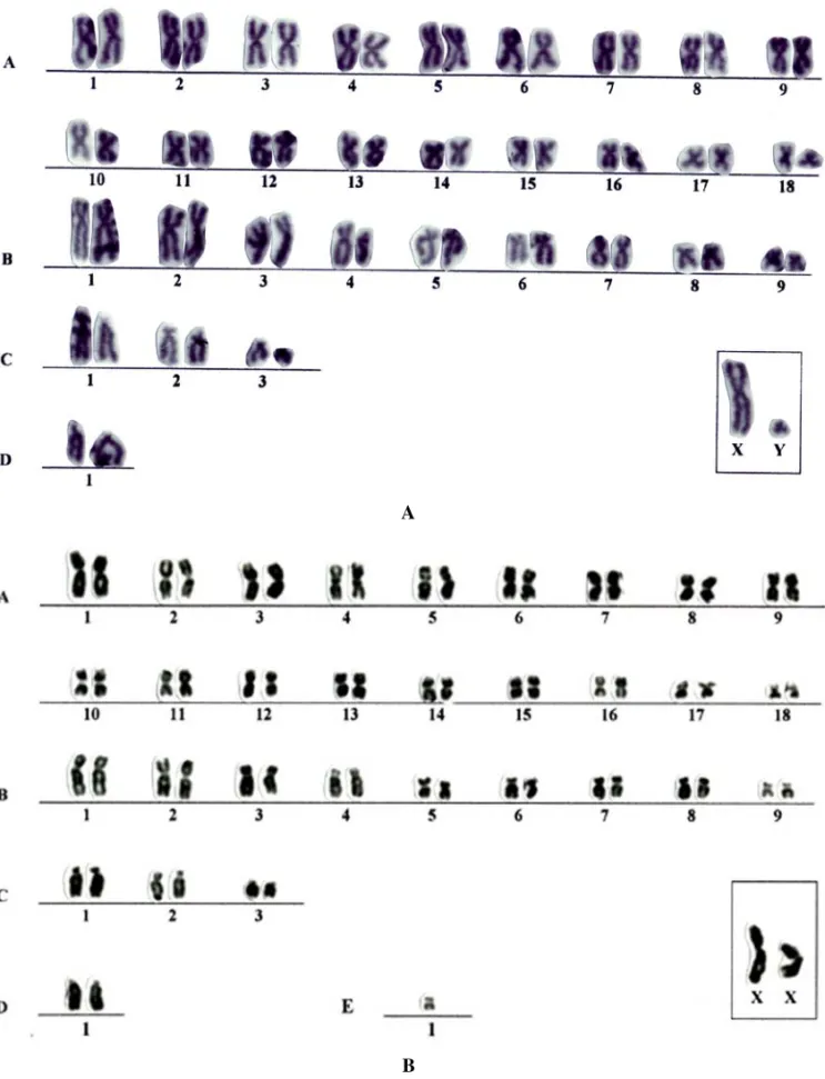
Fig. 1 – Karyotypes of the lineages 2n=64 and 2n=65 (lymphocytes culture) from the male sample (N=277) ofD. prymnolophain conventional Giemsa stain. A) lineage 2n=64; B) lineage 2n=65.
58 ROSEMAR S. L. RAMOS, WILLIAM G. VALE and FÁTIMA L. ASSIS
70% in cells with 2n = 64 and 30% in cells with 2n = 65, in the three different tissues: periph-eral blood in all species; bone marrow and spleen inD. prymnolophaandD. leporina(Tables II, III and IV). The lineage 2n = 65 presents a
super-numerary metacentric chromosome with size rang-ing between medium and small (Figures 1B, 2B and 3B) inD. prymnolopha, D. leporinaandDasyprocta sp., whereas inD. fuliginosait seems to be a sub-metacentric (Figure 4). Through the conventional Giemsa stain, the chromosomes of the lot A (2n= 64) showed the same morphology in all studied species. The karyotypes were characterized by the following model:
Group A – 18 pairs of metacentric chromosomes being theA1 pair the largest metacentric and theA18 pair, the smallest, with other pairs varying in size from large to small. The A18 pair presents, in the short arm, a morphology similar to a lower degree of spiralization looking like a secondary constriction, in conventional stain (Figure 2A).
Group B – nine pairs of submetacentric chromo-somes, ranging from large to small.
Group C – three pairs of subtelocentric ones, pairs C1 and C2 of medium size, and C3 pair with small size.
Group D – only one acrocentric pair with large size. X Chromosome – submetacentric with large size. Y Chromosome – small submetacentric, smaller than the B9 pair. This chromosome was observed in D. prymnolopha, D. leporinaandDasyproctasp.; however only one female ofD. fuliginosawas kary-otyped.
G band pattern was similar in all analyzed sam-ples (Figure 4). C banding technique allows the identification in all species, in the different analyzed tissues, of constitutive heterochromatin in the peri-centromeric region of the most of the autosomes, including the supernumerary chromosomes (Figure 5A).
In the X chromosome, the constitutive hete-rochromatin was restricted to pericentromeric
re-gion, whereas in the Y chromosome, was observed variation in the distribution of this chromatin. In the D. prymnolopha the constitutive heterochromatin was limited to pericentromeric region (Figure 5). InD. leporinaandDasyproctasp., the Y chromo-some was almost totally heterochromatic (Figures 5B and C). The A6 pair inD. prymnolopha, D. lepo-rinaandDasyproctasp. presented polymorphism in the amount of constitutive heterochromatin. One of the chromosomes showed the heterochromatic block twice the size of the other homologous of the pair (Figures 5A, B and C). InD. fuliginosakaryotype no polymorphism was observed (Figure 6). TheA18 pair inD. prymnolophaandDasyproctasp. kary-otypes showed constitutive heterochromatin in only one of the homologues (Figures 5A and C). This pair, in theD. leporinaandD. fuliginosakaryotypes did not show variation (Figures 5B and 6). D. fuligi-nosakaryotype showed uniformity in the standard distribution of constitutive heterochromatin in all of chromosomes (Figure 6).
The NORs were observed in the telomeric re-gion of D1 acrocentric pair short arm, in the kary-otypes obtained using different tissues of all speci-mens studied (Figure 7).
DISCUSSION
All specimens of D. prymnolopha, D. lepo-rina, Dasyproctasp. andD. fuliginosapresented two cellular lineages, with 2n = 64 and 2n = 65 chromosomes. No animal showed only the basic karyotype of 2n = 64, as described in the
litera-ture (Kasahara and Yonenaga-Yassuda 1984, Lima 1993, Lima and Langguth 1995, 1998). This vari-ation is because of the presence of one supernu-merary chromosome metacentric in theD. prym-nolopha, D. leporinaandDasyproctasp. and sub-metacentric inD. fuliginosa. Supernumerary chro-mosomes are considered common among the Hys-tricognathi, mainly in the Chinchillidae, Octodon-tidae, Caviidae, Ctenomyidae, Hydrochaeridae and Echimyidae (George and Weir 1974, Kasahara and Yonenaga-Yassuda 1984, Leal-Mesquita 1991a, b,
KARYOTYPIC ANALYSIS IN SPECIES OF THE GENUS Dasyprocta 59
A
B
Fig. 2 – Karyotype of the lineage 2n=64 (marrow bone) and 2n=65 (lymphocytes culture) ofD. leporinain conventional Giemsa stain. A) lineage 2n=64 from a male; B) lineage 2n=65 from a female.
60 ROSEMAR S. L. RAMOS, WILLIAM G. VALE and FÁTIMA L. ASSIS
A
B
Fig. 3 – Karyotypes of the lineages 2n=64 and 2n=65 (lymphocytes culture) from a male sample (N=283) ofDasyproctasp. in conventional Giemsa stain. A) lineage 2n=64; B) lineage 2n=65.
KAR
Y
O
TYPIC
AN
AL
YSIS
IN
SPECIES
OF
THE
GENUS
Dasypr
octa
61
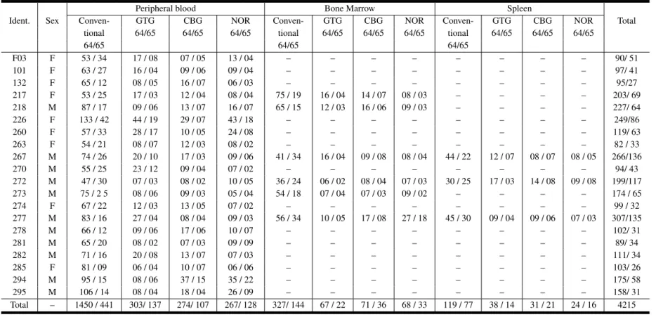
TABLE II
Number of 2n=64 and 2n=65 cells, collected from peripheral blood, bone marrow and spleen, inD. prymnolopha.
Peripheral blood Bone Marrow Spleen
Ident. Sex Conven- GTG CBG NOR Conven- GTG CBG NOR Conven- GTG CBG NOR Total tional 64/65 64/65 64/65 tional 64/65 64/65 64/65 tional 64/65 64/65 64/65
64/65 64/65 64/65
F03 F 53 / 34 17 / 08 07 / 05 13 / 04 – – – – – – – – 90/ 51
101 F 63 / 27 16 / 04 09 / 06 09 / 04 – – – – – – – – 97/ 41
132 F 65 / 12 08 / 05 16 / 07 06 / 03 – – – – – – – – 95/27
217 F 53 / 25 17 / 03 12 / 04 08 / 04 75 / 19 16 / 04 14 / 07 08 / 03 – – – – 203/ 69 218 M 87 / 17 09 / 06 13 / 07 16 / 07 65 / 15 12 / 03 16 / 06 09 / 03 – – – – 227/ 64
226 F 133 / 42 44 / 19 29 / 07 43 / 18 – – – – – – – – 249/86
260 F 57 / 33 28 / 17 10 / 05 24 / 08 – – – – – – – – 119/ 63
263 F 54 / 21 08 / 07 12 / 03 08 / 02 – – – – – – – – 82 / 33
267 M 74 / 26 20 / 10 17 / 03 09 / 06 41 / 34 16 / 04 09 / 08 08 / 04 44 / 22 12 / 07 08 / 07 08 / 05 266/136
270 M 55 / 25 23 / 12 09 / 04 07 / 02 – – – – – – – – 94/ 43
272 M 47 / 30 07 / 03 08 / 02 10 / 05 36 / 24 06 / 02 08 / 04 07 / 03 30 / 25 17 / 03 14 / 08 09 / 08 199/117 273 M 75 / 2 5 08 / 06 09 / 03 05 / 04 54 / 18 07 / 04 07 / 03 09 / 02 – – – – 174 / 65
274 F 67 / 22 12 / 03 13 / 05 07 / 02 – – – – – – – – 99 / 32
277 M 83 / 16 27 / 04 08 / 04 09 / 03 56 / 34 10 / 05 17 / 08 27 / 18 45 / 30 09 / 04 09 / 06 07 / 03 307/135
278 M 66 / 12 09 / 06 17 / 06 10 / 07 – – – – – – – – 102/ 31
281 M 65 / 20 08 / 02 07 / 03 09 / 09 – – – – – – – – 89/ 34
282 M 71 / 16 20 / 08 13 / 07 07 / 03 – – – – – – – – 111/ 34
285 F 81 / 09 06 / 04 10 / 07 06 / 06 – – – – – – – – 103/ 26
294 M 95 / 15 08 / 06 37 / 15 35 / 22 – – – – – – – – 175/ 58
295 M 106 / 14 08 / 04 18 / 04 26 / 09 – – – – – – – – 158/ 31
Total – 1450 / 441 303/ 137 274/ 107 267/ 128 327/ 144 67 / 22 71 / 36 68 / 33 119 / 77 38 / 14 31 / 21 24 / 16 4215
Ident. = Identification.
An
Acad
Br
as
Cienc
(2003)
75
62
R
OSEMAR
S.
L.
RAMOS,
WILLIAM
G.
V
ALE
and
FÁ
TIMA
L.
ASSIS
TABLE III
Number of 2n=64 and 2n=65 cells, collected from peripheral blood, bone marrow and spleen, inD. leporina.
Peripheral blood Bone Marrow Spleen
Ident. Sex Conven- GTG CBG NOR Conven- GTG CBG NOR Conven- GTG CBG NOR Total
tional 64/65 64/65 64/65 tional 64/65 64/65 64/65 tional 64/65 64/65 64/65
64/65 64/65 64/65
M01 M 58 / 29 31 / 27 06 / 04 17 / 09 – – – – – – – – 112 / 69
M02 M 59 / 29 08 / 04 14 / 06 09 / 01 – – – – – – – – 90 / 40
190 F 47 / 38 09 / 06 18 / 07 08 / 05 – – – – – – – – 82 / 56
216 M 34 / 06 09 / 03 05 / 03 03 / 01 10 / 05 09 / 05 06 / 03 08 / 05 08 / 02 07 / 04 06 / 02 04 / 04 109 / 43
220 M 22 / 11 10 / 05 15 / 08 16 / 04 27 / 17 09 / 06 07 / 03 05 / 04 14 / 07 05 / 02 03 / 01 04 / 03 137 / 71
261 M 47 / 23 15 / 05 09 / 03 11 / 05 – – – – – – – – 82 / 36
Total – 267 / 136 82 / 50 67 / 31 64 / 25 37 / 22 18 / 11 13 / 06 13 / 09 22 / 09 12 / 06 09 / 03 08 / 07 927
Ident. = Identification.
An
Acad
Br
as
Cienc
(2003)
75
KARYOTYPIC ANALYSIS IN SPECIES OF THE GENUS Dasyprocta 63
Fig. 4 – Banding G (GTG) standard of the lineage 2n=65 from a female sample ofD. fuliginosa. The arrow points to the banded supernumerary chromosome. Standing out, it appears the sexual pair: X and Y chromosomes, from a male ofD. prymnolopha.
TABLE IV
Number of 2n=64 and 2n=65 cells, collected from peripheral blood, bone marrow and spleen, inDasyproctasp. and peripheral blood, inD. fuliginosa.
Staining and Banding
Species Sex Conven- GTG CBG NOR Total
tional 64/65 64/65 64/65 64/65
01 F 63 / 17 10 / 08 13 / 10 12 / 09 98 / 44 D.sp. 283 M 84 / 08 32 / 13 09 / 06 08 / 04 133 / 31
284 M 79 / 16 32 / 27 20 / 10 15 / 05 146 / 58 Total 226 / 41 74 / 48 42 / 26 35 / 18 510 D.fulig. 53 F 74 / 21 43 / 17 15 / 06 17 / 07 149 / 51
D.sp. =Dasyproctasp.;D. fulig.=D. fuliginosa.
Gallardo 1991, 1992, Fagundes and Yassuda 1995, Moreira et al. 1995). The presence of lineage 2n= 65, with one supernumerary chromosome in our sample, can characterize a geographic variation, since all constants species of literature were derived
from different South America regions other than the Northern region.
InD. prymnolopha, D. leporinaandD. fuligi-nosawe detected a small variation in the A18 pair in relation toDasyproctasp. due to the fact that 100%
64 ROSEMAR S. L. RAMOS, WILLIAM G. VALE and FÁTIMA L. ASSIS
Fig. 5A
Fig. 5B
KARYOTYPIC ANALYSIS IN SPECIES OF THE GENUS Dasyprocta 65
Fig. 5C
Fig. 5 – Banding C (CBG) standard in the lineages 2n=64 and 2n=65. A) and C) the arrows point to one of the homologues from the pair A18, lacking the heterochromatical block (D. prymnolophaandDasyproctasp., respectively.); A), B) and C) one of the homologues from the pair A6 had an increasing of the heterochromatical block (D. prymnolopha, D. leporinaandDasyproctasp., respectively); A) and C) the supernumerary chromosome with a constitutive heterochromatin in the pericentromeric region (D. prymnolophaand Dasyproctasp., respectively). B) and C) the arrows point to the Y chromosome, almost totally heterochromatical (D. leporinaand Dasyproctasp., respectively).
of the analyzed cells showed one of the homologues with a secondary constriction and about 40% from the cells of both lineages present this morphology in two homologues. However, there is a necessity of a more detailed research to confirm this characteris-tic. The marking character of A18 chromosome of our samples was first described here. The remaining chromosome complement agrees with Lima (1993), Lima and Langguth (1995, 1998) researches inD. leporinaandD. fuliginosa.
The X chromosome of the four species here studied is submetacentric, different from the one found inD. leporinaandD. fuliginosadescribed as large metacentric (Kasahara and Yonenaga-Yassuda 1984, Lima and Langguth 1995, 1998). TheY
chro-mosome of D. prymnolopha, D. leporina and Dasyproctasp. is a small submetacentric, similar to the one described byD. variegata(Hungerford and Snyder 1964), but different from the described small metacentric chromosome ofD. leporina(Kasahara and Yonenaga-Yassuda 1984, Lima 1993, Lima and Langguth 1995, 1998).
G banding comparison between species show-ed several chromosomes similarities (Figure 8), sug-gesting that these species are conserved at the chro-mosome level. This finding is similar to that found in four species ofCtenomys(C. flamarioni, C. men-docinus, C. porteousiandC. australis), in relation to 1, 2, 4-8, 11-14, 16, 18, 21 and 23 pairs (Freitas 1994), and in two species ofCavia(C. apereaand
66 ROSEMAR S. L. RAMOS, WILLIAM G. VALE and FÁTIMA L. ASSIS
Fig. 6 – Banding C (CBG) standard of the lineage 2n=64 ofD. fuliginosa.
Fig. 7 – Localization of the Nucleolous Organizer Regions in the pair D1 in: A)D. prymnolopha; B)D. leporina; C)D. fuliginosa; D)Dasyproctasp.
C. aperea pamparum) that present similarity in the G banding standard in most of the autosomes (Maia 1984).
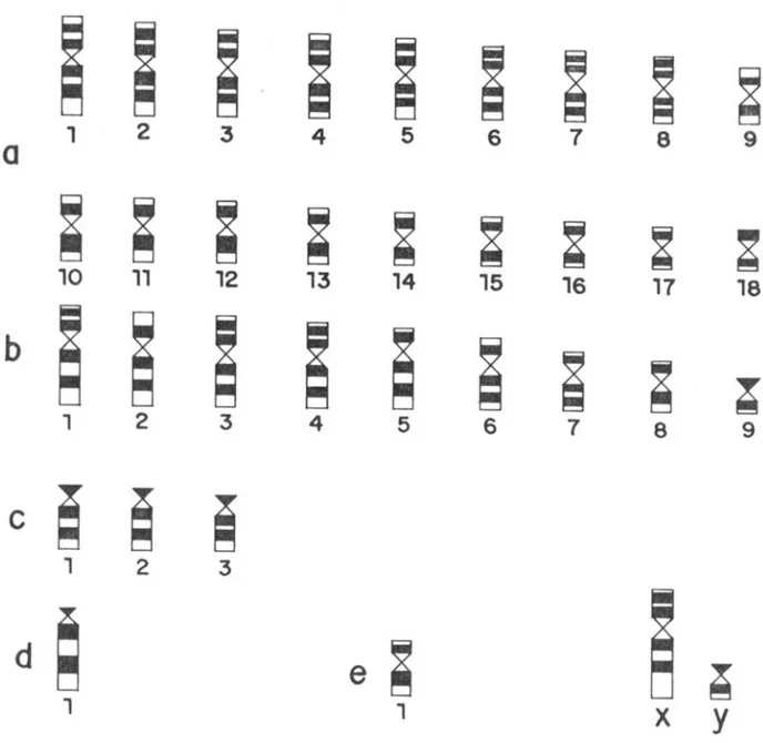
In spite of the difficulties in obtaining banded trypsin-Giemsa karyotypes ofD. leporina and D. fuliginosa, we presented an idiogram (Figure 9) where we suggest the interpretation of positive (dark) and negative (clear ) bands distribution like an essay of G banding standard inDasyprocta.
In C banding analysis, it was evident the peri-centromeric constitutive heterochromatin is all
chro-mosomes, including the supernumerary chromo-some and X andY sexual pair. This pattern is similar to the one found in other Hystricognathi asCavia porcellus (Natarajan and Raposa 1974); Clyomys laticeps laticeps (Souza and Yonenaga-Yassuda 1982) andP. longicaudatus(Maia et al. 1988). D. leporinaandDasyproctasp. presented the Y chro-mosome almost totally heterochromatic, as found in D. leporina(Lima 1993, Lima and Langguth 1995, 1998) and in some species fromCtenomysthat pre-sented Y chromosome varying from totally
KARYOTYPIC ANALYSIS IN SPECIES OF THE GENUS Dasyprocta 67
Fig. 8 – Comparisons of the banding standard G (GTG) in the four analized species. D.p –D. prymnolopha; D.l –D. leporina; D.sp – Dasyproctasp.; D.f –D. fuliginosa.
rochromatic to C banding negative standard (Frei-tas 1994). D. prymnolopha, D. leporina and Dasyproctasp. presented variation in the amount of heterochromatin in the A6 pair, similar to the ob-served by Reig et al. (1990) in the Ctenomyídeos (Hystricognathi). D. fuliginosadid not show varia-tion in C banding and probably, this karyotype rep-resents the basic standard of distribution and amount of constitutive heterochromatin inDasyprocta.
In the four species the NORs were located in telomeric region of the short arm of both homo-logues of D1 pair, without size variation or NORs absence in any analyzed cell. This results are similar to the ones described inD. leporinaandD. fuligi-nosa(Lima 1993).
ACKNOWLEDGMENTS
The authors thank Conselho Regional de Desen-volvimento Científico e Tecnológico (CNPq) for the financial support conceded and Museu Paraense Emílio Goeldi Zoobotanic Park and the vivarium of
the Federal University of Pará for the donation of animals that built up our samples.
RESUMO
Foram estudados citogeneticamente um total de 30 ani-mais das espéciesD. prymnolopha(N=20),D. leporina (N=6), D. fuliginosa (N=1) e Dasyproctasp. (N=3) (Dasyproctidae, Histricognathi). As preparações cromos-sômicas foram obtidas do cultivo de sangue periférico, além de medula óssea e baço emD. prymnolophaeD. leporina. O número diplóide foi de 64/65 em todos os exemplares. O cariótipo mostrou similaridade, não sendo detectado, através de coloração convencional de giemsa e de banda G, polimorfismo cromossômico em qualquer uma das espécies estudadas. A distribuição da hetero-cromatina constitutiva na região pericentromérica de to-dos os cromossomos foi similar nas quatro espécies. D. prymnolopha, D. leporinaeDasyproctasp. apresentaram variação no tamanho do bloco heterocromático em um dos homólogos do par A18. D. fuliginosaapresentou a heterocromatina uniformemente distribuída em todos os
68 ROSEMAR S. L. RAMOS, WILLIAM G. VALE and FÁTIMA L. ASSIS
Fig. 9 – Idiogram presenting the banding G standards inD. prymnolopha, D. leporina, Dasyproctasp. and D. fuliginosa, including the supernumerary chromosome (e1) of the last species.
cromossomos. Não houve variação no padrão das RONs entre as espécies estudadas.
Palavras-chave: Citogenética, Hystricognathi,
Dasy-procta, Cariótipo.
REFERENCES
Baker RJ, Haiduk MW, Robbins LW, Cadena A and
Koop BF. 1982. Chromosomal studies of South American bats and their systematic implications. In: M.A. Mares., H.H. Genoways(eds.) Mammalian Biology South America USA., p.303-306.
Eisenberg JF. 1989. Order Rodentia (Rodents,
Roden-tia). In: Mammals of the Neotropics. Chicago: Uni-versity of Chicago, p.329-418.
Emmons L. 1990. Large cavyalike rodents (Agoutidae, Dasyproctidae, Hydrochaeridae, Dynomyidae). In: Neotropical rainforest mammals. Chicago: Univer-sity of Chicago, p. 203-212.
Fagundes V and Yassuda YY. 1995. Dois carióti-pos de Trichomys aperoides (Echimyidae, Rodentia) com 2n=26 e 2n=30, investigados por hibridização “in situ” com sondas teloméricas. In: Congresso Nacional de Genética, 41◦, Caxambú-Mg, Re-sumo...Seção C-155 18(3): 486.
Ford CE and Hamerton JL. 1956. A colchicine
KARYOTYPIC ANALYSIS IN SPECIES OF THE GENUS Dasyprocta 69
tonic citrate squash sequence for mammalian chro-mosome. Stain Techn 31: 247-251.
Fredga K. 1966. Chromosome studies in five species of SouthAmerican rodents (Suborder Hystricomorpha). Mamm Chrom News 20: 45-46.
Freitas TRO. 1994. Geographical variation of hete-rochromatin in (Rodentia,Octodontidae) and its cyto-genetics relationships with other species of the genus. Cytogenet Cell Genet 67: 193-198.
Gallardo M. 1991. Karyotypic evolution inCtenomys (Rodentia, Ctenomyidae). J Mamm 72(1): 11-21.
Gallardo MH. 1992. Karyotypic evolution in octodon-tid rodents based on C-band analysis. J Mamm 73: 89-98.
George W and Weir BJ. 1974. Hystrycomorph chro-mosomes. Symp Zool Soc Lond 34: 79-108. Howell WM and Black DA. 1980. Controlled
silver-staining of nucleolus organizer regions with a protec-tive colloidal developer - - a 1-step method. Experi-entia 36: 1014-1015.
Hsu TC and Benirschke K. 1968. An atlas of mam-malian chromosomes. New York: Springer-Verlag, 2: 72-76.
Hungerford DA and Snyder RL. 1964. Karyotypes of two more mammals. Amer Natur 98: 125-127.
Kasahara S and Yonenaga-Yassuda Y. 1984. A progress report of cytogenetic data on brazilian ro-dents. Rev Bras Genet 7(3): 509-533.
Leal-Mesquita ERRBP. 1991a. Estudos citogenéticos em dez espécies de roedores brasileiros da família Echimyidae. Dissertação de Mestrado. São Paulo, Universidade de São Paulo, 167p.
Leal-Mesquita ERRBP. 1991b. Cytogenetics studies of 10 species of brazilian rodent of the family Echimyi-dae. Rev Bras Genet 14: 1094.
Lima JFS. 1993. Descrição de novos cariótipos em es-pécies de Sciuridae, Dasyproctidae e Erethizontidae com discussão da evolução cromossômica nos Cavio-morpha,. Dissertação de Mestrado, Universidade Fe-deral da Paraíba, 1993. 81p.
Lima JFS and Langguth A. 1995. Uma análise dos da-dos citogenéticos atuais da-dos Caviomorphos do Novo Mundo. In: Congresso Nacional de Genética, 41◦, 1995, Caxambú-Mg. Resumo... Seção C-162 18(3): 490.
Lima JFS and Langguth A. 1998. The karyotypes of three Brazilian species of the genusDasyprocta (Rodentia:Dasyproctidae). Iher Ser Zool 85: 141-145.
Maia V. 1984. Karyotypes of three species of Caviinae (Rodentia, Caviidae). Experientia 40: 564-566.
Maia V, Langguth A and Almeida AB. 1988. Ca-racterização cromossômica do roedor equimídeo Proechimys cuvierida região Amazônica (Rio Ua-tumã, AM). Cien Cult 40: 760.
Moorhead PS, Nowell PC, Nellman WJ, Battips DM
and Hungerford DA. 1960. Chromosome prepa-rations of leukocytes cultured from human peripheral blood. Exp Cell Res 20: 613-616.
Moreira R, Garcia MRS, Lopes LD, Santos CR and
Leal-Mesquita ER. 1995. Análise cariotípica em Echimys chrysurus (Echimyidae, Rodentia). In: Congresso Nacional de Genética, 41◦, Caxam-bú-Mg, Resumo..., Seção C-157 18(3): 487.
Natarajan AI and Raposa I. 1974. Repetitive DNA and constitutive heterochromatin in the chromosomes of Guinea pig. Hereditas 76: 145-147.
Reig OA, Busch C, Ortells MO and Contreras JR. 1990. An overview of evolution. Systematics, popu-lation biology and speciation inCtenomys. In:Nevo, E. and Reig, O.A. (eds.). Biology of Subterranean Mammals at the Organismal and Molecular Levels, New York, p. 71-96.
Seabright M. 1971. A rapid banding technique for hu-man chromosome. Lancet: 971-972.
Souza MJ and Yonenaga-Yassuda Y. 1982. Chromo-somal variability of sex chromosomes and NORs in Trichomys aperoides(Rodentia, Echimyidae). Cyto-genet Cell Genet 33: 197-203.
Sumner AT. 1972. A simple technique for demonstrating centromeric heterochromatin. Exp Cell Res 75: 304-306.