Diagnostic methods for identification of root-knot nematodes species from Brazil
Métodos diagnósticos usados na identificação de espécies do nematoide das galhas do Brasil Tiago Garcia da Cunha1 Liliane Evangelista Visôtto2* Everaldo Antônio Lopes1
Claúdio Marcelo Gonçalves Oliveira3 Pedro Ivo Vieira Good God1
ISSNe 1678-4596
INTRODUCTION
The genus Meloidogyne comprises more than 100 species and its host range exceeds 3000 species of plants (HUNT & HANDOO, 2009). These nematodes are spread worldwide, with a high diversity of species in tropical and subtropical regions. They cause damage to cash and subsistence crops all over the globe (LOPES & FERRAZ, 2016). RKN are sedentary endoparasites that induce the formation of giant cells in the roots, from which the nematode feed to complete its life cycle (BALDACCI-CRESP et al., 2015).The availability of water and nutrients to the plant decreases while the giant cells are located close to the root systems xylem and phloem. As the
giant cells are located close to the xylem and phloem, availability of water and nutrients to the plant decreases (SIDDIQUI et al., 2014). Morphological and biochemical alterations induced by nematode parasitism cause abnormal growth of plants, nutrient
deficiency symptoms, roots with galls, forking and
other deformations (MOENS et al., 2009).
In Brazil, the major species of the
root-knot nematode (RKN) are Meloidogyne incognita, M. javanica, M. arenaria, M. hapla, M. exigua, M. paranaensis, M. enterolobii (=Meloidogyne mayaguensis) and M. ethiopica (CARNEIRO et al.,
2016). Accurate identification of RKN species is
essential for implementing management strategies. For instance, the choice of resistant cultivars or cover
1Instituto de Ciências Agrárias, Universidade Federal de Viçosa (UFV), Campus Rio Paranaíba, Rio Paranaíba, MG, Brasil.
2Instituto de Ciências Biológicas e da Saúde, Universidade Federal de Viçosa (UFV), Campus Rio Paranaíba, 38810-000, Rio Paranaíba, MG,
Brasil. E-mail: [email protected]. *Corresponding author. 3Instituto Biológico, Campinas, SP, Brasil.
ABSTRACT: The accurate identification of root-knot nematode (RKN) species (Meloidogyne spp.) is essential for implementing management strategies. Methods based on the morphology of adults, isozymes phenotypes and DNA analysis can be used for the diagnosis of RKN. Traditionally, RKN species are identified by the analysis of the perineal patterns and esterase phenotypes. For both procedures, mature females are required. Over the last few decades, accurate and rapid molecular techniques have been validated for RKN diagnosis, including eggs, juveniles and adults as DNA sources. Here, we emphasized the methods used for diagnosis of RKN, including emerging molecular techniques, focusing on the major species reported in Brazil.
Key words: DNA, esterase, Meloidogyne, molecular biology, morphological pattern.
RESUMO: A identificação acurada de espécies do nematoide das galhas (NG) (Meloidogyne spp.) é essencial para a implementação de estratégias de manejo. Métodos baseados na morfologia de adultos, fenótipos de isoenzimas e análise de DNA podem ser usados para a diagnose do NG. Tradicionalmente, as espécies de NG são identificadas pela análise do padrão perineal e fenótipos de esterase. Em ambos os procedimentos, fêmeas maduras são necessárias. Nas últimas décadas, técnicas moleculares acuradas e rápidas têm sido validadas para a diagnose de NG, incluindo ovos, juvenis e adultos como fontes de DNA. Aqui, nós destacamos os métodos usados para a diagnose de NG, incluindo técnicas moleculares emergentes, focando nas principais espécies encontradas no Brasil.
Palavras-chave: DNA, esterase, Meloidogyne, biologia molecular, padrão morfológico.
crops for nematode control depends on the information
of which species are dominant in the field. The coffee
(Coffea arabica) cultivar ‘IAPAR 59’ is resistant to
M. exigua and susceptible to M. paranaensis and M. incognita. Velvet bean (Mucuna pruriens var. utilis) does not allow M. incognita reproduction, but the same behavior is not observed when the nematode is
M. javanica (FERRAZ et al., 2010).
For decades, observation of female perineal patterns was the routine method for RKN
identification. Currently, isozyme phenotypes are
used for diagnostics of RKN in many laboratories worldwide. However, in the last decade, several accurate and rapid molecular protocols for RKN diagnostics have been developed (SEESAO et al., 2017). Here, we discussed the morphological, biochemical and molecular methods used for the diagnosis of RKN, focusing on the major species reported in Brazil and including pros and cons of these techniques.
Diagnostic methods
Method based on morphology
Morphological characteristics of males
Head shape and stylet morphology of males are useful characteristics in the identification of some RKN species, such as
M. incognita, M. enterolobii, M. paranaensis
and M. javanica (Figure 1). For instance, male
M. paranaensis can be identified based on its
distinctive head morphology (Figure 1). For diagnostic purposes, specimens must be viewed in the lateral position. The distance from the dorsal esophageal gland orifice (DGO) to the stylet base of the males may also be used for the distinction between some species, such as
M. enterolobii and M. incognita (ALMEIDA et
al., 2008). The size and shape of the stylet also have a complementary taxonomical value for the identification of RKN species; although some RKN species share similarly sized stylets.
Figure 1 - Anterior region of males of Meloidogyne incognita, M. enterolobii, M. paranaensis and M. javanica. DGO* =
Perineal pattern of females
Traditionally, the perineal pattern of females has been the main technique for RKN
species identification. The shape and visual
aspect of the whole perineal region, dorsal arch, dorsal striae, lateral lines and phasmids are the
morphological characteristics used in identification.
This is an inexpensive technique as it only requires a microscope, microscopy slide, coverslip, lactic acid
and glycerin, but requires manual skills and it is time
consuming during slide preparation. Also mature females are required, which means that roots with females must be available for diagnosis (SEESSAO et al., 2016). For images of perineal pattern of several RKN species, see FERRIS (1999).
In the late 1940s, this method was
proposed as a tool for the identification of M. incognita, M. javanica, M. arenaria and M. hapla
(CHITWOOD, 1949). However, with the discovery of new species of the pathogen, this technique proved not to be accurate enough to distinguish some
species. The misidentification of M. enterolobii and
M. inornata as M. incognita is a notorious example of this situation, since the perineal patterns of these species are similar (CARNEIRO et al., 2016). Such as for the use of morphological characteristics of males, the diagnosis of RKN species using this technique demands specialized taxonomic
knowledge (OLIVEIRA et al., 2011).
Biochemical method Isozyme phenotypes
Isoenzyme phenotyping is the routine diagnostic test for RKN in many laboratories worldwide. It is based on the relative mobility of enzymes extracted from mature females on gel electrophoresis (BLOK & POWERS, 2009). The
whole procedure takes three to four hours, from
sample processing to gel revelation. Protein extract from M. javanica females is applied on the gel for use as reference phenotype. Details on the method can be found in CARNEIRO & ALMEIDA (2001) and FREITAS et al. (2016).
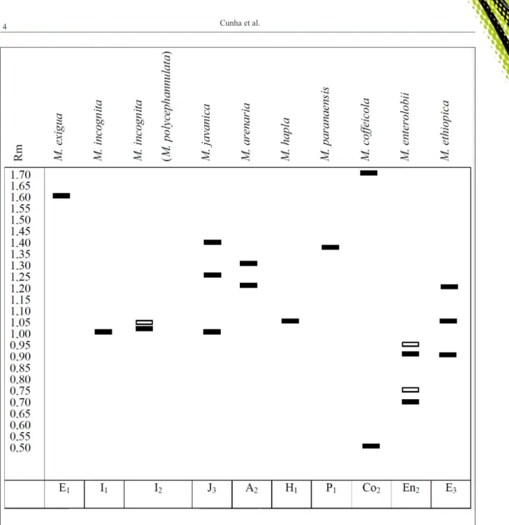
Esterase phenotype (EST) analysis is usually enough to identify Meloidogyne species (ESBENSHADE & TRIANTAPHYLLOU, 1990; CARNEIRO et al., 1996; CARNEIRO & ALMEIDA, 2001) (Figure 2); although, other enzymes can also provide complementary information, such as malate dehydrogenase (MDH), superoxide dismutase (SOD) and glutamate oxaloacetate transaminase (GOT) (FREITAS et al., 2016). In some cases, EST are similar between two species, such as M. naasi and
M. exigua. In this situation, MDH can be used to differentiate species (CARNEIRO et al., 2016).
Intraspecific variations may occur in M. javanica, M. arenaria, M. exigua and M. paranaensis
and other species, limiting the accuracy of the diagnosis (KUNIEDA et al., 1995; CARNEIRO et al., 1996; CARNEIRO et al., 2004; SALGADO et al., 2015). In addition, the procedure required the use of mature females and several individuals are needed in the case of species with small specimens, such as M. exigua (CARNEIRO et al., 2016).
Molecular methods
Molecular techniques rely on the occurrence of polymorphisms in DNA sequences among groups of nematodes, especially in nuclear ribosomal DNA (rDNA) and mitochondrial DNA (mtDNA). Diagrams of rDNA and mtDNA of some RKN species can be found in VERONICO et al. (2004) and GARCÍA & SÁNCHEZ-PUERTA
(2015). For identification and phylogenetic analysis
of plant-parasitic nematodes, the genes 18S, 28S, 5.8S and the spacer regions (internal transcribed spacer - ITS, external transcribed spacer – ETS and intergenic spacer- IGS) have been the most studied rDNA regions, while the gene cytochrome c oxidase subunits I (COI, CO1 or COX 1) and II (COII, COII or COX 2) have been the main targets of mtDNA (ROBERTS et al., 2016).
The multicopy base of the rDNA contains variation and stability for discrimination of the major RKN species (ROBERTS et al., 2016). The small subunit of ribosomal DNA (18S rDNA or SSU) has been used in studies on inter-genera relationships, due to its low evolution rate (ROBERTS et al., 2016).
Using a SSU as marker, 12 RKN species were grouped
in three clades: I - M. incognita, M. arenaria and M. javanica; II - M. hapla races A and B, M. duytsi and
M. maritima; III - M. exigua, M. graminicola and M.
chitwoodi (DE LEY et al., 2002). The large subunit rDNA (28S rDNA or LSU) has conserved domains and expansion segments, such as the D2-D3 expansion region. This region is very useful for designing
diagnostics for identification of species (ROBERTS et
al., 2016). Several molecular protocols for nematode
identification use spacer regions (ITS, ETS and IGS)
Figure 2 - Phenotypic profile of the esterase enzyme of the main Meloidogyne species reported in Brazil. Colorless boxes represent weak
bands, filled boxes represent strong bands. Rm: Migration ratio in relation to the slower band (Rm = 1.0) of M. javanica. E1, I1,
I2, J3, A2, H1, P1, Co2, En2 and E3: codes used to identify the esterase bands pattern of M. exigua, M. incognita, M. incognita
= M. polycephannulata, M. javanica, M. arenaria, M. hapla, M. paranaensis, M. coffeicola, M. enterolobii and M. ethiopica, respectively. Source: Adapted from CARNEIRO et al. (2016).
chitwoodi and M. fallax from other species, including
M. enterolobii, M. hapla, M. incognita, M. javanica
and M. arenaria (WISHART et al., 2002).
The mtDNA genes can be used in
phylogenetic studies and for identification of RKN
(KIEWNICK et al., 2014). Multiple copies of mtDNA
are found per cell, which facilitates its amplification.
In addition, it is highly conserved among animals and
has variable regions flanked by conserved domains
(ROBERTS et al., 2016). Despite these advantages as
a marker, mtDNA has not been as studied as rDNA
for the diagnosis of RKN.
required (OLIVEIRA et al., 2011). Furthermore, procedures used in molecular methods of nematodes
are like those applied to other organisms, and do not require advanced knowledge on taxonomy of
nematodes (BERRY et al., 2008).
The main molecular methods for the diagnosis of Meloidogyne species rely on the polymerase chain reaction (PCR), such as
species-specific PCR, multiplex PCR, real-time PCR (qPCR)
and RFLP (OLIVEIRA et al., 2011). Furthermore,
isothermal amplification of loop-mediated DNA
(LAMP), sequencing and DNA microarrays are useful for molecular diagnosis of RKN.
Species-specific PCR
In the last few decades, several
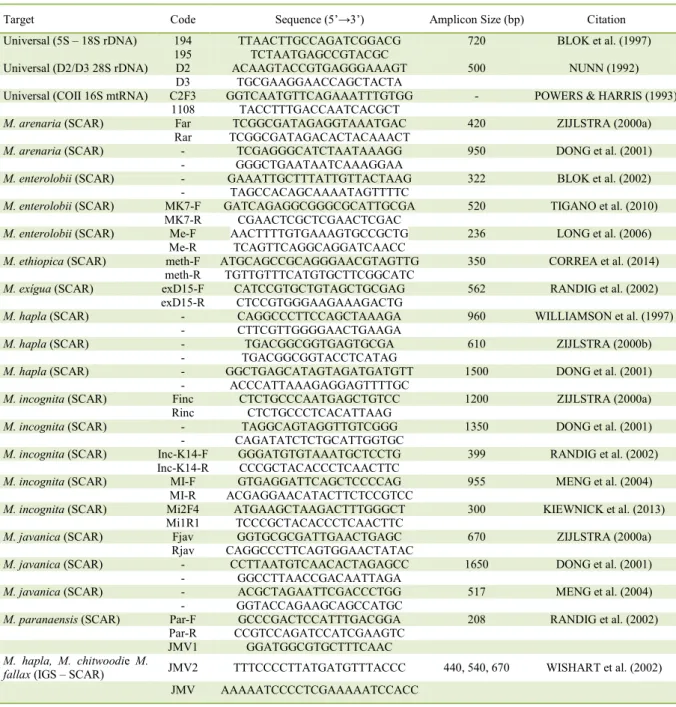
species-specific primer sets have been developed for RKN diagnosis (Table 1). Primer design is crucial for the
success of this approach. Species-specific primers must cover any intra-specific variation and not to
amplify non-target nematodes (ROBERTS et al.,
2016). Several primers designed for identification of
tropical RKN species are based on SCAR
(Sequence-characterized amplified region), including M. arenaria, M. incognita, M. javanica, M. paranaensis,
M. exigua and M. enterolobii (Table 1). SCAR
markers are developed from the characterization and
sequencing of polymorphic bands resulting from
RAPD (Random Amplified Polymorphic DNA)
analysis (CARNEIRO et al., 2016).
Multiplex PCR
Two or more primers sets for different targets can be included in the same PCR reaction.
As a result, amplification products of the distinct
targets are visualized in a single gel, decreasing time, labor and costs. However, this approach requires similar reaction conditions for all the primer sets. The ability to detect species with low population densities in the sample may also be inhibited when a high concentration of DNA from another target is present. In a multiplex reaction, the DNA of Xiphinema elongatum amplified better than M. javanica DNA
and, the latter, amplifies better than Pratylenchus
zeae (BERRY et al., 2008).
Some protocols for multiplex PCR have
already been developed for identification of RKN.
Meloidogyne incognita, M. enterolobii and M.
javanica, for example, can be identified by using
three pairs of specific primers and one universal (HU
et al, 2011). In this case, all species produce bands of approximately 500bp when universal primers
(D2/D3) were used, while specific primers for M.
incognita, M. enterolobii and M. javanica produced bands of approximately 1000, 200 and 700bp, respectively (HU et al., 2011). A multiplex assay can also identify tropical species M. incognita, M. javanica and M. arenaria (KIEWNICK et al., 2013).
Real-Time PCR (qPCR)
Conventional PCR is a qualitative method. A variation of this technique, real-time PCR or
quantitative PCR (qPCR), allows the identification and real-time quantification of target sequences.
qPCR is faster and more sensitive than conventional PCR, quantitative and does not require the preparation of gels. However, the high costs of equipment and reagents are still the main disadvantages of qPCR.
Real-time PCR detects and quantifies DNA based on the emission of fluorescence. The optical unit of the thermocycler monitors fluorescence emitted during cycling and the data is processed by a computer. The cycle threshold, that is, the number of cycles required to initiate the amplification of the target sequence, is then calculated. The two main systems used in the production of fluorescence in qPCR are hydrolysis probes (formerly Taqman®) and SYBR
Green® dye. So far, qPCR-based diagnostics are
available for M. arenaria, M. incognita, M. hapla,
M. enterolobii and M. minor detection (Table 2).
Despite its importance, no qPCR diagnostics is available for M. javanica.
Restriction fragment length polymorphism (RFLP)
In this method, DNA is amplified by PCR
using universal primers and then the products are digested by restriction endonuclease. As nucleotide sequences vary among species, restriction sites differ in their locations along the genome, resulting in fragments of different sizes. Finally, restriction products are separated by gel electrophoresis (SEESSAO et al., 2017). This technique allows
the identification of M. hapla, M. incognita and M. arenaria (HAN et al., 2004). First, the PCR reaction is carried out by using primers that amplify the region between COII and LrRNA of mitochondrial DNA.
Meloidogyne hapla sample will result in a 500bp band,
while a 1.7 kb band is formed for M. incognita and
M. arenaria DNA (HAN et al., 2004). To distinguish between the latter two species, the PCR products are cleaved with the restriction endonucleases Hinf I and Alu I, resulting in fragments of different sizes between M. incognita and M. arenaria, allowing
their identification (HAN et al., 2004). The possible
organisms is a disadvantage associated with PCR-RFLP (ROBERTS et al., 2016).
Loop-mediated isothermal amplification (LAMP)
The loop-mediated DNA amplification technique (LAMP) amplifies DNA with high specificity, sensitivity (up to 10 times more sensitive than PCR), efficiency and speed under isothermal
conditions (NOTOMI et al., 2000). This procedure requires the use of four to six primers (FIP, BIP, F3, B3, with the possibility of one or two additional
primers to increase the amplification efficiency - LB
and LF) and a DNA polymerase (Bst polymerase) with strand displacement activity. The whole procedure
takes from 30 minutes to less than 2 hours, depending
on the protocol, and isothermal conditions (57 - 65ºC) Table 1 - Universal and species-specific primers used in the identification of some Meloidogyne species through the polymerase chain
reaction.
Target Code Sequence (5’→3’) Amplicon Size (bp) Citation
Universal (5S – 18S rDNA) 194 TTAACTTGCCAGATCGGACG 720 BLOK et al. (1997)
195 TCTAATGAGCCGTACGC
Universal (D2/D3 28S rDNA) D2 ACAAGTACCGTGAGGGAAAGT 500 NUNN (1992)
D3 TGCGAAGGAACCAGCTACTA
Universal (COII 16S mtRNA) C2F3 GGTCAATGTTCAGAAATTTGTGG - POWERS & HARRIS (1993) 1108 TACCTTTGACCAATCACGCT
M. arenaria (SCAR) Far TCGGCGATAGAGGTAAATGAC 420 ZIJLSTRA (2000a)
Rar TCGGCGATAGACACTACAAACT
M. arenaria (SCAR) - TCGAGGGCATCTAATAAAGG 950 DONG et al. (2001)
- GGGCTGAATAATCAAAGGAA
M. enterolobii (SCAR) - GAAATTGCTTTATTGTTACTAAG 322 BLOK et al. (2002) - TAGCCACAGCAAAATAGTTTTC
M. enterolobii (SCAR) MK7-F GATCAGAGGCGGGCGCATTGCGA 520 TIGANO et al. (2010) MK7-R CGAACTCGCTCGAACTCGAC
M. enterolobii (SCAR) Me-F AACTTTTGTGAAAGTGCCGCTG 236 LONG et al. (2006) Me-R TCAGTTCAGGCAGGATCAACC
M. ethiopica (SCAR) meth-F ATGCAGCCGCAGGGAACGTAGTTG 350 CORREA et al. (2014) meth-R TGTTGTTTCATGTGCTTCGGCATC
M. exígua (SCAR) exD15-F CATCCGTGCTGTAGCTGCGAG 562 RANDIG et al. (2002) exD15-R CTCCGTGGGAAGAAAGACTG
M. hapla (SCAR) - CAGGCCCTTCCAGCTAAAGA 960 WILLIAMSON et al. (1997)
- CTTCGTTGGGGAACTGAAGA
M. hapla (SCAR) - TGACGGCGGTGAGTGCGA 610 ZIJLSTRA (2000b)
- TGACGGCGGTACCTCATAG
M. hapla (SCAR) - GGCTGAGCATAGTAGATGATGTT 1500 DONG et al. (2001)
- ACCCATTAAAGAGGAGTTTTGC
M. incognita (SCAR) Finc CTCTGCCCAATGAGCTGTCC 1200 ZIJLSTRA (2000a)
Rinc CTCTGCCCTCACATTAAG
M. incognita (SCAR) - TAGGCAGTAGGTTGTCGGG 1350 DONG et al. (2001)
- CAGATATCTCTGCATTGGTGC
M. incognita (SCAR) Inc-K14-F GGGATGTGTAAATGCTCCTG 399 RANDIG et al. (2002) Inc-K14-R CCCGCTACACCCTCAACTTC
M. incognita (SCAR) MI-F GTGAGGATTCAGCTCCCCAG 955 MENG et al. (2004)
MI-R ACGAGGAACATACTTCTCCGTCC
M. incognita (SCAR) Mi2F4 ATGAAGCTAAGACTTTGGGCT 300 KIEWNICK et al. (2013) Mi1R1 TCCCGCTACACCCTCAACTTC
M. javanica (SCAR) Fjav GGTGCGCGATTGAACTGAGC 670 ZIJLSTRA (2000a)
Rjav CAGGCCCTTCAGTGGAACTATAC
M. javanica (SCAR) - CCTTAATGTCAACACTAGAGCC 1650 DONG et al. (2001)
- GGCCTTAACCGACAATTAGA
M. javanica (SCAR) - ACGCTAGAATTCGACCCTGG 517 MENG et al. (2004)
- GGTACCAGAAGCAGCCATGC
M. paranaensis (SCAR) Par-F GCCCGACTCCATTTGACGGA 208 RANDIG et al. (2002) Par-R CCGTCCAGATCCATCGAAGTC
JMV1 GGATGGCGTGCTTTCAAC
M. hapla, M. chitwoodie M.
Table 2 - Primers for the identification of Meloidogyne species by real-time PCR (qPCR) and loop-mediated isothermal amplification (LAMP), including probes for qPCR.
Target Code Sequence (5’→3’) Citation
---qPCR---Ma* QareF TCCATTTCTCCTTGGGTTTG 1
QareR GCCATCCCTTCACACGTTAT
Mi* RKNf GCTGGTGTCTAAGTGTTGCTGATAC 2
RKNr GAGCCTAGTGATCCACCGATAAG
Forward TGGTTCAGGGTCATTTTTCTATAAAGT
Mh Reverse CAAATCGCTGCGTACCAACA 3
Probe FAM-CCATTGGCACTATAAC-MGB
Ment17F TGTGGTGGCTCATTTTCATTA
Me Ment17R AAAAACCCTAAAAATACCCCAAA 4
Probe AGGAGCTG
Mminor_f299 CCGTGACTGAATATGAGGTGA
Mm+ Mminor_r362 GAGGCTCATTAAGTCTTACGATTAT 5
Probe FAM-ATGTTAGGATTATCG-MGBNFQ
RKN* Melo-Rlong GGCCTCACTTAAGAGGCTCA 6
Melo-R short TATACAGCCACGGACGTTCA
---LAMP---RKN-F3 CTGCCCTTTGTACACACC
RKN-B3 GACACCAGCGACAGCCGTT
RKN RKN-FIP CTGCGATTAAATTGGTTTCCATCAACGGGACTGAGCCATTTCG 7
RKN-BIP GCTTGAACCGGGCAAAAGTCCATAAAGTAATGATCCAGCAGC
RKN-LB GTAACAAGGTAGCTGTAGGTGAAC
Mi-F3 TATGTCAGCCCCCGGTTC
Mi Mi-B3 GAGAAGGAAAAGAGTGCCAA 7
Mi-FIP CTTTCCTTGGAATTGGAACAGGGTCAATTGCTTTATATCAAACACC
Mi-BIP GGACGGAGAAGTATGTTCCTCTCTGGAAAAGAAAAATCAGTCTT
Me-F3 CCAAGTACTAAGGAAGCCC
Me-B3 ATCCTAATTTTYCTCCCACACA
Me Me-FIP ACAGTGATTACGACCATACCGCGTTCGTTGCTTAACTTGCCAGA 8
Me-BIP TCTAAGGCAAAGTGGGCGGAGCTCTYTTTGCCTTAAACCATTCCC
Me-LF AAGCACGCCATCCCGTC
Me-LB TGTTGTTCGCTGTTCGC
Mh-F3 GAATATGAGGTGACATGTTAGG
Mh-B3 TCAATGTTTCTGCAGTTCG
Mh Mh-FIP TGAAAAAAATATTGCTGGCGTCCACCTTAATCGGGTTTAAGACT 9
Mh-BIP TCTATCCTTATCGGTGGATCACTCCACAAATTATCGCAGTTAGCT
Mh-LB GGCTCGTGGATCCATGAAGAACG
*SYBR Green system. +Quarantine pest for Brazil. RKN: Universal sets for root-knot nematodes. Targets: Ma – M. arenaria; Me – M.
can be reached by using simple equipment, such as
water baths or heating blocks (NOTOMI et al., 2000). Amplification can be detected through visualization with naked eye, due to the formation of a white
precipitate of magnesium pyrophosphate or the change of color of the solution by using dyes (SYBR Green, calcein, HNB, picogreen). This technique
has been widely studied in many fields of science;
although, few LAMP-based diagnostics for RKN have been developed so far (Table 2).
Sequencing
The availability of complete or partial genome sequences of nematodes is crucial to the development of molecular diagnostic protocols. Partial genome sequences from several Meloidogyne
species have been published in open access databases,
such as GenBank (<http://www.ncbi.nlm.nih.gov>). Complete genomes of M. incognita (ABAD et al., 2008) and M. hapla (OPPERMAN et al., 2008) are also available to scientists. The advent of next generation sequencing (NGS) technologies has reduced cost-per-base and time to decode partial or entire genomes. The number of genomic data tends to grow rapidly, as the new sequencers (2nd, 3rd and 4th generations) are faster than Sanger sequencing.
DNA microarrays
In this technique, a probe (cDNA or
oligonucleotide) is hybridized with a
fluorophore-labeled target molecule, usually a cDNA, produced from the conversion of total RNA or mRNA extracted from the sample. Several targets can be detected simultaneously, without any problems related to
the competition of distinct targets for amplification, reduced specificity, occurrence of false-positives and difficulty of separation of the products in the gel, as
can occur with multiplex PCR (FRANÇOIS et al., 2006). Despite the limited number of studies on the use of DNA microarrays in plant nematology, this
technique has proven to be useful for the identification
of M. chitwoodi (FRANÇOIS et al., 2006) and M.
hapla (VAN DOORN et al., 2007).
CONCLUSION
The classical taxonomy, based on morphological and morphometric studies, and biochemical methods have been widely used in the
identification of RKN species. Due to the limited
number of taxonomic features and the declining interest in classical taxonomy, there is an increasing effort towards the development of molecular-based
diagnostics. In general, molecular approaches are accurate, rapid and do not require only females for diagnosis purposes, such as isozyme phenotypes and perineal patterns. It is hoped that molecular techniques will soon be used more regularly in laboratories worldwide. However, scientists should use the integrative taxonomy approach for accurate diagnosis of RKN species. Molecular, biochemical and morphological data should be combined to enhance the resolution and reliability of studies on phylogenetics and etiology. For practical purposes, the information gathered in this review are expected to be useful for scientists, practitioners, students and other professionals interested in diagnosing RKN. As
a consequence of an accurate identification, control
strategies can be recommended and the losses caused by RKN can be decreased.
ACKNOWLEDGMENTS
E. A. Lopes thanks Conselho Nacional de Desenvolvimento Científico e Tecnológico (CNPq) for the productivity grant (304663/2014-0).
REFERENCES
ABAD, P. et al. Genome sequence of the metazoan plant-parasitic nematode Meloidogyne incognita. Nature Biotechnology, v.8, p.909-915, 2008. Available from: <https://www.researchgate.net/ publication/33549325>. Accessed: Feb. 15, 2017. doi: 10.1038/nbt.1482.
AGUDELO, P. et al. Validation of a real-time polymerase chain
reaction assay for the identification of Meloidogyne arenaria.
Plant Disease, v.95, p.835-838, 2011. Available from: <https://doi. org/10.1094/PDIS-09-10-0668>. Accessed: Dec. 03, 2016. doi: 10.1094/ PDIS-09-10-0668.
ALMEIDA, E.J. et al. New records on Meloidogyne mayaguensis
in Brazil and comparative morphological study with M. incognita.
Nematologia Brasileira, v.32, p.236-241, 2008. Available from: <http://hdl.handle.net/11449/42698>. Accessed: Mar. 03, 2017.
BALDACCI-CRESP, F. et al. Maturation of nematode-induced galls in Medicago truncatula is related to water status and primary
metabolism modifications. Plant Science, v.232, p.77-85, 2015.
Available from: <http://dx.doi.org/10.1016/j.plantsci.2014.12.019>.
Accessed: Sept. 11, 2017. doi: 10.1016/j.plantsci.2014.12.019. BERRY, S.D. et al. Detection and quantification of root-knot nematode (Meloidogyne javanica), lesion nematode (Pratylenchus
zeae) and dagger nematode (Xiphinema elongatum) parasites of sugarcane using real-time PCR. Molecular and Cellular
Probes, v.22, n.3, p.168-176, 2008. Available from: <https://doi. org/10.1016/j.mcp.2008.01.003>. Accessed: Jan. 01, 2017. doi: 10.1016/j.mcp.2008.01.003.
BLOK, V.C. et al. Comparison of sequences from the ribosomal DNA intergenic region of Meloidogyne mayaguensis and other
major tropical root knot nematodes. Journal of Nematology, v.29,
BLOK, V.C. et al. Mitochondrial differences distinguishing
Meloidogyne mayaguensis from the major species of tropical
root-knot nematodes. Nematology, v.4, n.7, p.773-781, 2002. Accessed:
Mar. 09, 2017. doi: 10.1163/156854102760402559.
BLOK, V.C.; POWERS, T.O. Biochemical and molecular
identification. In: PERRY, N.R.; MOENS, M. STARR, J.L. Root-knot
nematodes. Wallingford: CABI Publishing, 2009. Cap.4., p.98-118.
CARNEIRO, R.M.D.G. et al. Enzyme phenotypes of Brazilian populations of Meloidogyne spp. Fundamental and Applied
Nematology, v.19, n.6, p.555-560, 1996. Available from:
<http://horizon.documentation.ird.fr/exl-doc/pleins_textes/
fan/010007637.pdf>. Accessed: Sept. 12, 2017.
CARNEIRO, R.M.D.G.; ALMEIDA, M.R.A. Electrophoresis technique used in the study of root-knot nematode enzymes to identify species Nematologia Brasileira, v.25, n.1 p.35-44, 2001. Available from: <http://docentes.esalq.usp.br/sbn/nbonline/ol%20 251/35-44%20gr.pdf>. Accessed: Dec. 07, 2017.
CARNEIRO, et al. Identification and genetic diversity of
Meloidogyne spp. (Tylenchida: Meloidogynidae) on coffee from Brazil, Central America and Hawaii. Nematology, v.6, n.2, p.287-298, 2004. Accessed: Sept. 12, 2017. doi: 10.1163/1568541041217942.
CARNEIRO, R.M.D.G. et al. Gênero Meloidogyne: diagnose através de eletroforese de isoenzimas e marcadores SCAR. In: OLIVEIRA, C.M.G.; SANTOS, M.A.; CASTRO, L.H.S.
Diagnose de fitonematoides. Campinas: Millennium Editora, 2016. Cap.3, p.47-72.
CHITWOOD, B.G. Root-knot nematodes - Part I.A revision of the genus Meloidogyne Goeldi, 1887. Proceedings of the
Helminthological Society of Washington, v.16, p.90-104, 1949. Available from: <http://docentes.esalq.usp.br/sbn/ajuda/chitw. pdf>. Accessed: Jun. 26, 2017.
CORREA, V.R. et al. Genetic diversity of the root-knot nematode
Meloidogyne ethiopica and development of a species-specific SCAR
marker for its diagnosis. Plant Pathology, v.63, n.2, p.476-483,
2014. Available from: <http://onlinelibrary.wiley.com/doi/10.1111/
ppa.12108/full>. Accessed: Jan. 17, 2017. doi: 10.1111/ppa.12108.
DE LEY, I.T. et al. Phylogenetic analyses of Meloidogyne small subunit rDNA. Journal of Nematology, v.34, n.4, p.319-327, 2002. Available from: <https://www.ncbi.nlm.nih.gov/pmc/ articles/PMC2620593/>. Accessed: Feb. 17, 2017.
DE WEERDT, M. et al. A real-time PCR assay to identify Meloidogyne minor. Journal of Phytopathology, v.159, p.80-84, 2011. Available
from: <https://doi.org/10.1111/j.1439-0434.2010.01717.x>.
Accessed: Dec. 03, 2017. doi: 10.1111/j.1439-0434.2010.01717.x.
DONG, K. et al. Development of PCR primers to identify species
of root-knot nematodes: Meloidogyne arenaria, M. hapla, M.
incognita and M. javanica. Nematropica, v.31, n.2, p.271-280, 2001. Available from: <http://journals.fcla.edu/nematropica/ article/viewFile/69633/67293>. Accessed: Mar. 03, 2017.
ESBENSHADE, P.R.; TRIANTAPHYLLOU, A.C. Isozyme
phenotypes for the identification of Meloidogyne species.
Journal of Nematology, v.22, p.10-15, 1990. Available from: <https://www.ncbi.nlm.nih.gov/pmc/articles/PMC2619005/>. Accessed: Sept. 12, 2017.
FERRAZ, S. et al. Manejo sustentável de fitonematoides. Viçosa: Editora UFV, 2010. 304p.
FERRIS, H. Nemaplex. 1999. Available from: <http:// plpnemweb.ucdavis.edu/nemaplex/Taxamnus/G076mnu.htm>. Accessed: Jun. 27, 2017.
FRANÇOIS, C. et al. Towards specific diagnosis of plant-parasitic nematodes using DNA oligonucleotide microarray technology: a case study with the quarantine species Meloidogyne chitwoodi.
Molecular and Cellular Probes, v.20, n.1, p.64-69, 2006. Available from: <https://doi.org/10.1016/j.mcp.2005.09.004>. Accessed: Feb. 03, 2017. doi: 10.1016/j.mcp.2005.09.004.
FREITAS, L.G. et al. Métodos em nematologia vegetal. In: ALFENAS, A.C.; MAFIA, R.G. Métodos em fitopatologia. Viçosa: Editora UFV, 2016. Cap.11, p. 257-296.
GARCÍA, L.E.; SÁNCHEZ-PUERTA, M.V. Comparative and evolutionary analyses of Meloidogyne spp.based on mitochondrial genome sequences. PLoS ONE, v.10, e0121142, 2015. Available from: <https://doi.org/10.1371/journal.pone.0121142>. Accessed: Apr. 19, 2017. doi: 10.1371/journal.pone.0121142.
HAN, H. et al. PCR-RFLP identification of three major Meloidogyne
species in Korea. Journal of Asia-Pacific Entomology, v.7, n.2, p.171-175, 2004. Available from: <https://doi.org/10.1016/S1226-8615(08)60212-5>. Accessed: Feb. 03, 2017.
HU, M.X. et al. Multiplex PCR for the simultaneous identification and detection of Meloidogyne incognita, M. enterolobii and M. javanica
using DNA extracted directly from individual galls. Phytopathology,
v.101, n.11, p.1270-1277, 2011. Available from: <http://apsjournals.
apsnet.org/doi/pdf/10.1094/PHYTO-04-11-0095>. Accessed: Jan. 09 2017. doi: 10.1094/ PHYTO-04-11-0095.
HUNT, D.J.; HANDOO, Z.A. Taxonomy, identification and principal species. In: PERRY, R.N.; MOENS, M.; STARR, J.L.
Root-knot nematodes. Wallingford: CAB International, 2009. Cap.3, p.55-88.
KIEWNICK, S. et al. Identification of the tropical
root-knot nematode species Meloidogyne incognita, M. javanica
and M. arenaria using a multiplex PCR assay. Nematology, v.15, p.891-894, 2013. Accessed: Feb. 17, 2017. doi: 10.1163/15685411-00002751.
KIEWNICK, S. et al. Comparison of two short DNA barcoding loci (COI and COII) and two longer ribosomal DNA genes (SSU & LSU rRNA) for specimen identification among quarantine root-knot nematodes (Meloidogyne spp.) and their close relatives. European
Journal Plant Pathology, v.140, p.97-110, 2014. Available from: <https://link.springer.com/content/pdf/10.1007/s10658-014-0446-1. pdf>. Accessed: Dec. 12, 2016. doi: 10.1007/s10658-014-0446-1.
KIEWNICK, S. et al. Development and validation of LNA-based quantitative real-time PCR assays for detection
and identification of the root-knot nematode Meloidogyne enterolobii in complex DNA backgrounds. Phytopathology, v.105, n.9, p.1245-1249, 2015. Available from: <http://dx.doi. org/10.1094/PHYTO-12-14-0364-R>. Accessed: Feb. 17, 2017. doi: 10.1094/PHYTO-12-14-0364-R.
LONG, H. et al. Development of a PCR diagnostic for the
root-knot nematode Meloidogyne enterolobii. Acta Phytopathologica
Sinica, v.36, p.109-115, 2006. Accessed: Mar. 05, 2017.
LOPES, E.A.; FERRAZ, S. Importância dos fitonematoides na agricultura. In: OLIVEIRA, C.M.G.; SANTOS, M.A.; CASTRO, L.H.S. Diagnose de fitonematoides. Campinas: Millennium Editora, 2016. Cap.1, p.1-10.
MENG, Q.P. et al. PCR assays for rapid and sensitive identification
of three major root-knot nematodes, Meloidogyne incognita, M.
javanica and M. arenaria. Acta Phytopathologica Sinica, v.34, p.204-210, 2004. Accessed: Mar. 11, 2017.
MOENS, M. et al. Meloidogyne species – A diverse group of novel and important plant parasites. In: PERRY, R.N.; MOENS, M.; STARR, J.L. Root-knot nematodes. Wallingford: CAB International, 2009. Cap.1, p.1-17.
NIU, J.H. et al. Evaluation of loop-mediated isothermal amplification (LAMP) assays based on 5S rDNA-IGS2 regions for detecting Meloidogyne enterolobii. Plant Pathology, v.61,
p.809-819, 2012. Available from: <https://www.researchgate.
net/publication/262890929>. Accessed: Jan. 15, 2017. doi: 10.1111/j.1365-3059.2011.02562.x.
NIU, J.H. et al. Rapid detection of Meloidogyne spp. by LAMP assay in soil and roots. Crop Protection, v.30, n.8, p.1063-1069, 2011. Available from: <https://doi.org/10.1016/j.cropro.2011.03.028>. Accessed: Feb. 17, 2017. doi: 10.1016/j.cropro.2011.03.028. NOTOMI, T. et al. Loop-mediated isothermal amplification of DNA. Nucleic Acids Research, v.28, n.12, p.e63, 2000. Available from: <https://www.ncbi.nlm.nih.gov/ pubmed/10871386>. Accessed: May 16, 2017.
NUNN, G. Nematode molecular evolution. An investigation of evolutionary patterns among nematodes based upon DNA
sequences. 1992. Ph.D. dissertation, University of Nottingham, UK, 1992. Available from: <http://ethos.bl.uk/OrderDetails. do?uin=uk.bl.ethos.334855>. Accessed: Mar. 15, 2017.
OLIVEIRA, C.M.G. et al. Morphological and molecular diagnostics for plant parasitic nematodes: working together to
get the identification done. Tropical Plant Pathology, v.36, n.2,
p.65-73, 2011. Available from:
<http://dx.doi.org/10.1590/S1982-56762011000200001>. Accessed: Apr. 04, 2017.
ONKENDI, E.M.; MOLELEKI, L.N. Detection of Meloidogyne enterolobii in potatoes in South Africa and phylogenetic analysis based on intergenic region and the mitochondrial DNA sequences.
European Journal of Plant Pathology, v.136, p.1-5, 2013. Available
from: <
https://link.springer.com/article/10.1007/s10658-012-0142-y>. Accessed: Sept. 13, 2017. doi: 10.1007/s10658-012-0142-y.
OPPERMAN, C.H. et al. Sequence and genetic map of Meloidogyne hapla: A compact nematode genome for plant parasitism. Proceedings
of the National Academy of Sciences, v.105, p.14802-14807, 2008. Available from: <http://www.pnas.org/content/105/39/14802.full. pdf>. Accessed: Feb. 15, 2017. doi: 10.1073/pnas.0805946105.
ORUI, Y. Species identification of Meloidogyne spp. (Nematoda:
Meloidogynidae) in Japan by random amplified polymorphic DNA
(RAPD-PCR). Japanese Journal of Nematology, v.29, p.7-15, 1999. Available from: <http://doi.org/10.3725/jjn1993.29.2_7>. Accessed: May 27, 2017.
PENG, H. et al. Rapid, simple and direct detection of Meloidogyne hapla from infected root galls using loop-mediated isothermal
amplification combined with FTA technology. Scientific Reports,
v.7, Article number 44853, 2017. Available from: <https://www. ncbi.nlm.nih.gov/pmc/articles/PMC5377304/>. Accessed: May 24, 2017. doi: 10.1038/srep44853.
POWERS, T.O.; HARRIS, T.S.A polymerase chain reaction
method for identification of five major Meloidogyne species.
Journal of Nematology, v.25, p.1-6, 1993. Available from: <https://www.ncbi.nlm.nih.gov/pmc/articles/PMC2619349/>. Accessed: Jan. 14, 2017.
RANDIG, O. et al. A species-specific satellite DNA family in the genome of the coffee root nematode Meloidogyne exigua: application to molecular diagnosis of the parasite. Molecular
Plant Pathology, v.3, p.431-437, 2002. Available from: <http:// onlinelibrary.wiley.com/doi/10.1046/j.1364-3703.2002.00134.x/ full>. Accessed: Dec. 12, 2016. doi: 10.1046/j.1364-3703.2002.00134.x.
ROBERTS, D. et al. Diagnose molecular de nematoides parasitos de plantas. In: OLIVEIRA, C.M.G.; SANTOS, M.A.; CASTRO, L.H.S. Diagnose de fitonematoides. Campinas: Millennium Editora, 2016. Cap. 14, p.281-324.
SALGADO, S.M.L. et al. Meloidogyne paranaensis e
Meloidogyne exigua in coffee plantations from the southern region of Minas Gerais. Coffee Science, v.10, n.4, p.475-481, 2015. Available from: <https://www.researchgate.net/ publication/283765581_Meloidogyne_paranaensis_and_ meloidogyne_exigua_in_coffee_plantations_from_the_south_ of_Minas_Gerais_State>. Accessed: Sept. 12, 2017.
SAPKOTA, R. et al. A TaqMan real-time PCR assay for detection of Meloidogyne hapla in root galls and in soil. Nematology, v.18,
p.147-154, 2016. Available from: <https://www.researchgate.net/
publication/284712140_A_TaqMan_real-time_PCR_assay_for_ detection_of_Meloidogyne_hapla_in_root_galls_and_in_soil>. Accessed: Jan. 01, 2017. doi: 10.1163/15685411-00002950.
SEESSAO, Y. et al. A review of methods for nematode identification.
Journal of Microbiological Methods, v.138, p.37-49, 2017. Available from: <http://dx.doi.org/10.1016/j.mimet.2016.05.030 0167-7012/>. Accessed: Sept.05, 2017. doi: 10.1016/j.mimet.2016.05.030.
SIDDIQUI, Y. et al. Histopathological changes induced by
Meloidogyne incognita in some ornamental plants. Crop
Protection, v.65, p.216-220, 2014. Available from: <http://dx.doi.
org/10.1016/j.cropro.2014.08.001>. Accessed: Sept.05, 2017.
TIGANO, M.S. et al. Genetic diversity of the root-knot nematode
Meloidogyne enterolobii and development of a SCAR marker
for this guava-damaging species. Plant Pathology, v.59, n.6, p.1054-1061, 2010. Available from: <https://www.researchgate.
net/publication/229721445_Genetic_diversity_of_the_root-knot_
nematode_Meloidogyne_enterolobii_and_development_of_a_
SCAR_marker_for_this_guava-damaging_species>. Accessed: Nov. 13, 2017. doi: 10.1111/j.1365-3059.2010.02350.x.
VAN DOORN, R. et al. Quantitative multiplex detection of plant pathogens using a novel ligation probe-base system coupled with universal, high-throughput real-time PCR and Open Arrays.
BMC Genomics, v.8, p.276, 2007. Available from: <http://www. biomedcentral.com/1471-2164/8/276>. Accessed: Feb. 15, 2017. doi: 10.1186/1471-2164-8-276.
VERONICO, P. et al. Molecular dissection of the rDNA array and of the 5S rDNA gene in Meloidogyne artiellia: phylogenetic and diagnostic implications. Molecular and Cellular Probes, v.18, n.3, p.177-183, 2004. Available from: <https://doi.org/10.1016/j. mcp.2003.12.003>. Accessed: Dec. 13, 2016. doi: 10.1016/j. mcp.2003.12.003.
WILLIAMSON, V.M. et al. A PCR assay to identify and distinguish single juveniles of Meloidogyne hapla and
M. chitwoodi. Journal of Nematology, v.29, n.1, p.9-15,
1997. Available from: <http://journals.fcla.edu/jon/article/
view/66873/64541>. Accessed: Dec. 03, 2016.
WISHART, J. et al. Ribosomal intergenic spacer: a PCR diagnostic for Meloidogyne chitwoodi, M. fallax and M. hapla. Phytopathology,
v.92, n.8, p.884-892, 2002. Available from: <http://apsjournals. apsnet.org/doi/pdf/10.1094/PHYTO.2002.92.8.884>. Accessed: Feb. 17, 2017. doi: 10.1094/PHYTO.2002.92.8.884.
ZIJLSTRA, C. et al. Differences between ITS regions of isolates
of root-knot nematodes Meloidogyne hapla and M. chitwoodi.
Phytopathology, v.85, p.1231-1237, 1995. Available from: <https://www.jstage.jst.go.jp/article/jjn1993/29/2/29_2_7/_pdf>. Accessed: Sept. 13, 2017.
ZIJLSTRA, C. et al. Identification of Meloidogyne incognita, M.
javanica and M. arenaria using sequence characterized amplified
regions (SCAR) based PCR assays. Nematology, v.2, n.8, p.847-853, 2000a. Accessed: Dec. 03, 2017. doi: 10.1163/156854100750112798.
ZIJLSTRA, C. Identification of Meloidogyne chitwoodi, M. fallax
and M. haplabased on SCAR–PCR: a powerful way of enabling reliable identification of populations or individuals that share common traits. European Journal of Plant Pathology, v.106,
n.3, p.283-290, 2000b. Available from: <https://link.springer.com/