GLUCAGON-LIKE PEPTIDE 1: BIOCHEMISTRY,
SECRETION AND MAIN PHYSIOLOGICAL EFFECTS
Amélia M. Silva
Assistant professor
Department of biology and environment – utAD
centre for the research and technology of Agro-environment and biological sciences – utAD [email protected]
Carla Martins Lopes
Assistant professor
Faculty of Health sciences – uFp
institute of biotechnology and bioengineering, center of genetics and biotechnology – utAD [email protected]
Stanley Misler
Associate professor
Department of internal medicine, Washington university school of medicine, st. louis, mo, usA [email protected]
Gregory D. Cooper
research biomedical engineer
Department of internal medicine, Washington university school of medicine, st. louis, mo, usA [email protected]
Tatiana Andreani
student
center of Health sciences, Department of pharmacy and pharmacology – university estadual do maringá, brazil [email protected]
Eliana B. Souto
Assistant professor
Faculty of Health sciences – uFp
105
RESUMO
o “glucagon-like peptide 1” (glp-1), hormona produzida nas células-l intestinais por proces-samento diferencial do proglucagon, é libertado após ingestão de alimentos. efeitos de glp-1 observam-se, essencialmente, a nível gastrintestinal e pancreático resultando da sua acção directa, ligação ao receptor nas células alvo, ou indirecta, por regulação parácrina. pelo seu papel na regulação da ingestão de alimentos e na secreção de insulina induzida por glicose, agonistas do receptor de glp-1 são alvo de estudos para a terapia da obesidade e diabetes.
PALAVRAS-CHAVE
Hormona peptídica; “glucagon-like peptide 1”, glp-1, incretina, Diabetes mellitus tipo 2, obesidade
ABSTRACT
the peptide hormone glucagon-like peptide 1 (glp-1), produced in the intestinal l-cells by differential processing of proglucagon, is secreted in response to meal intake. glp-1 affects various systems, the gastrointestinal and pancreatic systems being the best studied, either by direct binding to the glp-1 receptor, at the target-cells surface, or indirectly as a result of paracrine regulation. because of glp-1’s roles, in augmenting glucose-induced insulin secretion and modulating food intake, currently glp-1 receptor agonists are being studied for diabetes and obesity therapy.
KEYWORDS
peptide hormone; glucagon-like peptide 1, glp-1, incretin, Diabetes mellitus type 2, obesity
colleagues reported endocrine cells of the intestinal mucosa that stained with antibodies to glucagon (orci et al., 1968), while unger and colleagues reported that a glucagon-like immunoreactive material, physico-chemically and biologically distinct from glucagon, was secreted by the intestine in response to an oral glucose challenge (unger et al., 1968). by this time, solid evidence accumulated that up to twice as much insulin is secreted after increasing plasma glucose by ingestion of glucose than after a similar increase in plasma glucose in response to intravenous injection - the so-called “incretin effect of the gut” and evidence began to emerge that the “incretin effect” was severely blunted in type 2 diabetes mellitus (perley & Kipnis, 1967). by 1985, a glucagon-like peptide (glp), along with a second similar peptide gip, alternately called gastrin inhibitory or glucose-dependent insulinotropic polypeptide, were seriously considered as candidate incretins. Altogether, this suggested that a supra-physiological dose of glp might serve as a therapeutic enhancer of insulin secretion in type 2 diabetic patients who were those hyperglycemic in spite of apparently adequate insulin stores.
Here we review more recent studies characterizing the glucagon-like peptide 1 biochemi-cally and physiologibiochemi-cally and indicate its potential utility in the therapy of obesity as well as diabetes mellitus.
2.
BIOCHEMISTRY AND PHYSIOLOGY OF GLP-1
2.1.
PROGLUCAGON AND ITS PROCESSING BY
PANCREATIC AND INTESTINAL TISSUE
proglucagon is the main pro-hormone protein product of two distinct endocrine cell types, the pancreatic alpha-cell and the intestinal mucosa l-cell. During the maturation of these two cell types, a single proglucagon gene is activated. However with further cell differen-tiation, post-translationally the 160 amino acid proglucagon protein precursor undergoes differential proteolytic processing by secretory granules convertases at distinct dibasic resi-dues (see Figure 1). the alpha-cells cleave glucagon from the region spanning amino acids (aas) 33 to 61 and then release it along with the major proglucagon fragment (mpgF) (Holst et al., 1994). in contrast, l-cells cleave two structurally related glps from c-terminally located portions of the precursor molecule, namely glp-1, from the region spanning aas 78 to 107, and glp-2 from region spanning aas 126 to 158 (mojsov et al., 1986). l-cells also process and secrete glicentin from the region spanning aas 1-69, and oxyntomodulin, a c-terminally ex-tended glucagon, from the region spanning aas 33-69 (Ørskov, 1992; Holst, 2007).
107
Figure 1. posttranslational processing of proglucagon in mammalian pancreatic alpha-cells and small intestinal l-cells. the proglucagon is a 160-amino acid peptide (pg 1-160), where 1 indicates the n-terminus and 160 the c-terminus amino acid and the vertical lines indicate positions of the basic amino acid residues that are typical cleavage sites. the peptide products are represented in boxes and marked according to their position in the proglucagon sequence. (Adapted from Ørskov, 1992; Holst, 2007).
2.2.
GLP-1 DEGRADATION
the catalytic enzyme dipeptidyl peptidase iV (Dpp-iV; Dp iV; cD 26) is a 766 amino acid, membrane-associated ecto-peptidase that is widely distributed in numerous tissues (e.g. lumenal membranes of capillary endothelial cells, the apical membranes of kidney tubule cells, the plasma membranes of hepatocytes, blood). this enzyme also exists as a soluble circulating form in plasma and significant Dpp-iV-like activity is detectable in plasma from humans and rodents. Dpp-iV has substrate specificity for oligopeptides with a penultimate prolyl-, analyl-, or seryl-, residue at their n-termini. in the presence of this Dpp-iV the n-termi-nus dipeptide of a number of metabolic hormones and neuroendocrine factors are cleaved,
the order of catalytic efficiency being neuropeptide Y (npY)
>
peptide YY (pYY) > glp-1 > gip > glucagon (Drucker, 2003). since an intact n-terminus is obligatory for the biological activity of the members of the glucagon/Vip peptide family, Dpp-iV inactivates these pepti-de hormones. in the case of glp-1, the metabolites generated, namely glp-1 (9-36) amipepti-de from glp-1 (7-36) and glp-1 (9-37) from glp-1 (7-37), are not only inactive, they may act as competitive antagonists of the intact glp-1 at the glp-1 receptors (Knudsen & pridal, 1996).bably their interaction with the l-cells, stimulates glp-1 secretion. the l-cells response is dependent on the meal size and is highly correlated with the rate of the gastric emptying (Wachters-Hagedoorn et al., 2006; gribble, 2008). plasma glp-1 remains elevated for a consi-derable period of time after feeding cessation, indicating its continued secretion.
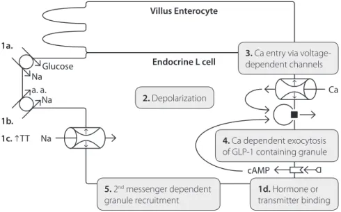
mechanistically, stimulus-secretion coupling in l-cells is unclear. However, cell lines derived from l-cells depolarize, fire action potentials and display ca2+-entry dependent exocytosis
of glp-1 (gribble, 2008). possible routes for stimulus-depolarization coupling include na+
coupled glucose and/or amino acid transport, glucose metabolism resulting in closure of Atp dependent K+ (KAtp) channels and increased luminal osmolarity causing cell shrinkage
and opening of stretch inactivated cation channels. more distally, depolarization-exocytosis coupling might be enhanced by signals from the proximal gut that may increase cytosolic cAmp and enhance the supply of release-ready glp-1 granules (see Figure 2).
Villus Enterocyte
Endocrine L cell Na
Ca Na
Na
cAMP Glucose
1a.
1b. a. a.
1c. TT
3. ca entry via voltage-dependent channels
1d. Hormone or transmitter binding
5. 2nd messenger dependent
granule recruitment
4. ca dependent exocytosis of glp-1 containing granule
2. Depolarization
Figure 2. two possible pathways for stimulus-secretion coupling in intestinal glp-1 secreting l-cells. left: apical glucose, amino acid and osmolar stimulation (1a-c) of depolarization (2) and triggering of ca2+-entry (3) and of
ca2+-dependent exocytosis of glp-1 containing granules (4). right: basolateral synaptic transmitter or hormone
109
2.4.
GLP-1 RECEPTORS AND CELL SIGNALING
the glp-1 receptor was first cloned in 1992 (thorens, 1992). it is a class b gpcr, i.e., one of the group of 15 receptors (in the human genome), including gip and the glucagon recep-tors, that are activated by intermediate sized peptides (typically ~30-40 amino acid resi-dues) (mayo et al., 2003). the glp-1 receptor is coupled, functionally, to the adenylate cyclase (Drucker et al., 1987) via the stimulatory g protein gs (mayo et al., 2003; thorens, 1992). in pancreatic beta-cells, the activation of the glp-1 receptor leads to the increase in the cyto-solic [cAmp] with a subsequent activation of the protein kinase A (pKA) and the cAmp-re-gulated guanine nucleotide exchange factor ii (cAmp-geFii, also known as epac2) leading to a plethora of events (e.g. altered ion channel activity, intracellular calcium handling, and enhanced exocytosis of insulin-containing granules (silva et al., 2009)) that culminate in an enhancement of glucose-induced insulin secretion (for review see Holst & gromada, 2004; mayo et al., 2003; Holst, 2007).
glp-1 receptors were first found in pancreatic islets, stomach and lung (on rat and rat insu-linoma cell-line, ins-1) (thorens, 1992). in 2003, glp-1 receptors were detected in hypotha-lamus and brain stem, heart and kidney, but not in liver, skeletal muscle or adipose tissue (mayo et al., 2003). more specifically, using fluorescence immunohistochemical microsco-py, glp-1 receptors have been selectively localized to beta-cells of the islet and pancreatic ducts, in a study on mice, rat and human tissues (tornehave et al., 2008).
2.4.1.
THE GLP-1 RECEPTOR PHARMACOLOGY
Agonists for the glp-1 receptor include glp-1(7–37), glp-1 (7–36)amide (kd = 0.3 nm), the heloderma suspectum peptides exendin-3 and exendin-4 (kd = 0.1 nm) (naturally occurring gila monster peptide from salivary secretion (eng et al., 1992)) and some labeled ligands (e.g. fluorescein-trp25-exendin-4, 125iglp-1, and tyr39-exendin-4). structurally related members
of the glucagon family such as glp-2, glucagon, and gip do not activate the glp-1 receptor at physiologically relevant concentrations (mayo et al., 2003; goke et al., 1993).
Antagonists for the glp-1 receptor include the truncated lizard peptide glp-1 receptor anta-gonist exendin-(9–39) (kd of 2.9 nm) (goke et al., 1993) and a small non-peptide ligand (t-0632), that binds the glp-1 receptor within the micromolar range, exhibiting about ~100-fold selecti-vity for the human versus the homologous rat glp-1 receptor (tibaduiza et al., 2001).
2.5.
MAIN PHYSIOLOGICAL EFFECTS OF GLP-1
humans, as well as does the truncated glp-1 (a naturally occurring peptide) being this more potent than glp-1 (schjoldager et al., 1989). it decreases and delays gastric emptying rate by stimulating antral churning while inhibiting pyloric propulsion and duodenal peristalsis (schirra et al., 2006). it inhibits, significantly, the postprandial pancreatic secretion of trypsin and lipase, an effect that seemed to be secondary to gastric emptying as truncated glp-1 did not affect the linear relationship that correlates pancreatic enzyme output to gastric emptying (Wettergren et al., 1993).
lastly, glp-1 suppresses appetite either by reducing gastric emptying and inducing stoma-ch fullness or by activating satiety centers on the arcuate nucleus of the hypothalamus or inhibiting the solitary tract nucleus of the brain stem (Holst, 2007).
111
3.
THERAPEUTIC USE IN TYPE 2 DIABETES
MELLITUS ASSOCIATED WITH OBESITY
the acute actions of glp-1 to slow absorption of ingested glucose, “amplify” glucose-indu-ced insulin secretion and inhibit glucagon secretion, combined with its chronic action to maintain beta-cell mass, suggest glp-1 as a useful agent in the treatment of type 2 diabetes mellitus (t2-Dm), where glucose insensitivity of both beta-cells and peripheral target cells produces chronic hyperglycemia and ongoing beta-cells injury. to overcome the impedi-ment of very short (~5 min) circulating half-life of glp-1, three new approaches are now in development / early clinical use: (i) gpl-1 agonists or mimetics = long acting recombinant glp-1 analogues (e.g., intravenous administered exenatide, a synthetic exendin-4); (ii) glp-1 enhancers = inhibitors of Dpp-iV (e.g., orally administered sitagliptin); and (iii) stimulators of glp-1 release by l-cells (Ar231453, in trial). in addition, the appetite suppressing effects of glp-1 might also treat obesity-related t2-Dm, where chronically increased caloric intake, overstuffs adipocytes which chronically over-release free fatty acids (ffas). When taken up beta-cells and their peripheral targets ffas further contribute to progressive glucose-insensi-tivity and beta-cells failure. in therapeutic trials, twice daily subcutaneous administration of exenatide has produced 2-5 kg weight loss for up to 3 years (DeFronzo et al., 2005).
4.
CONCLUSIONS
Hence, by the various effects on beta-cells function, glp-1, glp-1 mimetics or enhancers, show improvements on glucose homeostasis in t2-Dm when added to other oral hypogly-cemic agents. improved methods for administration of these agents may be the key to their expanded and more efficacious usage (chia & egan, 2008).
REFERENCES
CHIA, C.W. AND EGAN, J.M. (2008). incretin-based therapies in type 2 diabetes mellitus. in: the Journal of clinical endocrinology and Metabolism. 93 (10), pp. 3703-3716.
DEFRONZO, R.A., RATNER, R.E., HAN, J., KIM, D.D., FINEMAN, M.S. AND BARON, A.D. (2005). effects of exenatide (exendin-4) on glycemic control and weight over 30 weeks in metformin-treated patients with type 2 diabetes. in: Diabetes care, 28 (5), pp. 1092-1100. DRUCKER, D.J. (2003). therapeutic potential of dipetidyl peptidase iV inhibitors for the treat-ment of type 2 diabetes. in: expert opinion on investigational Drugs 12 (1), pp. 87-100. DRUCKER, D.J. AND ASA, S. (1988). glucagon gene expression in Vertebrate brain. in: Jour-nal of biological chemistry, 263 (27), pp. 13475-13478.
DRUCKER, D.J., PHILIPPE, J., MOJSOV, S. AND CHICK, W.L. (1987). glucagon-like peptide i stimulates insulin gene expression and increases cyclic Amp levels in a rat islet cell line. in: proceedings of the national academy of sciences usa, 84 (10), pp. 3434-3438.
ENG, J., KLEINMAN, W.A., SINGH, L. AND RAUFMAN, J.P. (1992). isolation and characteri-zation of exendin-4, an exendin-3 analogue from heloderma suspectum venom. in: Journal of biological chemistry, 267 (11), pp. 7402-7405.
HOLST, J.J. AND GROMADA, J. (2004). role of incretin hormones in the regulation of insulin secretion in diabetic and nondiabetic humans. in: american Journal of physiology, endocrino-logy and Metabolism, 287, pp. e199-e206.
HOLZ, G.G., KüHTREIBER, W.M. AND HABENER, J.F. (1993). pancreatic beta-cells are ren-dered glucose-competent by the insulinotropic hormone glucagon-like peptide-1(7-37). in: nature 361, pp. 362-365.
KNUDSEN, L.B. AND PRIDAL, L. (1996). glucagon-like peptide-1-(9-36) amide is a major metabolite of glucagon-like peptide-1-(7-36) amide after in vivo administration to dogs, and it acts as an antagonist on the pancreatic receptor. in: european of Journal pharmacology, 318 (2-3), pp. 429-435.
MAYO, K.E., MILLER, L.J., BATAILLE, D., DALLE, S., GOKE, B., THORENS, B. AND DRUCKER, D.J. (2003). international union of pharmacology. XXXV. the glucagon receptor Family. in: pharmacological reviews, 55 (1), pp. 167-194.
MOJSOV, S., HEINRICH, G., WILSON, I.B., RAVAZZOLA, M., ORCI, L. AND HABENER, J.F. (1986). preproglucagon gene expression in pancreas and intestine Diversifies at the level of post-translational processing. in: Journal of biological chemistry, 261 (25), pp. 11880-11889. MURLIN, J.R., CLOUGH, H.D., GIBBS, C.B.F. AND STOKES, A.M. (1923). Aqueous extracts of the pancreas. i. influence of the carbohydrate metabolism of depancreatized animals. in: Journal of biological chemistry, 56 (1), pp. 253-296.
ORCI, L., PICTET, R., FORSSMAN, W.G., RENOLD, A.E. AND ROUILLER, C. (1968). structural evidence for glucagon producing cells in the intestinal mucosa of the rat. in: Diabetologia, 4 (1), pp. 56-67.
ØRSKOV, C. (1992). glucagon-like peptide-l, a new hormone of the entero-insular axis. in: Diabetologia, 35 (8), pp. 701-711.
ØRSKOV, C., WETTERGREN, A. AND HOLST, J.J. (1996). secretion of the incretin hormones glucagon-like peptide-1 and gastric inhibitory polypeptide correlates with insulin secretion in normal man throughout the day. in: scandinavian Journal of Gastroenterology, 31 (7), pp. 665-670.
PERLEY, M.J. AND KIPNIS, DM (1967). plasma insulin responses to oral and intravenous glucose: studies in normal and diabetic subjects. in: Journal of clinical investigation 46, pp. 1954-1962.
SCHIRRA, J., NICOLAUS, M., ROGGEL, R., KATSCHINSKY, M., STORR, M., WOERLE, H.J. AND GOKE, B. (2006). endogenous glucagon-like peptide 1 controls endocrine pancreatic secretion and antro-pyloroduodenal motility in humans. in: Gut, 55(2), pp. 243-251. SCHJOLDAGER, B.T., MORTENSEN, P.E., CHRISTIANSEN, J., ORSKOV, C. AND HOLST, J.J. (1989). glp-1 (glucagon-like peptide 1) and truncated glp-1, fragments of human proglu-cagon, inhibit gastric acid secretion in humans. in: Digestive Diseases and sciences, 34 (5), pp. 703-708.
113
SUTHERLAND, E.W. AND DE DUVE, C. (1948). origin and distribution of thehyperglyce-mic-glycogenolitic factor of the pancreas. in: Journal of biological chemistry, 175 (2), pp. 663-674.
THORENS, B. (1992). expression cloning of the pancreatic b cell receptor for the gluco-in-cretin hormone glucagon-like peptide 1. in: proceedings of the national academy of sciences usa, 89 (18), pp. 8641-8645.
TIBADUIZA, E.C., CHEN, C. AND BEINBORN, M. (2001). A small molecule ligand of the glucagon-like peptide 1 receptor targets its Amino-terminal Hormone binding Domain. in: Journal of biological chemistry, 276 (41), pp. 37787-37793.
TORNEHAVE, D., KRISTENSEN, P., ROMER, J., KNUDSEN, L.B. AND HELLER, R.S. (2008). expression of the glp-1 receptor in mouse, rat, and Human pancreas. in: Journal of histoche-mistry and cytochehistoche-mistry 56 (9), pp. 841-851.
UNGER, R.H., OHNEDA, A., VALVERDE, I., EISENTRAUT, A.M. AND EXTON, J. (1968). cha-racterization of the responses of circulating glucagon-like immunoreactivity to intraduode-nal and intravenous administration of glucose. in: the Jourintraduode-nal of clinical investigation, 47 (1), pp. 48-65.
WACHTERS-HAGEDOORN, R.E., PRIEBE, M.G., HEIMWEG, J.A.J., HEINER, A.M., ENGLYST, K.N., HOLST, J.J., STELLAARD, F. AND VONK, R.J. (2006). the rate of intestinal glucose Ab-sorption is correlated with plasma glucose-Dependent insulinotropic polypeptide concen-trations in Healthy men. in: the Journal of nutrition 136 (6), pp. 1511-1516.
WETTERGREN, A., SCHJOLDAGER, B., MORTENSEN, P.E., MYHRE, J., CHRISTIANSEN, J. AND HOLST, J.J. (1993). truncated glp-1 (proglucagon 78-107-amide) inhibits gastric and pancreatic functions in man. in: Digestive Diseases and sciences, 38 (4), pp. 665-673.