Histopathology, parasite density and cell phenotypes of the
popliteal lymph node in canine visceral leishmaniasis
Rodolfo Cordeiro Giunchetti
a,b,*
, Olindo Assis Martins-Filho
c,
Cla´udia Martins Carneiro
a,d, Wilson Mayrink
e, Marcos Jose´ Marques
f,
Washington Luiz Tafuri
a, Rodrigo Correˆa-Oliveira
b, Alexandre Barbosa Reis
a,b,d,*
aLaborato´rio Imunopatologia, Nu´cleo de Pesquisas em Cieˆncias Biolo´gicas/NUPEB, Instituto de Cieˆncias Exatas e Biolo´gicas,
Universidade Federal de Ouro Preto, Ouro Preto, Minas Gerais, Brazil
bLaborato´rio de Imunologia Celular e Molecular, Instituto de Pesquisas Rene´ Rachou, Fundac¸a˜o Oswaldo Cruz, Belo Horizonte,
Minas Gerais, Brazil
cLaborato´rio de Doenc¸a de Chagas, Instituto de Pesquisas Rene´ Rachou, Fundac¸a˜o Oswaldo Cruz, Belo Horizonte,
Minas Gerais, Brazil
dDepartamento de Ana´lises Clı´nicas, Escola de Farma´cia, Universidade Federal de Ouro Preto, Ouro Preto, Minas Gerais, Brazil eLaborato´rio de Leishmanioses, Departamento de Parasitologia, Instituto de Cieˆncias Biolo´gicas,
Universidade Federal de Minas Gerais, Belo Horizonte, Minas Gerais, Brazil
fLaborato´rio de Biologia Molecular e Biotecnologia, Universidade Federal de Alfenas, Alfenas, Minas Gerais, Brazil
Received 6 May 2007; received in revised form 16 July 2007; accepted 18 July 2007
Abstract
While enlargement of popliteal lymph nodes (LN) is frequently described in canine visceral leishmaniasis (CVL), there are few histopathologic studies of lymph nodes during this chronic immunopathological condition. Besides a detailed histopathologic analysis, we have characterized the parasite load and major immunophenotypic features of the LN inLeishmania(Leishmania)chagasi-infected dogs. Our major histopathological findings highlight that hypertrophy/hyperplasia of LN cortical and medullary zones was the principal characteristic observed in asymptomatic dogs (AD), whereas atrophy of LN cortical zone was predominant in symptomatic animals (SD). The LN parasite density detected by anti-Leishmaniaimmunohistochemical assay or expressed as Leishman Donovan Units was also highly correlated with the skin parasitism, the most reliable parameter to decode the clinical status of CVL. The major LN immunophenotypic changes during ongoing CVL were an increased frequency of T-lymphocytes, particularly CD8+T-cells, up-regulation of MHC-II expression by lymphocytes and decreased levels of CD21+B-cells. Our findings further demonstrated that changes in the LN B-lymphocyte compartment exhibited a negative correlation with the skin parasite load. Conversely, we also showed evidence for a positive association between skin parasitism and LN T-cell-mediated immunity, suggesting that T-cells, especially CD8+ lymphocytes, may have a Type-2 immunological profile in this lymphoid tissue in response to CVL.
#2007 Elsevier B.V. All rights reserved.
Keywords: Canine visceral leishmaniasis;Leishmania chagasi; Lymph node; Histopathology; Parasitism; Lymphocyte subsets; Flow cytometry
1. Introduction
Leishmania(Leishmania) chagasi[synLeishmania
(Leishmania) infantum] infection is one of the most relevant zoonotic protozoa infection that affects humans and dogs in Europe and Latin America (Desjeux, 2004).
www.elsevier.com/locate/vetimm
* Corresponding authors at: Laborato´rio de Imunopatologia, Nu´cleo de Pesquisas em Cieˆncias Biolo´gicas, Universidade Federal de Ouro Preto, Campus Universita´rio, Morro do Cruzeiro, Ouro Preto CEP 35400-000, MG, Brazil. Tel.: +55 31 3559 1694;
fax: +55 31 3559 1680.
E-mail addresses:[email protected](R.C. Giunchetti),
[email protected](A.B. Reis).
0165-2427/$ – see front matter#2007 Elsevier B.V. All rights reserved.
Canine visceral leishmaniasis (CVL) is highly relevant from the epidemiological stand point, with an increas-ing incidence in recent decades. Indeed, in endemic areas of Brazil, elimination of infected dogs was associated with decreased prevalence of human disease
(Alencar, 1961; Palatnik-de-Sousa et al., 2001). It has
been postulated that the intense cutaneous parasitism reported in infected dogs, including asymptomatic carriers (Abranches et al., 1991; Giunchetti et al., 2006), represents the major feature that assign the dogs as the principal domestic reservoir for human infection
(Deane and Deane, 1962; Abranches et al., 1991).
Although CVL is known to be a severe systemic disease, there are few studies describing detailed histopatological features of distinct host compartments affected by the parasite. We have previously described that symptomatic dogs present an intense diffuse dermal inflammatory infiltrate and high parasitic burden when compared to the asymptomatic dogs (Giunchetti et al., 2006). Moreover, we have also reported that sympto-matic dogs display high frequency of hypertrophy/ hyperplasia of Ku¨pffer cells and enhanced hepatic parasitism when compared to asymptomatic carriers
(Giunchetti et al., 2007). Despite the fact that
general-ized lymphadenomegaly is a classical clinical feature frequently described in CVL, the histological aspects underlying the lymph node (LN) pathological condition is still poorly investigated (Keenan et al., 1984; Tafuri
et al., 2001; Lima et al., 2004). It has been demonstrated
that hypertrophy/hyperplasia of cortical and medullary zones are the most relevant findings, with no specific lymph node lesions in asymptomatic (AD), oligosymp-tomatic (OD) or sympoligosymp-tomatic animals (SD), suggesting that LN immunopathological condition is a common characteristic of diffuse chronic inflammation despite the clinical form of the disease (Lima et al., 2004).
Although LN are one of the most relevant lymphoid tissues involved in the parasite–host interface during the stages of L. chagasi infection, the cellular and molecular immune response in the LN is still poorly elucidated. We hypothesize that the major histopatho-logical changes in LN during CVL may reflect not only the profile of the host’s immune response but also the parasite burden intensity throughoutL. chagasi infec-tion. Aiming to further characterize the immunological events that take place during CVL, we have performed a detailed investigation focusing on major LN histo-pathological, parasitological and immunological aspects. Our findings highlight that LN histopatholo-gical features previously described may reflect the overall tissue parasitism and have close association with the T-cell subset distribution in the popliteal lymph
node. Together, our findings revealed a positive association between skin parasitism and the LN T-cell mediated immunity, suggesting that T-cells, especially CD8+ lymphocytes, may play a distinct role in this lymphoid tissue in addition to the previously described immunoprotective function of CD8+T-cells in response to CVL.
2. Materials and methods
2.1. Animals
Thirty-four mixed-breed adult dogs of both genders, 2–6 years old, were selected from the Control Zoonosis Center in Belo Horizonte City Council, Minas Gerais State, Brazil, in an endemic area for CVL. They were maintained in the kennels of the Institute of Biological Sciences of Federal University of Minas Gerais. Clinical pre-selection was carried out in the latter location. The dogs used in this study were stray or domiciled mongrel dogs, selected based on their serological results on indirect immunofluorescence assay test (IFAT), used as a ‘gold standard’ immuno-logical test for diagnosis of CVL. Animals presenting indirect immunofluorescence assay test (IFAT) titer
1:40 and positive parasitological diagnosis to
Leish-mania in at least one tissue smear (bone marrow, ear, skin, spleen, liver or popliteous lymph node) were enrolled into the group of infected dogs (ID). Animals with negative IFAT results at 1:40 and negative parasitological tests to Leishmania were included as uninfected control (UD).
This study was approved by the Ethical Committee for the use of Experimental Animals of the Federal University of Minas Gerais, Brazil (CETEA).
2.2. Clinical evaluation
Leishmania(Leishmania)chagasinaturally infected dogs (ID = 26) were clinically sub-divided according to
Mancianti et al. (1988)based on the presence/absence
of infection signs: asymptomatic (AD, n= 8), with no suggestive signs of disease; oligosymptomatic (OD,
n= 8), with a maximum of three clinical signs including opaque bristles and/or localized alopecia and/or moderate loss of weight; symptomatic (SD, n= 10), with characteristic clinical signs of visceral leishma-niasis, such as cutaneous lesions, onychogriphosis, opaque bristles, severe loss of weight, apathy and keratoconjunctivitis; and uninfected dogs (UD,n= 8), animals who were negative on serological and para-sitological examination for Leishmania.
2.3. Lymph node and ear skin specimen collection
Lymph node and ear skin specimen collection were carried out after euthanasia of the dogs with a barbiturate anesthesia (Thiopental1
at 30 mg/kg of body weight). LN fragments were stored at room temperature, in 10% neutral buffered formalin for routine histological procedures by hematoxylin–eosin staining (lymph node) and anti-Leishmania immuno-histochemical (lymph node and ear skin) examinations. Lymph node and ear skin imprints were performed on two microscopic slides and were air-dried. Samples were fixed in methanol, stained with Giemsa, and examined under optical microscopy for the identifica-tion ofLeishmaniaamastigote forms.
Lymph node sections (5mm) were stored on ice, up
to 12 h, in sterile RPMI-1640 for immunophenotyping by flow cytometry.
2.4. Histological evaluation
The hypertrophy/hyperplasia of cortical and medul-lary zones patterns were evaluated through routine histological HE-staining of 5mm sections under optical
microscopy. The hypertrophy/hyperplasia of cortical zone was evaluated by semi-quantitative analysis of major morphological aspects, including number of cells and germinal centers morphology, specifically the presence of large nuclei, branched chromatin and prominent nucleoli. The hypertrophy/hyperplasia of medullary zone was also evaluated by a semi-quantitative analysis of the number of plasma cells, macrophages and lymphocytes. Histological aspects were further graded as atrophy or absence of hypertrophy/hyperplasia ( ); light (+); moderate (++) and intense hypertrophy/hyperplasia (+++).
2.5. Parasite load assessment
Giemsa-stained LN impression smears were exam-ined under optical microscopy to determine the parasite density expressed as ‘‘Leishman Donovan Units’’ (LDU) as described by Stauber (1956) with some modifications. Briefly, Leishmania amastigote stages were counted and the results expressed as LDU index, equivalent to the number of amastigotes per 1000 nucleated cells. Parasite density assessed by LDU was categorized into tertiles, according Reis et al. (2006a) modified, as absent, LDU = 0; low, LDU = 1–2 and 1–9; medium, LDU = 3–24 and 10–130 and high parasitism, LDU = 25–616 and 131–7246 for lymph node and ear skin, respectively.
Additionally, parasite density was also evaluated by anti-Leishmania immunohistochemistry (IHC), as described byTafuri et al. (2004). Briefly, heterologous hyperimmune serum from aL. chagasinaturally infected dog with IFAT titers1:1280, diluted 1:100 in 0.01 M PBS, was used as primary antibody. Following incuba-tion and wash procedures, slides were treated with biotinylated goat anti-mouse and anti-rabbit antibody (Link-DAKO, LSAB2 Kit, Catalog # KO675-1; Carpin-teria, CA, USA), that display cross-reactivity with dog serum immunoglobulins, as previously reported by
Tafuri et al. (2004) and then incubated with the
streptavidin–peroxidase complex (DAKO, LSAB2 Kit, Catalog # K0675-1; Carpinteria, CA, USA). The reaction was developed with a 0.024% diaminobenzidine (DAB; Sigma, St. Louis, USA) solution and 0.16% hydrogen peroxide 40% (v/v). Slides were then dehydrated, cleared, counter-stained with Harris’ hematoxylin, and mounted with coverslips. Semi-quantitative analysis was per-formed under optical microscopy, and parasitism assessed as the number of immunolabeled amastigotes parasites and expressed as the mean number of amastigotes observed in five 400fields. Parasite density detected by IHC was graded as absent ( ), low (+), medium (++) and high (+++) parasitism corresponding to 0, 1–2, 3–24 and>24 amastigotes, respectively.
2.6. Flow cytometry analysis
2.6.1. Isolation of LN mononuclear cells
Lymph node fragments (5 mm) were immersed in cold RPMI-1640 in a Petri dish and placed on ice. The tissue was minced in tissue grinder and transferred to 2 ml of RPMI-1640 (GIBCO, Grand Island, NY, USA). The cell suspension was then filtered using stainless steel gauze to obtain a single cell suspension. The mononuclear LN cells were isolated by differential centrifugation (800g for 40 min at room tempera-ture(RT)) on Ficoll–Hypaque gradient (Histopaque1 1.077—Sigma Chemical Co.). The cell suspension was washed twice in RPMI-1640 and resuspended to the concentration of 107cells/ml.
2.6.2. Immunophenotyping procedures
Analysis of LN mononuclear cell phenotypes was performed by flow cytometry, as previously described
byReis et al. (2005). Briefly, 1 ml of cell suspension
was submitted to pre-fixation by slow addition of 5 ml of lysing pre-fix solution (FACS lysing solution (FLS) Becton Dickinson, Moutain View, CA) followed by incubation for 10 min at room temperature (RT). After centrifugation (450g, for 10 min, at RT), the pellet
was resuspended in 500ml phosphate buffered saline
supplemented with 10% of fetal bovine serum. In 96 wells ‘‘U’’ bottom plate (LIMBRO Biomedicals Inc., Aurora, Ohio), 30ml of pre-fixed leukocyte suspension
(approximately 6105cells) were incubated at RT for 30 min in the dark, with 30ml anti-canine cell surface
markers antibodies. Monoclonal antibodies (MAbs) that define canine cell phenotypes included purified rat anti-Thy-1 (Rat-IgG2b: Clone YKIX337.217), anti-CD5
(Rat-IgG2a: Clone YKIX322.3), anti-CD4 (Rat-IgG2a: Clone YKIX302.9), anti-CD8 (Rat-IgG1: Clone YCATE55.9), anti-MHC-II (Rat-IgG2b: Clone YKIX334.2), and FITC-labeled mouse anti-CD21 (Mouse-IgG1: Clone IOBla). These antibodies were used on indirect and direct immunofluorescence procedures. Unlabeled MAbs used in this study were purchased from SEROTEC (Oxford, UK) and anti-CD21 from Immunotech Co. (Marseilles, France).
R.C. Giunchetti et al. / Veterinary Immunology and Immunopathology 121 (2008) 23–33
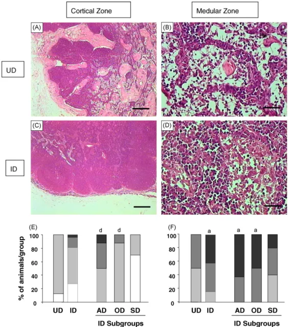
Fig. 1. Major histopathological features of popliteal lymph node from uninfected dogs (UD) andLeishmania chagasi-infected dogs (ID). Photomicroscopy of haematoxylin–eosin stained specimens highlighting the histological aspect of cortical (A; bar = 250mm) and medullary regions
(B; bar = 25mm) from UD in contrast to hypertrophy/hyperplasia of cortical (C; bar = 250mm) and medullary (D; bar = 25mm) regions observed in
ID (bar = 25mm). The graphs illustrate the intensity of cortical (E) and medullary hypertrophy/hyperplasia (F) in popliteous lymph nodes from UD
and ID sub-groups (AD, asymptomatic dogs; OD, oligosymptomatic dogs; SD, symptomatic dogs). (&) = atrophy or absence of hypertrophy/ hyperplasia; ( ) = light; ( ) = moderate and (&) = intense hypertrophy/hyperplasia. Significant results (P<0.05) are represented by letters ‘‘a’’
When purified MAbs were used, the cells were incubated at the same conditions in the presence of 60ml of previous diluted FITC-conjugated sheep
anti-rat IgG antibody. Before flow cytometric data collection and analysis, labeled cells were fixed for 30 min with 200ml of FACS FIX solution (10.0 g/l
paraformalde-hyde; 10.2 g/l sodium cacodylate and 6.65 g/l sodium chloride, pH 7.2).
Results were expressed as percentage of gated lymphocytes for Thy-1+ and CD5+T-cells; CD21+ B-cells; CD4+ and CD8+ T-cell subsets. Data regarding lymphocyte activation status were presented as the
R.C. Giunchetti et al. / Veterinary Immunology and Immunopathology 121 (2008) 23–33
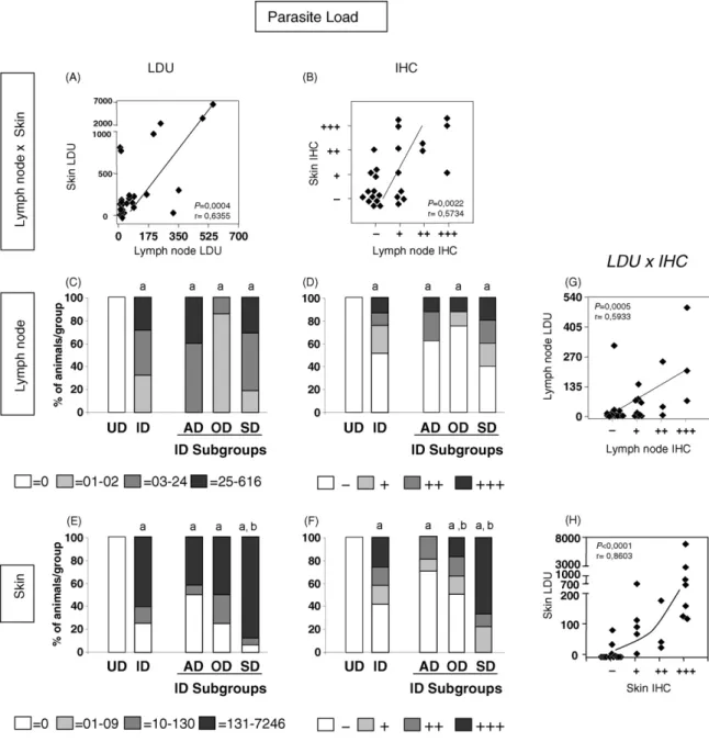
Fig. 2. Parasite density features in popliteal lymph node and ear skin expressed as Leishman Donovan Units (LDU) or anti-Leishmania
mean fluorescence intensity for MHC-II expression by gated cells.
2.7. Statistical analysis
Statistical analysis was performed using the Prism 4.1 software package (Prism Software, Irvine, CA, USA). Mann–Whitney test was used for comparative analysis of major histopathological and parasitological findings between UD and ID. Kruskal–Wallis followed by Dunns’ tests were used for multiple comparisons of major histopathological and parasitological features (UDADODSD). Analysis of LN immunophe-notypes was performed by the Mann–Whitney test to compare UD and ID groups. Spearman test was performed for different correlations strategies. Differ-ences were considered significant when the probabil-ities of equality,Pvalues were0.05.
3. Results
3.1. Hypertrophy/hyperplasia of LN cortical zone is the major characteristic of asymptomatic dogs whereas atrophy of LN cortical zone is the predominant histopathological finding in symptomatic animals
Histological analysis was performed by the routine hematoxylin–eosin (HE) procedure. Our findings revealed that depletion of follicular structures and lymphocytes which have been replaced mainly by macrophages are the major LN histological features at the cortical zone. The main findings observed in the LN cortical zone in uninfected and naturally L. chagasi -infected dogs are shown inFig. 1(left panels). Distinct from the histological aspect observed for UD (Fig. 1A) hypertrophy/hyperplasia of cortical region was com-monly observed in infected dogs (Fig. 1C). Our findings demonstrated intense hypertrophy/hyperplasia of cor-tical region in AD and OD group as compared to SD. On the other hand, atrophy of LN cortical zone was predominantly observed in SD (Fig. 1E).
3.2. Hypertrophy/hyperplasia of LN medullary zone was also the hallmark of asymptomatic disease
Histological aspects of cords and sinuses of LN medullary regions from uninfected and L. chagasi -infected dogs were evaluated by routine hematoxylin– eosin (HE) procedure and the major findings are presented in Fig. 1 (right panels). In contrast to the histology seen in UD (Fig. 1B), cellular infiltrates in the
medullary region of LN from L.chagasi-infected dogs were mainly characterized by the hypertrophy/hyper-plasia of plasmacytes when compared to the frequency of macrophages and lymphocytes (Fig. 1D). Data analysis demonstrated that ID presented higher LN medullary reactivity as compared to UD (Fig. 1F). Furthermore, only AD and OD showed marked LN medullary reactivity as compared to UD, characterized by prominent hypertrophy/hyperplasia of cords and sinus (Fig. 1F).
3.3. Despite the high correlation between skin and LN parasite burden observed in L. chagasi-infected dogs, the skin parasitism was the most reliable indicator of the clinical status of CVL
Lymph node and ear skin parasite load were assessed using two distinct approaches including Giemsa-stained microscopy to determine the Leishman Donovan Units (LDU) and anti-Leishmania immunohistochemical analysis (IHC; Fig. 2). Data analysis demonstrated positive correlation between the parasitism assessed by these different approaches (IHC and LDU;Fig. 2A and B) in both compartments (Fig. 2G and H). Additional analysis was performed by scoring popliteal lymph node and skin parasite load as absent, low, medium or high and reported for all clinical groups evaluated
(Fig. 2C–F). L. chagasi naturally infected dogs (ID)
were additionally sub-grouped as asymptomatic (AD), oligosymptomatic (OD) and symptomatic dogs (SD). Despite the fact that no difference was observed in LN parasite burden evaluation of different clinical groups
(Fig. 2C and D), the ear skin parasite load revealed
higher parasitism, expressed as LDU, in SD group when compared to both UD and AD groups (Fig. 2E). Analysis of parasite load by IHC showed better correspondence between parasite load and clinical status of CVL, highlighting the presence of higher ear skin parasite density in SD and OD when compared to UD and AD (Fig. 2F).
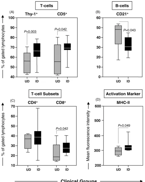
3.4. Increased frequency of T-lymphocytes, particularly CD8+cells, in addition to decreased levels of CD21+B-cells and up-regulation of MHC-II expression by lymphocytes represented the major LN immunophenotypic changes during ongoing CVL
Immunophenotypic analysis of LN cells was performed by direct and indirect flow cytometry immunofluorescence. The major LN immunophenoty-pic features are presented in Fig. 3. Data analysis
demonstrated higher frequency of T-lymphocytes (both Thy-1+and CD5+), mainly due the enhanced levels of CD8+ T-cells in addition to a lower percentage of CD21+ B-cells in naturally infected dogs (ID) in comparison to uninfected controls (UD) (Fig. 3A, C and B, respectively). No changes in the percentage of LN CD4+ T-cells were observed between ID and UD
(Fig. 3C).
Analysis of the activation status of LN cells was performed and the relative mean fluorescence intensity of the cell surface activation marker (MHC-II) is presented in Fig. 3D. Data analysis demonstrated differential expression of MHC-II in ID, leading to significantly increased fluorescence intensity on gated lymphocytes when compared to
UD (Fig. 3D).
R.C. Giunchetti et al. / Veterinary Immunology and Immunopathology 121 (2008) 23–33
R.C. Giunchetti et al. / Veterinary Immunology and Immunopathology 121 (2008) 23–33
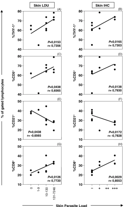
Fig. 4. Correlation between popliteal lymph nodes immunophenotypes and ear skin parasite load fromL. chagasi-infected dogs. Parasite load are expressed as Leishman Donovan Units (LDU) (left panels) and anti-Leishmaniaimmunohistochemical assay (IHC) (right panels), categorized as absent, LDU = 0 or IHC ( = 0); low, LDU (1–9) or IHC (+ = 1–2); medium, LDU (10–130) or IHC (++ = 3–24) and high parasitism, LDU (131– 7246) or IHC (+++ =>24). Cell phenotypes are expressed as percentage of Thy-1+(A, B), CD5+(C, D), CD21+(E, F) and CD8+(G, H) cells within
3.5. Positive association between the ear skin parasitism and LN T-cell mediated immunity suggests that LN CD8+lymphocytes may have a distinctive role in the immune response during CVL
Attempting to investigate whether LN immunophe-notypic features may be a reliable indicator of the ear skin parasite load, we have further characterized the level of association between these variables, as shown in
Fig. 4. Data analysis demonstrated that despite a
negative association between LN B-cells and ear skin parasitism was observed, a strong positive correlation between LN T-lymphocytes (both Thy-1+ and CD5+), particularly CD8+cells, are the hallmark ofL. chagasi
infection. Comparable correlation indexes were found using both parasitological approaches to detect the parasite burden, including LDU (left panels) and IHC (right panels). No significant correlation indexes were observed when ear skin parasite load was paired at individual levels with CD4+ cell frequency (data not shown).
4. Discussion
The lesions observed inL. chagasi-infected dogs are similar in many respect to those described for human visceral leishmaniasis, with major gross lesions including hepatosplenomegaly and lymphadenopathy
(Keenan et al., 1984). Despite the significant changes
observed in the lymphoid tissues in response to L. chagasiinfection, there are few studies focusing on the LN histopathology during CVL (Keenan et al., 1984; Martinez-Moreno et al., 1993; Tafuri et al., 2001; Lima
et al., 2004). In CVL, lymphadenopathy is defined as an
increase in LN size (enlargement of LN), usually described as a localized or generalized alteration
(Rogers et al., 1993). It has been demonstrated that
LN from L. chagasi-infected dogs display chronic lymphadenitis, independently of the anatomical region analyzed, with hypertrophy/hyperplasia of cortical and medullary zones (Lima et al., 2004).
In the present study, we confirmed most of these previous observations, reporting that depletion of follicular structures and lymphocytes, which are mainly replaced by macrophages, are the major LN histo-pathogical features in the cortical zone during CVL. On the other hand, the hypertrophy/hyperplasia of plasma cells, compared to the frequency of lymphocytes and macrophages, has been identified as the most relevant histological finding in the medullary region. These features have been also demonstrated in human visceral leishmaniasis (Veress et al., 1977), experimental
infections in hamsters (Corbett et al., 1992) and confirm previous reports in experimental and natural CVL
(Keenan et al., 1984; Martinez-Moreno et al., 1993).
It has been suggested that no significant differences of LN histopathological features are observed between asymptomatic and symptomatic dogs (Lima et al., 2004). However, we have found that hypertrophy/hyperplasia of cortical and medullary zones are distinctly observed in AD and SD. While intense hypertrophy/hyperplasia of cortical zone is observed in AD and OD groups, atrophy of cortical zone is more frequent in SD group. Moreover, only AD and OD groups exhibited a higher intensity of medullary reactivity as compared to UD group. Our data suggests that the decreased number of cells in germinal centers might be due to immunoregulatory mechanisms, such as apoptosis, would be one of the most relevant events underlying the cortical atrophy as observed in SD group. Further investigations are still required to test this hypothesis. Taken together, these findings supported the hypothesis that an intense LN immune response is important in controlling the clinical status during ongoing CVL.
Aiming to better understand the events underlying the compartmentalized immune response in the LN, we have performed a detailed analysis of LN parasite load and characterization of the phenotypic profile of major LN lymphocyte populations. Our previous paper reported that the skin is the major site of high parasite density during ongoing canine visceral leishmaniasis
(Reis et al., 2006a,c). To further investigate the
relationship between LN and skin we have performed a parasite load association. Analysis of parasite load in CVL in different tissues is a recent approach using
anti-Leishmania immunohistochemical and/or LDU index
(Giunchetti et al., 2006, 2007; Lima et al., 2004; Reis
et al., 2006a,b,c). Our findings demonstrated that
despite a positive correlation between LN and skin parasitism, only skin parasitism was associated with the clinical status of CVL. Therefore, skin parasitism should be considered the best site for parasitological analysis as a reliable indicator of the severity of clinical disease in CVL. Indeed, we have previously reported that symptomatic dogs showed a large number of cutaneous changes exemplified by an intense inflam-matory infiltrate, extracellular matrix changes and extreme parasite load (Giunchetti et al., 2006). The lack of association between the LN parasite burden scoring status and the clinical manifestation of CVL are in agreement with previous reports from Lima et al.
(2004). Importantly, the SD group displayed lower
frequency of negative IHC results in comparison to AD and OD (data not shown). Indeed, previous reports from
Reis et al. (2006a), taking into account the frequency of distinct degrees of LN parasite density using LDU index, have demonstrated that the SD group presented a lower percentage of animals displaying low LN parasitism.
No previous investigation has reported on the LN immunophenotypic profile duringL. chagasiinfection. Our findings demonstrated that the LN lymphocyte population of L. chagasi-infected dogs (ID) is characterized by a significant increase in Thy-1+ and CD5+ T-cells, particularly CD8+ T-cells, when com-pared to uninfected dogs (UD). CD8+ T-cells are thought to play an important role in the development of an effective immune response to Leishmania sp., possibly through cytotoxic mechanisms which act as potent protective events during CVL (Pinelli, 1997). Indeed, several studies have correlated the level of CD8+ T-cells with protection during L. chagasi
infection (Reis et al., 2006b; Pinelli, 1997). Notorious of importance was our finding that LN CD8+ T-cells displayed a positive correlation with skin parasite load, with the highest levels of CD8+ T-cell observed in animals bearing the highest skin parasitism. Typically, high CD8+ T-cells counts have been observed in the peripheral blood of asymptomatic dogs and are associated with low bone marrow parasitism (Reis
et al., 2006b). Therefore, we postulate that LN CD8+
T-cells may present a distinct activation status during CVL, possibly associated with immunomodulatory or suppressor cell activity. In support of this hypothesis,
Peruhype-Magalha˜es et al. (2006) showed in human
active VL that circulating CD8+T-cells showed a mixed cytokine pattern characterized by elevated levels of both intracellular IFN-gand IL-10. Similar results implying
a mixed pattern of cytokine mRNA in canine VL has been published (Lage et al., 2007). It is possible that a selective migration of Type-2 CD8+T-cells to the LN, committed with the synthesis of IL-10, represents a relevant immunological feature for suppressing the cell mediated immune response observed in dogs with high parasite load. We hypothesize that the elevated functional activation state towards a Type-2 immunolo-gical profile may account for the role of LN CD8+T-cells inL. chagasi-infected dogs. In addition, CD8+cells may mediate protection not only through a quantitative enhancement of cell numbers but also through a qualitative change in functional capacity towards a Type-1 immune response that increases circulating T-cells migration to distinct host tissues. Specific studies focusing this issue, through characterization of the cytokine pattern of LN CD8+ T-cells during CVL are currently under investigations in our laboratory.
In the present study, we have observed a significant decrease in LN CD21+ B lymphocytes in ID as compared to UD group. Lower levels of CD21+ B lymphocytes have been also reported in the peripheral blood during CVL, especially in symptomatic dogs
(Bourdoiseau et al., 1997; Reis et al., 2006b). The
intense LN plasmacyte infiltration observed in our histopatological analysis is consistent with the hypoth-esis that B-cell migrates from peripheral blood to LN during CVL. Indeed, CD21+ B cell differentiation toward plasmacytes may lead to the loss of the CD21 cell marker and therefore lower CD21+ B-cell frequency in LN. Consistent with this hypothesis, our data revealed a negative correlation between the LN CD21+ B-cell frequency and skin parasite load, since lower B-cell counts and higher parasite load are usually observed in SD and/or animals displaying higher anti -Leishmaniaantibody levels (Reis et al., 2006a,b,c).
It has been proposed that increased expression of MHC-II may reflect an antigenic-priming-related immunological event (Reis et al., 2006b). The evalua-tion in lymphocyte activaevalua-tion status showed significant increase in expression of MHC-II in ID compared to UD. Additional analysis showed that the level of MHC-II expression by LN lymphocytes was negatively associated with the skin parasite density evaluated as LDU index (P<0.0368/r= 0.7619). Together, our findings support the hypothesis that lymphocyte activation in the lymph nodes may favor cell migration and control of the parasite burden in parasitized organs. Consistent with this hypothesis, Reis and colleagues have demonstrated that asymptomatic dogs display enhanced activation status (expression of MHC-II) of circulating lymphocytes (Reis et al., 2006b) besides lower overall tissue parasitism (Reis et al., 2006a).
In conclusion, the histopathological profile of the asymptomatic disease was characterized by hypertro-phy/hyperplasia of LN cortical zone and intense cell reactivity in LN medullary zone, with hypertrophy/ hyperplasia of cords and sinus. Cell phenotype analysis revealed decreased levels of LN CD21+B-cells and an increased frequency of T-cells, particularly CD8+ T-cells, associated with higher ear skin parasitism, suggesting a Type-2 immunological profile for LN CD8+ T-cells. Further investigations focusing on LN cytokine profile, especially of CD8+ T-cells, are necessary to validate this hypothesis.
Acknowledgments
This work was supported by the CNPq/BR/grant: 521124/98-0; FAPEMIG/BR/grant: CBB 901/06 and
FIOCRUZ/PAPES IVB/2006. We are thankful to the Center for Zoonosis Control—Regional District of Belo Horizonte, Minas Gerais for the special dedication to this work. We also thank Dr. Erika Lamb of Microbiology and Immunology Department, USUHS, Bethesda, MD, USA for the critical reading of the manuscript, editorial suggestions and changes.
References
Abranches, P., Silva-Pereira, M.C.D., Conceic¸a˜o-Silva, F., Santos-Gomes, G.M., Jans, J.G., 1991. Canine leishmaniasis: pathological and ecological factors influencing transmission of infection. J. Parasitol. 77, 561–577.
Alencar, J.E., 1961. Profilaxia do kala-azar no Ceara. Brazil. Rev. Inst. Med. Trop. Sa˜o Paulo 3, 175–180.
Bourdoiseau, G., Bonnefont, C., Magnol, J.P., Saint-Anre´, I., Cha-banne, L., 1997. Lymphocyte subset abnormalities in canine leishmaniasis. Vet. Immunol. Immunopathol. 56, 345–351. Corbett, C.E., Pinto-Paes, P., Laurenti, R.A., Andrade Jr., M.D.,
Duarte, M.I.S., 1992. Histopathology of lymphoid organs in experimental leishmaniasis. Inst. J. Exp. Pathol. 73, 417–433. Deane, L.M., Deane, M.P., 1962. Visceral leishmaniasis in Brazil.
Geographical distribution and transmission. Rev. Inst. Med. Trop. Sa˜o Paulo 4, 149–212.
Desjeux, P., 2004. Leishmaniasis: current situation and new perspec-tives. Comp. Immunol. Microbiol. Infect. Dis. 27, 305–318. Giunchetti, R.C., Mayrink, W., Genaro, O., Carneiro, C.M.,
Correˆa-Oliveira, R., Martins-Filho, O.A., Marques, M.J., Tafuri, W.L., Reis, A.B., 2006. Relationship between canine visceral leishma-niosis and theLeishmania(Leishmania)chagasiburden in dermal inflammatory foci. J. Comp. Pathol. 135, 100–107.
Giunchetti, R.C., Mayrink, W., Carneiro, C.M., Correˆa-Oliveira, R., Martins-Filho, O.A., Marques, M.J., Tafuri, W.L., Reis, A.B., 2007. Histopathological and immunohistochemical investigations of the hepatic compartment associated with parasitism and serum biochemical changes in canine visceral leishmaniasis. Res. Vet. Sci., in press,doi:10.1016/j.rvsc.2007.04.020.
Keenan, C.N., Hendricks, L.D., Lightner, L., Johnson, A.J., 1984. Visceral leishmaniasis in a German shepherd dog II. Pathol. Vet. Pathol. 21, 80–86.
Lage, R.S., Oliveira, G.C., Buzek, S.C.U., Guerra, L.L., Giunchetti, R.C., Correˆa-Oliveira, R., Reis, A.B., 2007. Analysis of the cytokine profile in spleen cells from dogs naturally infected by
Leishmania chagasi. Vet. Immunol. Immunopathol. 115, 135–145. Lima, W.G., Michalick, M.S.M., Melo, M.N., Tafuri, W.L., Tafuri, Wg.L., 2004. Canine visceral leishmaniasis: a histopathological study of lymph nodes. Acta Trop. 92, 43–53.
Mancianti, F., Gramiccia, M., Gradoni, L., Pieri, S., 1988. Studies on canine leishmaniasis control I. Evolution of infection of different clinical forms of canine leishmaniasis following antimonils treat-ment. Trans. R. Soc. Trop. Med. Hyg. 82, 566–567.
Martinez-Moreno, A., Martinez-Cruz, M.S., Hernandez-Rodriguez, S., 1993. Immunological and histological study of T- and B-lymphocyte activity in canine visceral leishmaniosis. Vet. Para-sitol. 51, 49–59.
Palatnik-de-Sousa, C.B., Santos, W.R., Franc¸a-Silva, J.C., da Costa, R.T., Reis, A.B., Palatnik, M., Mayrink, W., Genaro, O., 2001. Impact of canine control on the epidemiology of canine and human visceral leishmaniasis in Brazil. Am. J. Trop. Med. Hyg. 65, 510–517.
Peruhype-Magalha˜es, V., Martins-Filho, O.A., Prata, A., Siva, L.A., Rabello, A., Teixeira-Carvalho, A., Figueiredo, R.M., Guimar-a˜es-Carvalho, S.F., Ferrari, T.C.A., Van Weyenbergh, J., Correa-Oliveira, R., 2006. Mixed inflammatory/regulatory cytokine profile marked by simultaneous raise of interferon-gamma and interleukin-10 and low frequency of tumour necrosis factor-alpha monocytes are hallmarks of active human visceral leishmaniasis due toLeishmania chagasiinfection. Clin. Exp. Immunol. 146, 124–132.
Pinelli, E., 1997. Cytokines in Canine Visceral Leishmaniasis. In: Schijns, V.E.C.J., Horzinek, M.C. (Eds.), Cytokines in Veterinary Medicine. Utrecht University, Netherlands, pp. 217–247. Reis, A.B., Carneiro, C.M., Carvalho, M.G., Teixeira-Carvalho, A.,
Giunchetti, R.C., Mayrink, W., Genaro, L.L, Correˆa-Oliveira, R., Martins-Filho, O.A., 2005. Establishment of a microplate assay for flow cytometric assessment and it is use for the evaluation of age-related phenotypic changes in canine whole blood leukocytes. Vet. Immunol. Immunopathol. 103, 173–185.
Reis, A.B., Teixeira-Carvalho, A., Vale, A.M., Marques, M.J., Giunchetti, R.C., Mayrink, W., Guerra, L.L., Andrade, R.A., Correˆa-Oliveira, R., Martins-Filho, O.A., 2006a. Isotype pat-terns of immunoglobulins: hallmarks for clinical status and tissue parasite density in Brazilian dogs naturally infected by
Leishmania (Leishmania) chagasi. Vet. Immunol. Immuno-pathol. 112, 102–116.
Reis, A.B., Teixeira-Carvalho, A., Giunchetti, R.C., Guerra, L.L., Carvalho, M.G., Mayrink, W., Genaro, O., Correˆa-Oliveira, R., Martins-Filho, O.A., 2006b. Phenotypic features of circulating leucocytes as immunological markers for clinical status and bone marrow parasite density in dogs naturally infected byLeishmania chagasi. Clin. Exp. Immunol. 146, 303–311.
Reis, A.B., Martins-Filho, O.A., Teixeira-Carvalho, A., Carvalho, M.G., Mayrink, W., Franc¸a-Silva, J.C., Giunchetti, R.C., Genaro, O., Correˆa-Oliveira, R., 2006c. Parasite density and impaired biochemical/hematological status are associated with severe clinical aspects of canine visceral leishmaniasis. Res. Vet. Sci. 81, 68–75.
Rogers, K.S., Barton, C.L., Landis, M., 1993. Canine and feline lymph nodes. Part II. Diagnosis, evaluation and lymphadenopathy. Com-pendium 15, 1493–1501.
Stauber, L.A., 1956. Resistance to the Khartoum strain ofLeishmania donovani. Rice Inst. Pamphlet. 45, 80–96.
Tafuri, W.L., De Oliveira, M.R., Melo, M.N., Tafuri, W.L., 2001. Canine visceral leishmaniasis: a remarkable histopathological picture of one case report from Brazil. Vet. Parasitol. 3, 203–212. Tafuri, Wg.L., Santos, R.L., Arantes, R.M.E., Gonc¸alves, R., Melo, M.N., Michalik, M.S.M., Tafuri, W.L., 2004. An alternative immunohistochemical method for detectingLeishmania amasti-gotes in paraffin-embedded canine tissues. J. Immunol. Methods 292, 17–23.
Veress, B., Omer, A., Satir, A.A., El Hassan, A.M., 1977. Morphology of the spleen and lymph nodes in fatal visceral leishmaniasis. Immunology 33, 605–610.