Profile of anti-
Leishmania
antibodies related to clinical picture in
canine visceral leishmaniasis
José Cláudio Carneiro de Freitas
a,⇑, Belarmino Eugênio Lopes-Neto
a, Cyntia Rafaelle Amaral de Abreu
a,
Wendel Coura-Vital
b, Samuel Leôncio Braga
b, Alexandre Barbosa Reis
b,
Diana Célia Sousa Nunes-Pinheiro
aaPrograma de Pós-Graduação em Ciências Veterinárias, Faculdade de Veterinária, Universidade Estadual do Ceará (UECE), Avenida Paranjana, 1700, Campus do Itaperi, Serrinha,
CEP 60740-000, Fortaleza, CE, Brazil
bLaboratório de Imunopatologia, Núcleo de Pesquisas em Ciências Biológicas, Universidade Federal de Ouro Preto (UFOP), Campus Universitário Morro do Cruzeiro, CEP 35400-000,
Ouro Preto, MG, Brazil
a r t i c l e
i n f o
Article history:
Received 19 July 2011 Accepted 5 December 2011
Keywords:
Canine visceral leishmaniasis Anti-Leishmaniaantibodies Clinical picture
Leishmania chagasi
a b s t r a c t
This research investigated the profile of anti-Leishmaniaantibodies in different clinical forms of canine visceral leishmaniasis (CVL). Naturally infected dogs were divided into two groups: subclinical dogs (SD,n= 10) and clinical dogs (CD,n= 68). Non-infected dogs (ND,n= 7) comprised the negative control group. The humoral response was evaluated by the profile of total IgG, IgG1, IgG2, IgM, IgA and IgE, deter-mined by ELISA. Infected animals showed increased levels of total IgG, IgA and IgE in addition to IgG1 and IgG2 in groups SD and CD, when compared with group ND. Furthermore, it was observed that IgG2 and IgM were correlated with symptomatology, while total IgG, IgG1 and IgA were negatively cor-related and IgE showed no correlation. It follows that serum levels of IgG2 anti-Leishmaniaare correlated with typical clinical signs of disease. Furthermore the determination of specific anti-Leishmania antibod-ies could be an important tool in monitoring CVL clinical picture.
Crown CopyrightÓ2011 Published by Elsevier Ltd. All rights reserved.
1. Introduction
The leishmaniasis are a complex of infectious diseases caused by different species of protozoa of the genusLeishmania. The infec-tion is transmitted by the bite of infected insects of the genus Phle-botomusand Lutzomyiain the New and Old World, respectively (Peters and Sacks, 2006). In Brazil, the Programme for the Control of Visceral Leishmaniasis recommends early diagnosis and treat-ment of human cases, vector control and the detection and eutha-nasia of seropositive dogs (Brasil, 2006).
Although humans can also act as reservoir of the agent and plays a role in the transmission cycle, the dog is considered one of the most important links in the epidemiological chain of leish-maniasis (Ribeiro, 2007). Studies conducted in São Paulo showed a prevalence of visceral leishmaniasis up to 40% in the canine pop-ulation (Ikeda et al., 2003). However, the prevalence in endemic areas may reach higher levels, as demonstrated byFreitas et al. (2010), through an epidemiological survey in Fortaleza, whose highest and lowest prevalence was 80.2% and 64% respectively.
Dogs with clinical leishmaniasis (CD) are characterized by clin-ical signs and/or clinclin-ical pathologclin-ical abnormalities, with infection
confirmed by specific tests, while dogs with subclinical leishman-iasis (SD) are characterized by no clinical signs or clinicopatholog-ical abnormalities, but with confirmed infection (Solano-Gallego et al., 2009).
The onset of clinical signs in canine visceral leishmaniasis (CVL) involves a number of factors, and these are associated with the ani-mal’s immune response (Ciaramella and Corona, 2003). In (CVL), the immune response mediated by Th1 lymphocytes, that secrete stimulatory cytokines (IFN-
c
), activates macrophages infected,effectively controlling the infection (Miranda et al., 2007). On the other hand, when the immune response is mediated by Th2 lym-phocytes, IL-4 secretors, there is a high production of antibodies that are associated with severe clinical manifestations (Miranda et al., 2007). It is noteworthy that when the immune response is mediated by regulatory T lymphocytes, producing IL-10, Th1 is inhibited and the infection is worsen (Miyara and Sakaguchi, 2007; Belkaid and Tarbell, 2009).
Studies report the role of antibodies in the CVL, relating the clin-ical picture with the presence of different classes and subclasses of immunoglobulins involved in the inflammatory response ( Trotz-Williams and Gradoni, 2003; Almeida et al., 2005). The soluble im-mune complexes are deposited in various organs and tissues such as kidneys, blood vessels, joints, among others, favoring the appearance of various symptoms such as epistaxis, polyuria and
0034-5288/$ - see front matter Crown CopyrightÓ2011 Published by Elsevier Ltd. All rights reserved.
doi:10.1016/j.rvsc.2011.12.009
⇑ Corresponding author. Tel.: +55 85 31019859.
E-mail address:joseclaudiocarneiro@yahoo.com.br(J.C.C. de Freitas).
Contents lists available atSciVerse ScienceDirect
Research in Veterinary Science
polydipsia, uveitis, conjunctivitis and episcleritis immune-medi-ated, skin ulcers and tips of ears, hyperkeratosis and limping by poliartritre (Ciaramella and Corona, 2003).
Given the great importance of the disease, this study aims to evaluate the profile of anti-Leishmaniaantibodies in different clin-ical pictures of CVL with emphasis on correlation with the clinclin-ical symptoms.
2. Material and methods
2.1. Animals
Adult dogs (n= 85) were used, varying in age, weight and breeds (including cross-breed). The seropositive dogs were cap-tured by the Zoonosis Control Center of Fortaleza (CCZ), as a CVL control measure. This study was approved by the Ethics Committee for Animal Use of the State University of Ceará (CEUA/UECE), pro-tocol number 08622833-1.
2.2. Immunofluorescence assay (IFA)
In all animals the immunofluorescence assay (IFA) was per-formed for canine visceral leishmaniasis, being considered sero-positive dogs the ones with titers above of 1:40, according to recommendations of the Ministry of Health of Brazil. Serologic testing was performed at CCZ using Bio-Manguinhos (FIOCRUZ-RJ) kits, following the manufacturer’s recommendations.
2.3. Parasitological diagnosis
After anesthetizing the dog with Xylazine (2 mg/kg) and Keta-mine (10 mg/kg) bone marrow aspiration for making imprints on microscope slides was performed. This material was fixed with methanol and stained with Panótipo fast dye. The stained imprints were observed under an optical microscope, and samples where the presence of amastigotes ofLeishmania chagasias revealed were considered positive
2.4. Clinical classification
All dogs were examined by observing the typical clinical signs of canine visceral leishmaniasis as onychogryphosis, hepatosplen-omegaly, cachexia, lymphadenopathy, keratoconjunctivitis, skin ulcers, apathy, alopecia.
The dogs were divided into three groups, according to Solano-Gallego et al. (2009). Negative dogs (ND = 7), which do not show clinical and laboratory alterations (hematology and biochemistry) and negative for visceral leishmaniasis, by serology and parasitol-ogy; subclinical dogs (SD = 10) which do not show clinical and lab-oratory alterations and positive forLeishmania chagasiinfection or as clinical dogs (CD = 68) which show clinical and laboratory alter-ations for routine testing and have infection confirmed by serolog-ical and parasitologserolog-ical diagnosis.
2.5. Collection of blood samples
Blood samples were collected by jugular venipuncture with a sterile syringe on dogs of different groups which were placed into a tube containing gel separation, without anticoagulant to obtain serum. Sera samples were stored at 20°C for further test.
2.6. Immunoenzymatic reaction – ELISA
To determine the profile of anti-Leishmaniaantibodies, ELISA as-say was performed, using soluble antigen (MHOM/BR/1972/BH46)
from promastigotes ofL. chagasi(SLA) from the axenic culture in LIT medium (Reis et al., 2006).
96-well microplates (Maxisorp™ Nunc International Nalgas, USA) were coated with SLA at a concentration of 2 mg/well, over-night at 4°C. After incubation, the plates were washed four times with phosphate buffer solution (PBS) containing 0.05% Tween 20 and blocked for 45 min at 37°C with 100 mL of fetal bovine serum (5%) in PBS per cell. Then, serum samples were added at a dilution of 1:80 for IgG, IgG1, IgG2, IgM and IgE and 1:40 for IgA. After this procedure, they were washed and added peroxidase conjugate (Bethyl Laboratories Inc., Montgomery, TX, USA) previously diluted as follows: anti-dog IgG1 (anti-heavy chain specific), 1:8000; IgM (anti-m chain specific), 1:1000; IgA (anti-a chain specific), 1:500; IgE (anti-e chain specific), 1:500, or anti-dog IgG and IgG2 (both anti-heavy chain specific), 1:16.000. After four washings (as de-scribed above), the reaction was started by adding 100 mL of 0.1 M citrate solution (pH 5.0) containing 0.03%
a
-phenylenedi-amine and 0.012% H2O2, followed by incubation at 37°C for 10 min. The reaction was stopped by adding 32
l
L of H2SO4 2.5 M and the absorbance (492 nm) was measured with a plate reader-ELISA ELX800 (Biotek InstrumentsÒVT, USA).
2.7. Statistical analysis
Statistical analysis was performed using the software GraphPad Prism 5.0. To compare the absorbance values of anti-Leishmania
antibodies among groups in different clinical forms, analysis of var-iance (ANOVA) one-way was used, followed by Tukey test. To iden-tify the association between the profile of anti-Leishmania
antibodies and the clinical symptoms, we used the Spearman cor-relation test (r). In all cases the differences were considered signif-icant atP60.05.
3. Results
The results of the evaluation of typical clinical signs of CVL were expressed in percentage (%) and are shown inTable 1. The more frequent clinical signs were cachexia (77.94%), keratoconjunctivitis (61.76%) and lymphadenopathy (55.88%), and 86.76% of the ani-mals showed more than one typical clinical sign of CVL.
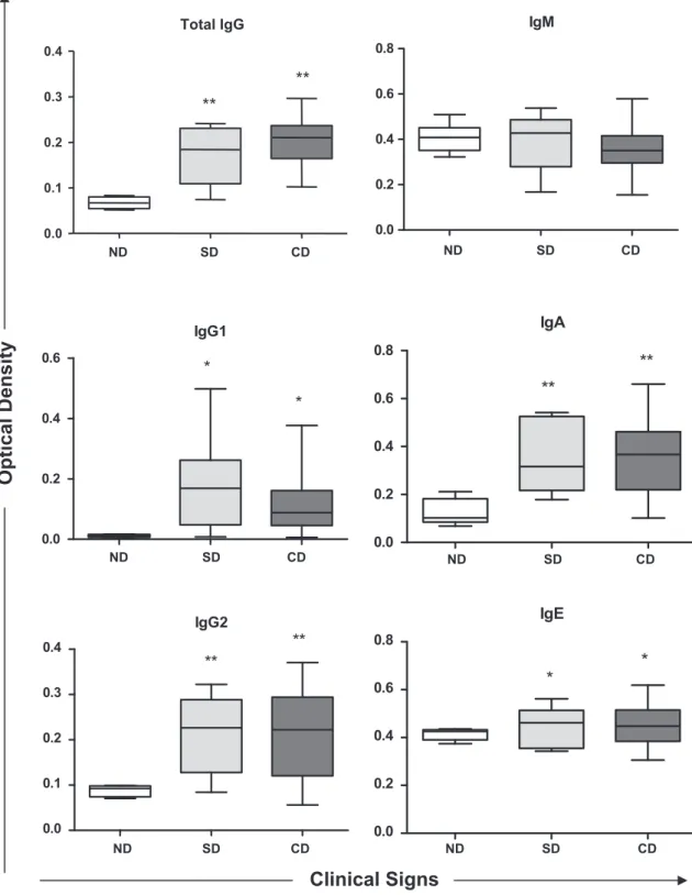
The profile of anti-Leishmaniaserum antibodies (total IgG, IgG1, IgG2, IgM, IgA and IgE) from naturally infected dogs in relation to different clinical pictures are shown inFig. 1. The correlation coef-ficient values (r) between the two factors are presented inTable 2. Increase in levels of IgG, IgG1, IgG2, IgA and IgE anti-Leishmania
in SD and CD groups was observed compared to control group (ND). As for IgM, there was no change in the response profile among the evaluated groups. When evaluating the association be-tween the profile of anti-Leishmaniaantibodies and the different clinical pictures, it was observed that serum levels of IgG2 (r= 0.12) and IgM (r= 0.38) showed positive correlation with the clinical signs (Table 2). It was also observed negative correlation between serum levels of total IgG (r= 0.28), IgG1 (r= 0.13)
Table 1
Clinical signs observed in dogs, in group CD, naturally infected byLeishmania chagasi.
Clinical sign n %
Onychogryphosis 23 33.82
Hepatosplenomegaly 31 45.59
Cachexia 53 77.94
Lymphadenopathy 38 55.88
Keratoconjunctivitis 42 61.76
Skin ulcers 24 35.29
Apathy 19 27.94
Alopecia 21 30.88
and IgA (r= 0.62) and the clinical signs. Moreover, IgE levels showed no correlation between SD and CD (r= 0.0), i.e., in these animals the increases of serum IgE are not correlated with the clin-ical changes evident in infected animals.
4. Discussion
It has been reported that the appearance of clinical signs char-acteristic of visceral leishmaniasis is mainly as consequences of the
host immune response associated with deposition of immune com-plexes soluble in different tissues (Quinnell et al., 2003). In this direction, it could be interesting to assess the serum levels of anti-Leishmaniaantibodies verifying the classes and subclasses of immunoglobulins involved, and their association with clinical signs shown by infected animal.
In our study, we found that dogs naturally infected by Leish-mania chagasi (CD and SD) showed results of total IgG, IgG1, IgG2, IgA and IgE anti-Leishmania significantly increased when compared to ND (Fig. 1). Furthermore, we observed a negative
IgM
0.0 0.2 0.4 0.6 0.8
IgG1
0.0 0.2 0.4 0.6
*
*
IgA
0.0
0.2
0.4
0.6
0.8
**
**
IgG2
0.0 0.1 0.2 0.3 0.4
**
**
IgE
0.0
0.2
0.4
0.6
0.8
*
*
Optical
Density
Clinical Signs
Total IgG
ND SD CD
0.0 0.1 0.2 0.3 0.4
**
**
ND SD CD
ND SD CD ND SD CD
ND SD CD ND SD CD
Fig. 1.Anti-Leishmaniaantibodies profiles in dogs naturally infected byLeishmania chagasiand showing different clinical forms. SD (subclinical dogs), CD (clinical dogs) and ND (negative dogs).⁄Represents difference with a significance level of 5% and⁄⁄represents difference with a significance level of 1%.
correlation between total IgG, IgG1 and IgA and symptomatology (r= 0.28, r= 0.13 and r= 0.62, respectively), demonstrating that, the increase in these immunoglobulins is not associated with clinical signs of visceral leishmaniasis (Table 2).
Almeida et al. (2005)found that the titers of IgG anti-Leishmania
in symptomatic dogs increased significantly when compared to asymptomatic and the control group. This fact was also found by Vercammen et al. (2002)that linked the increased levels of total IgG with progression from onset of symptoms.
In our study, the values of IgG1 anti-Leishmaniashowed a negative correlation coefficient with the clinical condition of the animals (r= 0.13), and also their average serum levels decreased in animals showing clinical signs. These data corroborate the findings of Vercam-men et al. (2002)andCordeiro da Silva et al. (2003). With respect to IgG2 subclass, it happened differently, because the average serum absorbance values are almost the same as in all infected animals, not differing between CD and SD. However, these values are corre-lated with typical clinical signs of visceral leishmaniasis (r= 0.12), a fact which is mainly due to the findings of the CD group.
It has been reported that the clinical signs are directly related to IgG1/IgG2 relationship, where the titers of the subclass IgG2 are strongly correlated with the symptoms of the animals. In this case, the titers of the subclass IgG1 also show increased, however with lower values than those found in IgG2 (Solano-Gallego et al., 2001; Leandro et al., 2001; Almeida et al., 2005).
Reis et al. (2006)found that IgG1 anti-Leishmaniahas a negative correlation with the clinical status of animals with visceral leish-maniasis, with their values diminished with clinical outcome, dem-onstrating association with the maintenance of the chronic and asymptomatic disease. With respect to IgG2, the same authors found that the values did not differ significantly between the in-fected animals, and only differed from the uninin-fected control group (Reis et al., 2006). These findings are in agreement with those ob-tained by other authors previously (Vercammen et al., 2002; Cordeiro da Silva et al., 2003).
In the present study, serum levels of IgM, anti-Leishmaniafrom naturally infected dogs (SD and CD), remained with no significant differences compared to the control group (ND). However, with re-spect to the association with the symptomatology, there was a po-sitive correlation coefficient between the groups and the clinical picture (r= 0.38). Yet, according to the one proposed by Reis et al. (2006), serum levels of IgM, in theL. chagasiinfection, remain high until the chronic phase of disease. It is noteworthy, that this immunoglobulin is strongly associated with acute forms of para-sitic diseases and that it does not present significant correlation with the clinical picture of CVL. Despite the above, little is known about the profile of IgM in CVL, because it is easily inactivated by reagents used in the main techniques of serology (Schallig et al., 2002), making it difficult to detect more accurate findings.
In relation to IgA anti-Leishmania, in our study, infected dogs showed negative correlation with the onset of the clinical picture
of CVL (r= 0.62), despite serum IgA levels were significantly in-creased (P= 0.0025) when compared to ND. Few studies emphasize the importance of IgA in the CVL. However,Reis et al. (2006) dem-onstrated a positive correlation of patterns of immunoglobulin-A with the clinical status of naturally infected animals. However, these data differ from our results. Whilst there are few data in lit-erature,Day (2007) in his review reported that the levels of IgA anti-Leishmania were increased in the mucosa of infected dogs showing symptomatology, suggesting a source of aggravation of the infectious process.
It is noteworthy that dogs infected byLeishmania chagasidevelop immune response mediated by Th1 lymphocytes, they may be capa-ble of preventing the spread of the parasite to the mucosal surface and, consequently, produce lower levels of specific IgA, with no dif-ferentiation with the clinical progression of CVL (Rodriguez-Cortes et al., 2007). Glomerulonephritis is a main clinical finding in the CVL, and the main triggering event of this abnormality has been attributed to the accumulation and deposition of immune com-plexes mediated by IgA in renal glomeruli (Nieto et al., 1992). More-over, in post-mortem analysis, it was found that this clinical disorder is the leading cause of death of infected animals (Feitosa et al., 2000). Regarding the levels of IgE anti-Leishmania, the results differed significantly between the infected animals (CD and SD) compared to non-infected (P= 0.0103). Still, when we evaluated the profile of IgE in the ND group, it was observed high absorbance values in some animals, which may represent false-negative results, by the diagnostic technique used, with possible evolution to a subclin-ical canine disease in these animals. These observations were also suggested byAlmeida et al. (2005), who associated the IgE levels with the development of symptomatic cases of CVL. In this case, the predominant immune response is mediated by Th2 lympho-cytes, which induce a decrease in the synthesis of IFN-
c
, within-creased expression of IL-4 and IL-10, with data obtained by measuring the levels of mRNA (Quinnell et al., 2001).
In our study there was no correlation between the titers of IgE anti-Leishmania with symptomatology (r= 0.00), similar titers were detected in groups SD and CD. From these data we can con-sider that the appearance of symptoms is due to action of other immunoglobulins, and other elements of the immune response that were not objects of study. However works carried out by Ini-esta et al. (2005)andReis et al. (2006)observed a strong correla-tion between the titers of IgE and the clinical status of animals.
5. Conclusion
Our data demonstrate that dogs naturally infected with Leish-mania chagasipresent high titers of IgE, IgA and IgG2 anti-Leishmania
and indicate that serum levels of IgG2 anti-Leishmaniaare correlated with typical clinical signs of disease, but serum levels of IgE anti -Leishmaniado not correlate with the disease progression. Further studies are needed to finally demonstrate that the determination of specific anti-Leishmaniaantibodies are an important tool to pre-dict the clinical course of the disease as suggested by this study.
Acknowledgments
The authors would like to express their appreciation to the Fun-dação Cearense de Apoio ao Desenvolvimento Científico e Tec-nológico (FUNCAP) for the financial help granted to the first author, which provided subsidies for the implementation of the project.
References
Almeida, M.A.O., Jesus, E.E.V., Sousa-Atta, M.L.B., Alves, L.C., Berne, M.E.A., Atta, A.M., 2005. Antileishmanial antibody profile in dogs naturally infected with
Table 2
Pandrvalues of anti-Leishmaniaantibodies profiles. WherePcompares differences in absorbance of immunoglobulins in different clinical forms (SD and CD) with the negative control group (ND) of naturally infected dogs byLeishmania chagasi.rvalue determines the correlation coefficient between immunoglobulins profiles and the clinical signs (CD) of naturally infected dogs byLeishmania chagasi.
Immunoglobulin Pvalue rvalue
NDSD NDCD SDCD
Total IgG 0.009 0.007 0.12 0.28
IgG1 0.02 0.04 0.08 0.13
IgG2 0.009 0.008 0.11 0.12
IgM 0.08 0.11 0.09 0.38
IgA 0.009 0.008 0.09 0.62
Leishmania chagasi. Veterinary Immunology and Immunopathology 106, 151– 158.
Belkaid, Y., Tarbell, K., 2009. Regulatory T cells in the control of host–microorganism interactions. Annual Reviews of Immunology 27, 551–589.
Brasil, M.S., 2006. Manual de Vigilância e Controle da Leishmaniose Visceral, first ed. Editora MS, Brasília, pp. 11–18.
Ciaramella, P., Corona, M., 2003. Canine leishmaniasis: clinical and diagnostic aspects. VetLearn 25, 358–368.
Cordeiro da Silva, A., Cardoso, L., Araújo, N., Castro, H., Tomas, A., Rodrigues, M., Cabral, M., Vergnes, B., Sereno, D., Ouaissi, A., 2003. Identification of antibodies toLeishmaniasilent information regulatory 2 (SIR2) protein homologue during canine natural infections: pathological implications. Immunology Letters 86, 155–162.
Day, M.J., 2007. Immunoglobulin G subclass distribution in canine leishmaniasis: a review and analysis of pitfalls in interpretation. Veterinary Parasitology 147, 2–8. Feitosa, M.M., Ikeda, F.A., Luvizotto, M.C., Perri, S.H.V., 2000. Aspectos clínicos de cães com leishmaniose visceral no município de Araçatuba – São Paulo (Brasil). Clínica Veterinária 28, 36–44.
Freitas, J.C.C., Nunes-Pinheiro, D.C.S., Abreu, C.R.A., 2010. Geographical variation in clinical signs and prevalence of Leishmania sp. infection among dogs in Fortaleza, Ceará State, Brazil. Acta Scientiae Veterinariae 38, 293–297. Ikeda, F.A., Luvizotto, M.C.R., Goçalves, M.E., Feitosa, M.M., Ciarlini, P.C., Lima, V.M.F.,
2003. Perfil hematológico de cães naturalmente infectados porLeishmania chagasino município de Araçatuba, São Paulo (Brasil): um estudo retrospectivo de 191 casos. Clínica Veterinária 47, 42–48.
Iniesta, L., Gallego, M., Portus, M., 2005. Immunoglobulin G and E responses in various stages of canine leishmaniosis. Veterinary Immunology and Immunopathology 103, 77–81.
Leandro, C., Santos-Gomes, G.M., Campino, L., Romão, P., Cortes, S., Rolao, N., Gomes-Pereira, S., Rica Capela, M.J., Abranches, P., 2001. Cell mediated immunity and specific IgG1 and IgG2 antibody response in natural and experimental canine leishmaniosis. Veterinary Immunology and Immunopathology 79, 273–284.
Miranda, S., Martorell, S., Costa, M., Ferrer, L., Ramis, A., 2007. Characterization of circulating lymphocyte subpopulation in canine leishmaniasis throughout treatment with antimonials and allopurinol. Veterinary Parasitology 144, 251–260. Miyara, M., Sakaguchi, S., 2007. Natural regulatory T cells: mechanisms of
suppression. Trends in Molecular Medicine 13, 108–116.
Nieto, C.G., Navarrete, I., Habela, M.A., Serrano, F., Redondo, E., 1992. Pathological changes in kidneys of dogs with naturalLeishmania infections. Veterinary Parasitology 45, 33–47.
Peters, N., Sacks, D., 2006. Immune privilege in sites of chronic infection:Leishmania
and regulatory T cells. Immunological Reviews 213, 159–179.
Quinnell, R.J., Courtenay, O., Shaw, M.A., Day, M.J., Garcez, L.M., Dye, C., Kaye, P.M., 2001. Tissue cytokine responses in canine visceral leishmaniasis. Journal of Infectious Disease 183, 1421–1424.
Quinnell, R.J., Courtenay, O., Garcez, L.M., Kaye, P.M., Shaw, M.A., Dye, C., Day, M.J., 2003. IgG subclass responses in a longitudinal study of canine visceral leishmaniasis. Veterinary Immunology and Immunopathology 91, 161–168.
Reis, A.B., Teixeira-Carvalho, A., Vale, A.M., Marques, M.J., Giunchetti, R.C., Mayrink, W., Guerra, L.L., Andrade, R.A., Corrêa-Oliveira, R., Martins-Filho, O.A., 2006. Isotype patterns of immunoglobulins: hallmarks for clinical status and tissue parasite density in brazilian dogs naturally infected byLeishmania(Leishmania)
chagasi. Veterinary Immunology and Immunopathology 112, 102–116. Ribeiro, V.M., 2007. Leishmaniose visceral canina: aspectos de tratamento e
controle. Clínica Veterinária 71, 66–76.
Rodriguez-Cortes, A., Fernandez-Bellon, H., Ramis, A., Ferrer, L., Alberola, J., Solano-Gallego, L., 2007.Leishmania-specific isotype levels and their relationship with specific cell-mediated immunity parameters in canine leishmaniasis. Veterinary Immunology and Immunopathology 116, 190–198.
Schallig, H.D.F.H., Schoone, G.J., Beijer, E.G.M., Kroon, C.C.M., Ozbel, Y., Ozensoy, S., Da Silva, E.S., Cardoso, L.M., Da Silva, E.D., 2002. Development of a fast agglutination screening test (FAST) for detection of anti-Leishmaniaantibodies in dogs. Veterinary Parasitology 109, 1–8.
Solano-Gallego, L., Riera, C., Roura, X., Iniesta, L., Gallego, M., Valladares, J.E., Fisa, R., Castillejo, S., Alberola, J., Ferrer, L., Arboix, M., Portus, M., 2001.Leishmania infantum-specific IgG, IgG1 and IgG2 antibody responses and healthy and ill dogs from endemic areas. Evolution in the course of infection and after treatment. Veterinary Parasitology 96, 265–276.
Solano-Gallego, L., Koutinas, A., Miró, G., Cardoso, L., Pennisi, M.G., Ferrer, L., Bourdeau, P., Oliva, G., Baneth, G., 2009. Directions for diagnosis, clinical staging, treatment and prevention of canine leishmaniosis. Veterinary Parasitology 165, 1–18.
Trotz-Williams, L., Gradoni, L., 2003. Disease risks for the travelling pet Leishmaniasis. In Practice 25, 190–197.
Vercammen, F., Fernandez-Perez, F.J., Del Amo, C., Alunda, J.M., 2002. Follow-up of
Leishmania infantum naturally infected dogs treated with allupurinol: immunofluorescence antibody test, ELISA and Western-blot. Acta Tropica 84, 175–181.