Cyanogenesis and the onset of tapping panel dryness in rubber tree
Elisabeth de Faÿ(1), Larissa Alexandra Cardoso Moraes(2) and Vicente Haroldo de Figueiredo Moraes(3)
(1)Nancy Université, IFR 110 EFABA, UMR UHP‑INRA 1136 IAM, BP 70239, 54506 Vandœuvre‑lès‑Nancy Cedex, France. E‑mail: [email protected]‑nancy.fr (2)Embrapa Amazônia Ocidental, Rodovia AM 010, Km 29, Caixa Postal 319, CEP 69 010‑970, Manaus, AM, Brazil. E‑mail: [email protected] (3)In memoriam
Abstract – The objective of this work was to study the influence of cyanogenesis on the onset of irreversible tapping panel dryness (TPD) and the physiological and histological aspects of secondary phloem in the trunk (tapping panel) of rubber trees (Hevea spp.). Two cyanogenic compounds, linamarin and KCN, were applied separately on the trunk bark of healthy mature trees belonging to two Brazilian clones (Fx 4098 and Fx 3899). Changes in histology, latex pressure potential (ΨP) and cyanogenic potential (HCNp) were followed in the
trunk inner barks. In addition, the HCNp levels were determined in TPD-affected plants of both clones. The applications of linamarin or KCN in healthy plants decreased latex ΨP, and formed tylosoids associated with
in situ coagulation of latex. The clone Fx 4098 had the higher HCNp and showed the quicker and stronger responses to the cyanogenic compounds. Plants with TPD syntoms had a higher HCNp than the untreated healthy ones. Since histological changes are also structural markers of early TPD, it can be inferred that excessive release of cyanide can induce it in sensitive rubber clones.
Index terms: Hevea, clonal sensitivity, cyanogenic potential, latex pressure potential, linamarin, secondary phloem.
Cianogênese e estabelecimento do secamento de painel
de sangria em seringueira
Resumo – O objetivo deste trabalho foi estudar a influência da cianogênese no estabelecimento do secamento do painel de sangria (TPD) e os aspectos fisiológicos e histológicos do floema secundário do tronco (painel de sangria) em seringueira (Hevea spp.). Dois compostos cianogênicos, linamarina e KCN, foram aplicados separadamente na casca do tronco de árvores sadias de dois clones adultos (Fx 4098 e Fx 3899). Alterações na histologia, potencial de pressão de látex (ΨP) e potencial cianogênico (HCNp) foram monitoradas. Além
disso, os níveis HCNp foram determinados em plantas afetadas por TPD, em ambos os clones. A aplicação de linamarina ou KCN em plantas saudáveis causou redução em ΨP e formação de tilosoides associados à
coagulação do látex in situ. O clone Fx 4098 apresentou o maior nível de HCNp, e respostas mais intensas e rápidas aos compostos cianogênicos. Plantas com sintomas de TPD têm maior HCNp de que as sadias. Como as alterações histológicas observadas são reconhecidos marcadores estruturais precoces de TPD, pode-se inferir que a liberação excessiva de cianeto pode induzi-las em clones de seringueira sucetíveis.
Termos para indexação: Hevea, sensibilidade clonal, potencial cianogênico, potencial de pressão de látex, linamarina, floema secundário.
Introduction
Species of the genus Hevea, particularly
H. brasiliensis, are intensively cultivated and exploited in modern plantations for the latex. Rubber tree is an important industrial crop, natural rubber representing almost half (42% in 2005) of total world rubber production. As in other crops, various plant physiological conditions and pathogenic diseases
influence rubber production. The tapping panel dryness
(TPD) is one of the most serious threats to natural rubber yield: it is estimated that TPD contributes to
15–20% loss of the annual rubber production, with an incidence of 20–50% of productive trees affected by TPD, in almost every rubber-growing regions.
The TPD syndrome – first known as brown bast – is
detected early by bark dryness upon tapping, i.e. partial
or ultimately complete stoppage of latex flow on the
tapping cut. The late macroscopic symptoms are brown spots in the barks, bark thickening, bark cracking and desquamations and, sometimes, bark deformations (Petch, 1921; de Faÿ, 1981; de Faÿ & Jacob, 1989b),
which make the affected trees finally unsuited for latex
Since the 1900s a great deal of effort has been invested to understand the nature and mechanism of TPD. It was initially thought that TPD might be caused by pathogens, but aetiological investigations failed
so far to confirm any biotic causal agent (Rubber
Board, 2010). Several lines of evidence appear to support the alternative hypothesis that TPD is a physiological disorder resulting from abiotic stress linked to overexploitation (Jacob et al., 1994). Latex and, to a lesser extent, barks of rubber trees have been extensively studied to characterize TPD biochemically, and the most recent investigations aimed at identifying the proteins and genes related to TPD (Krishnakumar et al., 2001; Sookmark et al., 2002; Venkatachalam et al., 2009). Barks of affected trees were also characterised histologically, and structural markers
of early stages of TPD were found(de Faÿ & Hébant,
1980; de Faÿ, 1981; de Faÿ & Jacob, 1989b). Whatever that may be, the immediate cause of TPD remains still unknown. Moreover, according to some researchers
(Clément-Demange et al., 2007), the term TPD covers
two syndromes: “tapping cut dryness” and “brown bast” (irreversible TPD).
The hypothesis that TPD might be directly related to cyanogenesis originated in Brazil, where clonal incompatibility has been detected through symptoms resembling some aspects of TPD, mainly bark dryness (Moraes et al., 2001, 2002). The idea that impaired cyanide metabolism causes damage in hevea barks was also taken up by other researchers (Chrestin et al., 2004; Sookmark et al., 2004) who study the “bark
necrosis” syndrome of rubber tree. Hevea spp. contains
cyanogenic glucosides, similarly to Manihot esculenta
(cassava), another member of the Euphorbiaceae family. The most abundant is linamarin, a β cyanoglucoside synthesized by leaves, stored in vacuoles and transported in the form of the β diglucoside linustatin (Lieberei, 1986; Gruhnert et al., 1994; Kongsawadworakul et al., 2009). Normally, rubber trees also contain β
glucosidases and β diglucosidases, which gradually
degrade the cyanogenic glucosides, which results in the release of cyanide, but also the key-enzyme of
cyanide detoxification β cyanoalanine synthase (CAS)
(Lieberei, 1986; Gruhnert et al., 1994; Moraes et al., 2002). Applications of linamarin and KCN to the bark were shown to cause bark dryness in clones in which
β CAS activity is low (as low as in noncyanogenic
plants), and β glucosidases and β diglucosidases
relatively high (Moraes et al., 2001, 2002), which suggests a relationship of TPD with cyanogenesis.
The objective of this work was to study the influence
of cyanogenesis on the onset of irreversible tapping panel dryness (TPD), and the physiological and histological aspects of secondary phloem in the trunk (tapping panel) of rubber trees.
Materials and Methods
Rubber plants were field-grown in the experimental
site of Embrapa Amazônia Ocidental (3º8' S, 59º52'W), near Manaus (Brazil), and belonged to two Brazilian
clones, Fx 4098 (H. brasiliensis) and Fx 3899
(H. benthamiana x H. brasiliensis), chosen for their proneness to spontaneous and experimental dryness of bark. The two clones are prone to TPD, Fx 3899 to a greater extent than Fx 4098. The latter was also shown to dry up in response to treatment with high concentrations of KCN or linamarin (Moraes et al., 2001).
The region’s climate is an Af type according to
the Köppen classification, i.e., humid tropical with
relatively abundant rainfall throughout the year, and annual average precipitation, temperature and air humidity are 2,500 mm, 26°C and 85%, respectively, with the period of greater precipitation between January and April, and the period of lower precipitation between July and September (monthly precipitation is always over 60 mm).
The experiment was carried out during the period of greater precipitation, in February 2005. The plants were newly exploited healthy mature trees (they had been opened for one month), or trees affected by TPD syndrome. In the latter case, dryness had started in the
first week of exploitation and trees were totally dry at the end of the first month of tapping, just before the
experiment.
Two tests were carried out. The first one consisted
in applying one of the following solutions to the trunk bark of three recently opened trees of the same stand
per clone: KCN solution – 1.25 g L-1 KCN in 0.01 mol
L-1 phosphate buffer [2/1 (v/v) Na
2HPO4/NaH2PO4],
pH 6.5, 0.005% Tween 20, 0.005% dimethylsulfoxide
(DMSO); linamarin solution – 0.75 g L-1 linamarin in
0.01 mol L-1 phosphate buffer, pH 6.5, 0.005% Tween
20, 0.005% DMSO; control solution – 0.01 mol L-1
DMSO. Five milliliters of solution were put down in every one of four vertical grooves made in the bark, 5 cm below the tapping cut and 6–7 cm apart from each other (length, 12 cm; width, 1 cm; depth, 5–7 mm, i.e. as far as approximately 3.0 mm from the cambium). The solution was maintained in the grooves with a plate made of modelling clay stuck to the bark. After three days, the remaining volume of the solution was removed to avoid a continuous soaking of the bark.
Three identical applications were made with a five-day
interval, after which partial bark dryness was detected when tapping (trees were tapped as usual in a half spiral cut, in alternate days, without stimulation). As soon
as latex flew slowly, or no longer flew at all from a
part of the cut surface, what commonly occurred at the 10th day after the beginning of the experiment, bark
samples were collected. A second sampling series (one sample per tree, every time) was made two weeks later, i.e., on the 25th day. Samples comprising periderm,
phloem and the cambium were stamped out between the grooves, 3–6 cm below the tapping cut, and with a 3 cm-diameter punch. The six specimens were intended for the microscopic study.
The second test had some minor changes: the three linamarin or KCN applications that triggered partial
bark dryness were made on alternate days and in five
individuals per clone, and the controls were untreated healthy trees. Besides, the old solutions were removed just before applying the new solutions, and the trunks were sampled above the grooves, at a 1.2 m height from the soil. These bark samples were intended to determine cyanogenic potentials, i.e., cyanide (CN) per gram of fresh matter, but the same plants were also used to measure the latex pressure potentials (ΨP). Sampling and treatment were made as follows:
day one, sampling intended for HCNp determination and measuring of latex ΨP; day two, no activities; day three, first application of linamarin or KCN; day four, no activities; day five, second application of
linamarin or KCN; day six, no activities; day seven, third application of linamarin or KCN; day eight, no activities; day nine, sampling intended for HCNp determination and measuring of latex ΨP. Note that bark and latex flow were examined every day on fresh
tapping cuts and from punctures made below, and that partial bark dryness was detected on day eight. In
addition, five individuals per clone affected by TPD
were also sampled from the same stand in order to determine their cyanogenic potential.
Cyanogenic potentials (HCNp) were thus determined
in five plants with TPD and five untreated healthy
plants per clone, before and after induction of partial dryness by applications of linamarin. The collected samples (one per individual) were washed in tap water and dried with absorbent paper. Edges of the bark disks were cut off with a sharp knife and then the samples were reduced to 1.0 g of fresh tissue from the approximately 2 mm-thick innermost phloem. Cyanogenic potentials were measured according to the method described by
Lieberei (1986), and with the modifications proposed
by Moraes et al. (2002). To provoke the total release
of cyanide, it was used the enzyme β-glucosidase
(linamarase), extracted from rubber plant leaflets at
the development stages B and C.
Latex pressure potentials (latex ΨP) were measured at
the level of tapping panels in ten untreated healthy trees per clone, and measured again in the same ones after induction of partial dryness by application of linamarin
(five trees) or KCN (five trees). Measurements (one per
individual) were made early in the morning, just before tapping, with a capillary bubble manometer adapted for the latex pressure (Buttery and Boatman type), constructed and used as in Milburn & Ranasinghe (1996).
The bark disks stamped out for microscopic
examination were immediately immersed in
chromic-acetic-formalin solution I (Sass, 1958). When
tissue fixation was achieved, "hard" barks – the outer
part of the disks rich in stone cells (sclereids) and very
poor in laticifer mantles – were removed, and "soft"
barks – the inner part rich in laticifers – were cut
transversely with a freezing microtome. Forty µm-thick
sections were treated with 1.0% Alcian blue in 1.0% acetic acid to stain blue the acidic polysaccharides of phloem primary walls, and with 1.0% Oil Red O in 90% isopropyl alcohol to stain red the latex. Sections could also be post-treated with I2/ KI solution to stain black
the starch. Moreover, the combination of oxidative polymerization and formaldehyde condensation
reactions, taking place in the fixating liquid, produced
insoluble brown phenolic derivatives (tannins), making them visible in the tissues.
Analyses of variance and nonparametric tests were
first performed. The Wilcoxon nonparametric test was
and after treatment of healthy trees with KCN or linamarin (10 pairs of samples every time). Two-factor ANOVA was used to test differences of HCNp (20 independent samples) between the two clones and between healthy and TPD-affected trees, and the differences of latex ΨP – expressed in relative
percent difference, 100 (ΨP before -ΨP after)/ ΨP before, (20
independent values) – between clones and between cyanogenic compounds. When ANOVA or the Wilcoxon test rejected the overall null hypothesis, the protected t-test was used for pairwise comparison of means between clones and health status of the trees. The
95% confidence limits for the means were calculated to represent the confidence intervals in graphs.
Results and Discussion
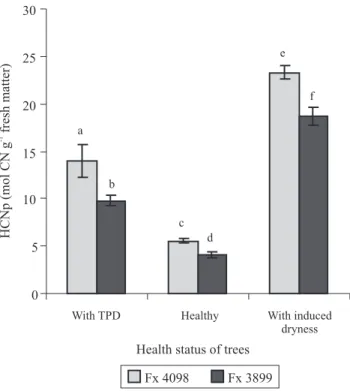
HCNp levels in the trunk inner barks were statistically different depending on tree health status and clone (Figure 1). The levels was relatively low in untreated healthy trees, and it was higher in plants with TPD. When partial dryness occurred following applications
of linamarin, the HCNp was very highly increased,
particularly in the clone Fx 4098.
The first important thing is that HCNp of healthy
rubber trees was low in the trunk bark compared to what is known in the literature for plants containing linamarin. Fokunang et al. (2001) found 530 µg of CN g-1 fresh matter in cassava roots, and Miller &
Conn (1980) 552 µg of CN g-1 fresh matter in flax aerial
parts, which are much higher than the 144 and 107 µg
of CN g-1 fresh matter observed here, respectively for
the Fx 4098 and Fx 3899 clones. However, it must be taken into account that HCNp also varies with the organ and the clone considered (Niedzwiedz-Siegien, 1998; Hydayat et al., 2002). In rubber tree, a clonal variability in HCNp on leaves was reported among Brazilian and between two Thai clones (Moraes et al., 2002; Kongsawadworakul et al., 2009). Moreover, Kongsawadworakul et al. (2009) reported that the levels of HCNp (expressed as mmol L-1 of cyanide)
are approximately four times lower in the trunk inner barks than in the young mature leaves. Therefore, the HCNps found in the tapping panel inner barks of healthy rubber trees were in the normal physiological
range for tissues that do not synthesize linamarin "de novo".
Secondly, temperature, moisture, light intensity and phosphorus nutrition are reported to affect
HCNp in white clover (Vickery et al., 1987), and temperature and light to affect the level of
cyanoglucosides and linamarase activity in flax
seedling (Niedzwiedz-Siegien & Gierasimiuk, 2001). But, climatic and nutritional factors are presumed to be inconsiderable in the examined rubber trees because humid tropical climate prevails at the Embrapa plantation, and there was only an eight-day interval between the measurements before and after treatment. Therefore, partial bark dryness probably took place in the trunk barks of the rubber trees because of the linamarin applications that caused a large increase of HCNp and, presumably, an excessive cyanogenesis in the inner phloem.
Thirdly, it was recently proved that tapping reduces
HCNp of Hevea trunk inner barks, at least during the
first three years of exploitation (Kongsawadworakul
et al., 2009). When TPD was detected in the Embrapa plantation, the trees had no longer been tapped as usual, but that only lasted around a fortnight until the end of the
experiment. Cessation of tapping in the TPD-affected trees could not be the principal cause of the difference of HCNp levels between these trees and the healthy ones or the trees with induced dryness. At the trunk inner barks, HCNp had increased with the occurrence of TPD in the trees exhibiting spontaneous bark dryness, and it increased also in trees with bark dryness induced by linamarin because of the treatment.
The pressure potential of the trunk laticifers (latex
ΨP) varied statistically depending on tree health status
and clone (Figure 2). Initially, the positive latex ΨP
was approximately 1.19 MPa in untreated healthy trees, with clonal difference: slightly lower in Fx 4098 than in Fx 3899. Just after the induction of partial dryness by applications of linamarin or KCN, it was reduced by more than 25% whatever the tree (average reduction of 30% in Fx 4098, and 36% in Fx 3899) and fell to approximately 0.79 MPa, regardless the clone and the cyanogenic compound applied.
Latex ΨP diurnal variations are reported in field-grown
rubber trees in Sri Lanka (Milburn & Ranasinghe, 1996): maximum values of 1.4 MPa occurred in the early morning, decreasing progressively to 0.35 MPa towards midday, then again increasing towards the end of the day. These variations only exist in non-wintering
(leafy) trees, they are associated with changes in the xylem sap pressure potentials and suggested to be caused by transpirational pull withdrawing water from the laticifers. Given the present experimental conditions (measurements performed just before tapping, in the early morning, with an eight-day interval, under humid tropical climate), the reduction of latex pressure potentials was probably caused by treatments. Furthermore, according to Pakianathan et al. (1982), latex ΨPs of less
than 7 to 8 atm (0.7–0.8 MPa) are frequently associated with TPD. Since partial bark dryness occurred when trunk bark HCNp was highly increased by the linamarin treatment, and since linamarin had the same effect as KCN, we deduced that excessive release of cyanide caused a fall of latex pressure potential in barks, down to approximately 0.79 MPa, resulting in partial dryness.
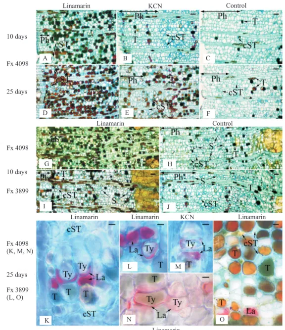
Structural and histochemical changes appeared in the inner part of the trunk secondary phloem following the cyanogenic treatments (Figure 3). When partial dryness occurred on the tapping cuts of treated trees (from the 10th day on after the beginning of the
experiment), tannin cells were more abundant than in the control trees whatever the compound (linamarin or KCN) used (Figure 3 A–J), and their amount increased with time, more rapidly with linamarin than with KCN (Figure 3 A–F), and more in Fx 4098 than in Fx 3899 (Figure 3 G–J). In parallel, the conducting phloem width, characterised by largely opened sieve tubes, was reduced (crushed sieve tubes found closer to the cambium), and the starch granules were depressed in number (Figure 3 A–J and G–J, respectively). Parenchyma cell outgrowths into adjacent laticifers
called tylosoids were found from the 10th day on, and
were always associated with in situ coagulated latex (Figure 3 K–O). Moreover, the number of tylosoids increased with time, and became higher with linamarin than with KCN, and in Fx 4098 rather than in Fx 3098
(Table 1). Other abnormalities were found from the 25th
Figure 2. Pressure potentials of the trunk laticifers (latex
ΨP) in the Fx 4098 and Fx 3899 rubber tree clones, two days
before (healthy, untreated) and after applications of linamarin or KCN on the trunk bark. Means followed by equal letters do not differ significantly at 99% confidence level by Student’s t-test. Bars show the 95% confidence interval.
Table 1. Tylosoid abundance(1) in the trunk secondary phloem
of healthy trees treated or not with cyanogenic compounds, according to the clone and the day of sampling.
(1)Number of tylosoids on cross sections stained with Alcian blue-Oil
Red O, in an area derived from the functioning of 1.5 mm-long cambial segment greater than 6 mm2. DAE, days after the beginning of the experiment.
Treatment Clone Fx 3899 Clone Fx 4098 10th DAE 25th DAE 10th DAE 25th DAE
Linamarin 0 8 13 44
KCN 0 1 0 12
day on in Fx 3899, notably deposits of material naturally coloured in yellow golden within the cell walls and intercellular spaces (Figure 3 O).
Healthy trees treated with control solution had normal secondary phloem (Figure 3 C, F, H and J), identical to
that of healthy trees previously studied (Hébant & de
Faÿ, 1980; de Faÿ & Jacob, 1989a; Hao & Wu, 2000; Sando et al., 2009). Therefore, the control solution and experimental wounds made for its application had no observable effect, and the applied cyanogenic compounds (linamarin or KCN) did induce the histological changes detected at the occurrence of partial dryness. The
abnormalities specific to the laticifers – tylosoids and
associated coagulated latex – were previously observed only in hevea barks. Laticifers containing tylosoids and in situ coagulated latex together are always present in dry barks, typical of irreversible TPD, but also at the dry/non-dry interfaces of the barks, or laterally in the partly dry band at the limit of the dry areas (de Faÿ &
Hébant, 1980; de Faÿ, 1981; de Faÿ & Jacob, 1989b).
It must be stressed that increases in tylosoid amount and associated coagulated latex with time, in trees treated with linamarin or KCN, should logically result
in total cessation of latex flow (total bark dryness).
Furthermore, these histological abnormalities are produced in dry barks induced by overtapping or by local compression of the trunk (de Faÿ, 1981; de Faÿ & Jacob, 1989b). Local enriching in phenolic compounds, wound gum-like material secretion and anarchic cell growth (hyperplasic cells) are general phloem degeneration phenomena, previously found
in barks affected with TPD (de Faÿ & Hébant, 1980;
de Faÿ, 1981; de Faÿ & Jacob, 1989b). Loss of functional sieve tubes by collapse is a normal seasonal phenomenon happening when sieve tubes are ageing, and it characterises the non-conducting phloem. However, in the treated trees collapses occurred in younger sieve tubes (closer to the cambium), so that the band of conducting phloem tended to disappear. Decreases in the phloem conducting area occur in dry rubber trees after overtapping (de Faÿ, 1981). Complete disappearance of functional sieve tubes is reported in the case of TPD, but only in the rare dry barks to the cambium, and the phenomenon is regarded as temporary (de Faÿ, 1981; de Faÿ & Jacob, 1989b).
Reduction of starch reserves in the inner part of the non-conducting phloem is probably directly linked to the loss of functional sieve tubes. However, total
sugar and starch contents of soft bark tissues of Hevea
brasiliensis are relatively higher in TPD-affected trees than in healthy ones (Krishnakumar et al., 2001). The difference between the histological approach (present work) and the biochemical approach above cited may be due to the fact that the rubber trees were studied
at two distinct reaction stages, at most 25 days after the beginning of cyanogenic treatments and at an advanced stage of TPD, respectively. Amylolysis may be transitory, and storage of carbohydrates would occur when biosynthesis of rubber became low in the tapping panel, due to major in situ coagulation of latex. Finally, the partially dry barks induced by cyanogenic treatment had a deteriorated phloem, notably displaying tylosoids associated with in situ coagulated latex, such as totally dry barks of trees irreversibly affected by TPD, dry barks induced by overtapping or as highly compressed barks.
The clone Fx 4098 had the higher HCNp and the quicker and stronger responses to the cyanogenic compounds, notably forming more tylosoids. However, it is known to be less prone to TPD than Fx 3899. Tylosoids associated with in situ coagulated latex are the
first-formed histological abnormalities able to cause bark
dryness through blockage of laticiferous vessels, but they are not the only ones. Golden yellow deposits resembling wound gums appear afterwards (in this experiment, on the 25th day in Fx 3899). They are lignified and present
in abundance in TPD-affected trees, particularly within the laticifer (cell wall and protoplasm) having their latex coagulated (de Faÿ, 1981; de Faÿ & Jacob, 1989b), and in the base of tapping panel when bark dryness spreads rapidly following overtapping (de Faÿ, 1981; de Faÿ
& Jacob, 1989b). These lignified deposits may cause
a rapid vertical spread of in situ coagulation of latex. Moreover, they are likely responsible for the brownish of bark at the macroscopic level, at least in part. Fx 3899 might be more prone than Fx 4098 to secrete the wound gum-like material, which would explain its greater proneness to TPD.
Conclusions
1. Excessive release of cyanide in the trunk bark of rubber trees decreases latex ΨP, resulting in partial
dryness of the bark, and increases HCNp level.
2. It also induces histological and histochemical changes in the secondary phloem, such as increased biosynthesis of tannins, early collapse of sieve tubes, and formation of tylosoids associated with in situ coagulated latex.
3. Tylosoids and the associated in situ coagulated latex are more precisely the structural markers of TPD early stages, and their presence is the microscopic sign
4. The clone Fx 4098, having the higher initial level of HCNp in the tapping panel phloem, is more prone to form these structural markers of early irreversible TPD.
References
CHRESTIN, H.; SOOKMARK, U.; TROUSLOT, P.; PELLEGRIN, F.; NANDRIS, D. Rubber tree (Hevea brasiliensis) bark necrosis syndrome. III. A physiological disease linked to impaired cyanide metabolism. Plant Disease, v.88, p.1047, 2004.
CLÉMENT-DEMANGE, A.; PRIYADARSHAN, P.M.; HOA, T.T.T.; VENKATACHALAM, P. Hevea rubber breeding and genetics. In: JANICK, J. (Ed.). Plant breeding reviews. Hoboken: John Wiley & Sons, 2007. v.29, p.177-283.
DE FAŸ, E. Histophysiologie comparée des écorces saines et pathologiques (maladie des encoches sèches) de l’Hevea
brasiliensis. 1981. 66p. Thesis (Ph.D.) – Université des Sciences
et Techniques du Languedoc, Montpellier.
DE FAŸ, E.; HEBANT, C. Étude histologique des écorces d’Hevea brasiliensis atteint de la maladie des encoches sèches. Comptes Rendus de l’Académie des Sciences de Paris. Série D, v.291, p.865-868, 1980.
DE FAŸ, E.; JACOB, J.-L. Anatomy and cytology of the laticiferous system of Hevea brasiliensis. Anatomical organization of the laticiferous system in the bark. In: D’AUZAC, J.; JACOB, J.-L.; CHRESTIN, H. (Ed.). Physiology of rubber tree latex. Boca Raton: CRC Press, 1989a. p.3-14.
DE FAŸ, E; JACOB, J.-L. The bark dryness “disease” (brown bast) of Hevea: symptomatology, histological and cytological aspects. In: D’AUZAC, J.; JACOB, J.-L.; CHRESTIN, H. (Ed.).
Physiology of rubber tree latex. Boca Raton: CRC Press, 1989b. p.407-430.
FOKUNANG, C.N.; TOMKINS, P.T.; DIXON, A.G.O.; TEMBE, E.A.; SALWA, B.; NUKENINE, E.N.; HORAN, I. Cyanogenic potential in food crops and its implication in cassava (Manihot esculenta Crantz) production. Pakistan Journal of Biological Science, v.4, p.926-930, 2001.
GRUHNERT, C.; BIEHL, B.; SELMAR D. Compartmentation of cyanogenic glucosides and their degrading enzymes. Planta, v.195, p.36-42, 1994.
HAO, B.-Z.; WU, J.-L. Laticifer differentiation in Hevea brasiliensis: induction by exogenous jasmonic acid and linolenic acid. Annals of Botany, v.85, p.37-43, 2000.
HÉBANT, C.; DE FAŸ, E. Functional organization of the bark of
Hevea brasiliensis (rubber tree): a structural and histoenzymological study. Zeitschrift für Pflanzenphysiologie, v.97, p.391-398,
1980.
HYDAYAT, A.; ZURAIDA, N.; HANARIDA, I. The cyanogenic potential of roots and leaves of ninety nine cassava cultivars.
Indonesian Journal of Agricultural Science, v.3, p.25-32, 2002.
JACOB, J.-L.; PRÉVÔT, J.-C.; LACROTTE, R. Tapping panel dryness in Hevea brasiliensis. Plant Research and Development, v.2, p.15-21, 1994.
KONGSAWADWORAKUL, P.; VIBOONJUN, U.; ROMRUENSUKHAROM, P.; CHANTUMA, P.; RUDERMAN, S.; CHRESTIN, H. The leaf, inner bark and latex cyanide potential of Hevea brasiliensis: evidence for involvement of cyanogenic glucosides in rubber yield. Phytochemistry, v.70, p.730-739, 2009.
KRISHNAKUMAR, R.; ANNAMALAINATHAN, K.; SIMON, S.P.; JACOB, J. Tapping panel dryness syndrome in Hevea
increases dark respiration but not ATP status. Indian Journal of Natural Rubber Research, v.14, p.14-19, 2001.
LIEBEREI, R. Cyanogenesis of Hevea brasiliensis during infection with Microcyclus ulei. Journal of Phytopathology, v.115, p.134-146, 1986.
MILBURN, J.A.; RANASINGHE, M.S. A comparison of methods for studying pressure and solute potentials in xylem and also in phloem laticifers of Hevea brasiliensis. Journal of Experimental Botany, v.47, p.135-143, 1996.
MILLER, J.M.; CONN, E.E. Metabolism of hydrogen cyanide by higher plants. Plant Physiology, v.65, p.1199-1202, 1980.
MORAES, L.A.C.; MORAES, V.H. de F.; CASTRO, P.R.C. Aplicação de KCN e linamarina e a incompatibilidade de enxertia por translocação no clone de seringueira IPA 1. Scientia Agricola, v.58, p.717-723, 2001.
MORAES, L.A.C.; MORAES, V.H. de F.; MOREIRA, A. Efeito da cianogênese na incompatibilidade entre clones de copa de seringueira e o clone de painel IPA1. Pesquisa Agropecuária Brasileira, v.37, p.925-932, 2002.
NIEDZWIEDZ-SIEGIEN, I. Cyanogenic glucosides in Linum usitatissimum. Phytochemistry, v.49, p.59-63, 1998.
NIEDZWIEDZ-SIEGIEN, I.; GIERASIMIUK, A. Environmental factors affecting the cyanogenic potential of flax seedlings. Acta Physiologiae Plantarum, v.23, p.383-390, 2001.
PAKIANATHAN, S.W.; SAMSIDAR, H.; SIVAKUMARAN, S.; GOMEZ, J.B. Physiological and anatomical investigation on long-term ethephon stimulated trees. Journal of the Rubber Research Institute of Malaysia, v.30, p.63-79, 1982.
PETCH, T. The diseases and pests of the rubber tree. London: Macmillan, 1921. 351p.
RUBBER BOARD. FAQs, rubber cultivation, diseases and pests, parasitic and non‑parasitic maladies, tapping panel dryness. Available at: <http://rubberboard.org.in/Faq. asp?Id=203&Fid=147>. Accessed on: 14 Oct. 2010.
SANDO, T.; HAYASHI, T.; TAKEDA, T.; AKIYAMA, Y.; NAKAZAWA, Y.; FUKUSAKI, E.; KOBAYASHI, A. Histochemical study of detailed laticifer structure and rubber biosynthesis-related protein localization in Hevea brasiliensis using spectral confocal laser scanning microscopy. Planta, v.230, p.215-225, 2009.
SASS, J.E. Botanical microtechnique. Iowa: Iowa State University, 1958. 336p.
SOOKMARK, U.; PUJADE-RENAUD, V.; CHRESTIN, H.; LACOTE, R.; NAIYANETR, C.; SEGUIN, M.; ROMRUENSUKHAROM, P.; NARANGAJAVANA, J. Characterization of polypeptides accumulated in the latex cytosol of rubber trees affected by tapping panel dryness syndrome. Plant and Cell Physiology, v.43, p.1323-1333, 2002.
VENKATACHALAM, P.; THULASEEDHARAN, A.; RAGHOTHAMA, K. Molecular identification and characterization
of a gene associated with the onset of tapping panel dryness (TPD) syndrome in rubber tree (Hevea brasiliensis Muell.) by mRNA differential display. Molecular Biotechnology, v.41, p.42-52, 2009.
VICKERY, P.J.; WHEELER, J.L.; MULCAHY, C. Factors affecting the hydrogen cyanide potential of white clover (Trifolium repens L.). Australian Journal of Agricultural Research, v.38, p.1053-1059, 1987.