www.ccarevista.ufc.br ISSN 1806-6690
In vitro
propagation and acclimatization of
Lippia rotundifolia
, an
endemic species of Brazilian Campos Rupestres
1Propagação
in vitro
e aclimatização de
Lippia rotundifolia
, uma espécie endêmica dos
Campos Rupestres Brasileiros
Cristiano Ferrara de Resende2, Ricardo Ernesto Bianchetti2, Aline Mystica Silva de Oliveira2, Virgínia Fernandes
Braga2 e Paulo Henrique Pereira Peixoto2*
ABSTRACT - The importance in folk medicine, combined to threats in their environment, becomes necessary to carry out studies involving large-scale propagation ofLippiagenus. Although the tissue culture propagation is widely disseminated for medicinal plants, forL. rotundifolia any article was published yet. The present study aimed to establish an efficient protocol for micropropagation ofL. rotundifolia. Nodal segments, taken from plants collected in the Espinhaço Range, were disinfected, and cultures were initiated on MS medium with PVPP (1 g L- 1), sucrose (3%) and agar (0.7%). The culture were maintained in a
growth room at controlled conditions. Disinfestation procedures and the supply of PVPP on culture media resulted in both reduced contamination and phenol oxidation rates, with more than 90% of viable cultures. In the multiplication phase were tried different BAP and NAA combinations supplied to the MS medium. The treatment that resulted in highest multiplication rates was 0.33 M BAP. The effects of NAA were evaluated forin vitrorooting. At 0.44 M, rooting was 70% higher than that observed in the control. The acclimatization was held in trays with substrate, coated with translucent plastic and kept under shade. The plantlets were transferred to the greenhouse after 15 days and transplanted to plant beds after 30 days. The acclimatized plantlets bloomed one year after the transference to field conditions, showing that thein vitro culture did not affect the vegetative and reproductive development, which confirms the potential of micropropagation to reduce the extinction risk ofL. rotundifolia.
Key words: Micropropagation.In vitro multiplication.In vitro rooting.Ex vitro acclimatization. Biodiversity conservation.
RESUMO - A importância na medicina popular, assim como as ameaças em seu ambiente, tornam necessária a realização de estudos envolvendo a propagação em larga escala de plantas do gêneroLippia. Embora a propagaçãoin vitro seja amplamente disseminada para plantas medicinais, nenhum artigo foi publicado ainda comL. rotundifolia. O presente estudo teve como objetivo estabelecer um protocolo eficiente para a micropropagação dessa espécie. Segmentos nodais, retirados de plantas coletadas na Cadeia do Espinhaço, foram desinfetados antes da inoculação em meio MS suplementado com PVPP (1 g L-1), sacarose (3%) e ágar (0,7%). Os tubos de
ensaio foram mantidos em sala de crescimento sob condições controladas. O tratamento de desinfestação e a adição de PVPP aos meios de cultura resultaram na redução da infecção microbiológica assim como nas taxas de oxidação fenólica, proporcionando mais de 90% de culturas assépticas e viáveis . Na fase de multiplicação foram testadas diferentes combinações de BAP e ANA adicionadas ao meio MS. O tratamento que resultou em maiores taxas de multiplicação foi de 0,33 M de BAP. Os efeitos do ANA foram avaliados no enraizamentoin vitro. A 0,44 M de ANA, o enraizamento foi 70% superior ao observado no controle. A aclimatização foi realizada em bandejas com substrato, revestidas com plástico translúcido e mantidas à sombra. As plantas foram transferidas para casa de vegetação após 15 dias e transplantadas para canteiros após 30 dias. As plantas aclimatizadas floresceram após um ano da transferência para condições de campo, demonstrando que os procedimentos de cultivoin vitro não afetaram o desenvolvimento vegetativo e reprodutivo das plantas, o que confirma o potencial da micropropagação para redução dos riscos de extinção deL. rotundifolia.
Palavras-chave: Micropropagação. Multiplicaçãoin vitro. Enraizamentoin vitro. Aclimatizaçãoex vitro. Conservação da biodiversidade.
DOI: 10.5935/1806-6690.20150041 *Autor para correspondência
1Recebido para publicação em 22/11/2013; aprovado em 25/03/2015
Parte do Trabalho de Iniciação Científica do segundo autor desenvolvido com bolsa do Programa BIC-UFJF
2Departamento de Botânica, Instituto de Ciências Biológicas/Universidade Federal de Juiz de Fora, Rua José Lourenço Kelmer, s/n, Martelos,
INTRODUCTION
The Verbenaceae, one of the five most important
eudicotyledonous families of theCampos Rupestres (dry,
rocky grasslands), is distributed in almost all terrestrial
ecosystems (GIULIETTI et al., 2000). In the family,
the genera Lippia, Lantana and Stachytarpheta are
considered the most important (SALIMENA-PIRES,
1991). The genusLippia belongs about 200 species that
occur in South and Central America and tropical Africa. The most number of species, about 150, is found in Brazil, with a higher incidence in the Espinhaço Range, in Minas Gerais, and in Chapada Diamantina, in the state of Bahia (SALIMENA-PIRES, 1991). Species
of genus Lippia present economic importance due
to the wide use of its essential oils and its medicinal
properties (PASCUAL et al., 2001;
SALIMENA-PIRES, 1991). In the Tropical America, Verbenaceae is quite used for its gastrointestinal properties and against respiratory diseases. However, in countries such Brazil and Guatemala, the Verbenaceae are used against skin diseases, burns and ulcers. In most cases, their leaves and flowers are used as a source of herbal drugs
(PASCUALet al., 2001).
Lippia rotundifolia Cham. is a Verbenaceae shrub
with underground xylopodium root system and oval leathery leaves. The plants present corymbs inflorescence with corolla lilacs strongly aromatic, containing many
glandular trichomes. Leaves ofL. rotundifolia are rich in
monoterpenes. Limonene is also found in inflorescences and myrtenol, your derivative, is accumulated over in the
leaves (LEITÃOet al., 2008). Limonene is known for its
medicinal properties, including action against tumors in mammals, and important drug in cancer therapies. To the myrtenol is also attributed the anticancer activity due to the inhibitory action of hepatocellular carcinoma and role in protecting membranes from damage caused by free radicals (BABU; PERUMAL; BALASUBRAMANIAN, 2012).
Considering that the anthropogenic perturbations in
Campos Rupestres are increasing strongly in recent decades, that little is known about the impact of these actions on the plant populations, and that the risk of extinction of some
endemic species are high, studies aiming at Lippia species
conservation become necessary (VITTA, 2002). The tissue culture technique has been widely used in recent years for the propagation of medicinal plants (PARVEEN; SHAHAZAD,
2011). Thein vitro clonal propagation through the axillary bud
may to produce lots of plants in a limited space, regardless of the season and under controlled environment conditions
(VARSHNEY; ANIS, 2012). For the success of in vitro
procedures, it is essential to establish protocols for large-scale propagation, rooting and acclimatization of these
plants. However, it is surprising that few in vitro studies
have been published with plants of the Lippia genus. In
the literature, studies they were found only for Lippia
junelliana (JULIANIet al., 1999),Lippia alba (GUPTA;
KHANUJA; SUSHIL, 2001),Lippia filifolia (PEIXOTO
et al., 2006) andLippia sidoides (COSTAet al., 2007).
Due to the endemism and the pharmacological activities of the Verbenaceae, this study aimed to develop
an efficient protocol forin vitro propagation andex vitro
acclimatization ofL. rotundifolia, from the perspective of
reducing the risk of extinction of this species in its natural environment and its future use in pharmacological studies.
MATERIAL AND METHODS
In vitroEstablishment:
Plants ofLippia rotundifolia Cham. were collected
in the Espinhaço Range (Minas Gerais, Brazil) and established by vegetative propagation in the Experimental Station of Plants Propagation at the Federal University of Juiz de Fora (Minas Gerais, Brazil). Fertile specimens were deposited in the Herbarium of the Federal University of Juiz de Fora (CESJ) under number 31,376. Aiming to
establish in vitro cultures, plants maintained under
field conditions were subjected to surface sterilization with 1.72 mM Benomyl (DuPont®, USA), sprayed once a week for 30 days. Thirty nodal segments were collected from the plants and washed in running tap water added drops of commercial detergent for 1 h. Then, the explants were immersed in 70% ethanol (v/v) for 30 minutes and after in a solution of 1.72 mM Benomyl for 10 minutes. Finally, the nodal segments were immersed in commercial bleach solution (2% of active chlorine) at 30% (v/v) dilution, added of Tween 20 (20 L) for 15 minutes. Subsequently, the explants were washed five times in distilled and autoclaved water and then inoculated on MS medium (Murashige; Skoog, 1962) at half strength salts and vitamins, without growth regulators. In order to control the phenolic oxidation,
polyvinylpolypyrrolidone (PVPP, 1 g L-1) was also supplied
to the culture media. The occurrence of bacterial and fungal contamination as well as phenolic oxidation in tissues and culture media were evaluated daily.
In vitro culture conditions: All cultures were kept in test tubes 2.5 x 15 cm. The culture media was the MS medium supplemented with 3% sucrose (w/v) and 0.7% agar (w/v). The pH of the culture media was adjusted to 5.7 ± 0.1 before autoclaving, performed for 20 min at 120 °C and 1 atm pressure. The tubes were capped with polyethylene autoclavable closures and sealed with 15 µM PVC film. Cultures were maintained in a growth chamber with controlled temperature (26 ± 1 °C),
In vitro multiplication: Thirty days after the
establishment of aseptic cultures, studies concerning in
vitro multiplication were performed. Nodal segments, measuring 2-3 cm were inoculated vertically on culture media in full strength of MS salts and vitamins, supplied with 6-benzylaminopurine (BAP: 0; 0.33; 0.66 or 1 µM) and -naphthalene acetic acid (NAA: 0; 0.001; 0.01 or 0.1 µM) in all combinations, for a total of 16 different treatments, with 10 replicates each. After 40 days of inoculation, we assessed the shoot and the root numbers and the height of the largest shoot.
In vitro rooting: The in vitro plantlets rooting was evaluated using nodal segments, measuring 2-3 cm, previously stabilized and maintained on MS medium, without growth regulators. The nodal segments were inoculated in MS medium at the total strength of the salts and vitamins and supplied of NAA (0; 0.11; 0.22; 0.33 or 0.44 µM), for a total of 5 different treatments, with 10 replicates each. After 45 days, the cultures were evaluated considering of the number and size of shoots and roots.
Ex vitro acclimatization: Fifty rooted plantlets were removed from the test tubes and their roots were washed in tap water. Then the material was transplanted into polystyrene trays filled with substrate Plantmax HT®, covered with clear plastic. The plants were kept under shade and watered for 15 days until complete wetting of the substrate. Subsequently, the trays were transferred to a greenhouse with automated micro-sprinkler irrigation. After 45 days, the seedlings were transferred to beds filled with soil/sand/cattle manure, in the proportion 3:2:1 (v/v/v) and irrigated weekly.
Statistical Analyses: In all trials, the count data were
normalized using (x + 0.5), and the measurement data employing log (x + 1). The experiments were performed in a completely randomized design (CRD) and the data analyzed by ANOVA. Means were compared with Tukey’s test at 5% probability using the software SAEG (version 9.1).
RESULTS AND DISCUSSION
In vitro establishment: The pharmacological
potential (PASCUALet al., 2001) and the anthropogenic
threats in their natural environment (GIULIETTIet al.,
2000; VITTA, 2002), makes it necessary to perform studies concerning alternative techniques of culture and
propagation of Lippia rotundifolia. Tissue culture is a
propagation procedure that enables rapid achievement of clones, regardless of adverse environmental influences. The success of micropropagation depends
primarily on in vitro establishment of aseptic cultures
free of phenolic oxidation. The PVPP showed high efficiency in the control of phenolic oxidation avoiding
tissue and culture medium browning. Less than 2% of explants showed browning caused by the action of polyphenol oxidases. The efficiency of PVPP in the control of phenolic oxidation is documented in the literature. The PVPP is an amorphous polymer without
physiological activity (BASHA, 2011), used forin vitro
establishment of genotypes that is prone to phenolic
oxidation (BUSSELEZet al., 2012). The PVPP acts by
complexing phenolic and alkaloids compounds released in response to tissue injury, reducing the toxic effects on
the cells (FOLCH-CANO; OLEA-AZAR; SPEISKY,
2013; MALIKet al., 2010; AMIN; JAISWAL, 1988).
The spraying of Benomyl in the plants maintained
ex situ and also their inclusion in the culture medium
coupled with the others aseptic procedures performed in the laboratory were quite effective, resulting in more than 90% cultures free of fungi and bacteria colonies.
The methodology used in disinfestation method of L.
rotundifolia showed high efficacy, which was similar to that
used successfully with other Verbenaceae such asL. filifolia
(PEIXOTOet al., 2006),Verbena litoralis (BRAGAet al.,
2012) andBouchea fluminensis (RESENDEet al., 2014).
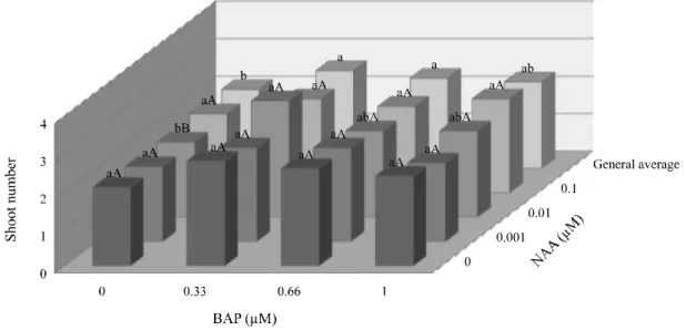
In vitro multiplication: In the multiplication phase, the explants showed highest propagation rates when inoculated on MS medium supplied with 0.33 µM BAP, with average of 2.6 shoots/explant (Figure 1). The higher concentrations of this growth regulator resulted in reduction in the shoots multiplication, suggesting sensitivity of this species to cytokinins. The supply of NAA does not significantly stimulated the proliferation of shoots, beyond that auxin had not significantly interacted with BAP.
In spite of BAP is widely reported in the literature
for Verbenaceae micropropagation (BRAGA et al.,
2012; PEIXOTO et al., 2006; VIDYA et al., 2012;
RESENDE et al., 2014), for L. rotundifolia a tendency
of phytotoxicity was observed in response to increasing in BAP concentration, which resulted in reduction in the number and in the size of shoots, besides to the occurrence of hyperhydricity. Typically, BAP is more efficient than
cytokinins derived from adenine onin vitro propagation
phase (SHIVA PRAKASH; PENTAL;
BHALLA-SARIN, 1994; SHUKLAet al., 2012; THIYAGARAJAN;
VENKATACHALAM, 2012). In in vitro cultures of L.
alba, elongated shoots were observed when BAP was
added to the culture medium (GUPTA; KHANUJA;
SUSHIL., 2001).
Figure 1 - Averages of shoot number in cultures of L. rotundifoliain response to BAP and NAA, 40 days afterin vitroinoculation. Means followed by the same lower-case letters (comparing BAP doses) and upper-case letters (comparing NAA doses) are not different according to Tukey’s test at 5% of probability
but also on its auxin interaction. In this phase of micropropagation, the highest multiplication rate was observed in response to 0.33 µM BAP + 0.01 µM NAA (Figure 1), however, without significant differences in relation to other auxin concentrations. Even so, the
multiplication rate found for L. rotundifolia (3.1 shoots/
explant) contrasts with the results for other Verbenaceae. ForLippia filifolia, about nine fold more shoots/explant
were found (PEIXOTOet al., 2006). ForLippia juneliana,
the maximum shoot number obtained was also higher than
in the present study (JULIANIet al., 1999). These results
demonstrate specificity in response toLippia genotypes,
which suggest the necessity for adjustment of protocols
to improve the efficiency ofin vitro propagation for each
species.
Besides presenting a relatively reduced propagation rate, callus formation and hyperhydricity were also
observed in cultures of L. rotundifolia in the treatments
where BAP was supplied. Although the hyperhydricity
and calli occurrence were generally observed in vitro
only at higher cytokinins concentrations (SHUKLA et
al., 2012), in vitro cultures of L. rotundifolia showed
that physiological abnormality in every one of BAP concentration (Figure 2a).
Among other adverse effects, the hyperhydricity may result in weakly rooting seedlings and in abnormal plantlets, with low stomatal efficiency and reduced amount of chlorophyll, which sometimes results in reduced seedling survival on acclimatization
(KEVERSet al., 2004). To avoid the effects caused by
hyperhydricity, BAP at 0.33 µM seems to be the most
suitable concentration for in vitro multiplication of L.
rotundifolia. According Shiva Prakash; Pental; Bhalla-Sarin (1994), the relative efficiency of cytokinins on shoot proliferation follows the order BAP > kinetin > zeatin > adenine sulfate. Recent studies demonstrating the existence of more powerful sources of this class of plant growth regulators such as N-(2-Chloro-4-pyridyl)-N-phenylurea (4-CPPU) and, specially, Thidiazuron
(TDZ) (ROLLI et al., 2012). The TDZ, besides acting
as a synthetic cytokinin, promotes the overexpression of natural cytokinins, being successfully used for some species to stimulate shoots multiplication at concentrations bellow than 1 µM (VARSHNEY; ANIS,
2012). Although the in vitro propagation observed
for L. rotundifolia have been relatively small, any other method of sexual or vegetative reproduction overcomes the micropropagation when comparing the multiplication rate/explant, which emphasizes the importance of tissue culture as an efficient method for large-scale propagation of this species. In addition, the
in vitro germplasm conservation is possible regardless of biotic and edaphoclimatic conditions. Although not assessed in this study, the TDZ might be the only cytokinin which stimulates a higher shoots proliferation in L. rotundifolia. However, in the literature, there are several studies that found a direct correlation between TDZ and hyperhydricity (HUETTEMAN; PREECE,
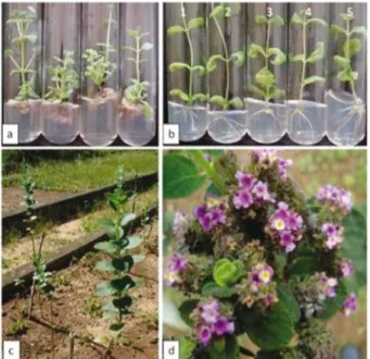
Figure 2 - In vitro propagation and ex vitro acclimatized Lippia rotundifolia. a. Multiplication phase showing shoots proliferation, hyperhydricity and basal callus on microcuttings in response to MS + BAP (0.33, 0.66 or 1 µ M). b. Rooting phase: 1 - MS (control); 2 - MS + NAA 0.1 µ M; 3 - MS + NAA 0.2 µM; 4 - MS + NAA 0.3 µM; 5 - MS + NAA 0.4 mM. c. Plants after completedex vitro acclimatization. d. Detail of inflorescence in one year acclimatized plant of Lippia rotundifolia
Significant interaction between BAP and NAA were observed in relation to the height of the largest shoot and also for the number of roots. In the multiplication phase, shoots more elongated were observed in response to 0.33 µM BAP + 0.001 µM NAA (Figure 3). In higher concentrations of both growth regulators, reduction in
the size of shoots was observed, suggesting that, for L.
rotundifolia, the optimal concentration range is below to
the limits assessed. In this phase of micropropagation,in
vitro root formation was observed only in the absence of BAP, with best results in response to 0.1 M NAA (Figure 4).
Besides to the rooting inhibiting, the addition of BAP stimulated the development of callus at the shoots base and also the occurrence of hyperhydricity (Figure 2a).
This response is typical for the effects of cytokinins onin
vitro adventitious root formation, which occurs in response to the imbalance of the endogenous ratio between natural auxins and cytokinins (SANTOS; ARRIGONI-BLANK;
BLANK, 2012). These results show that in vitrorooting
phase should not be suppressed from theL. rotundifolia
micropropagation protocol.
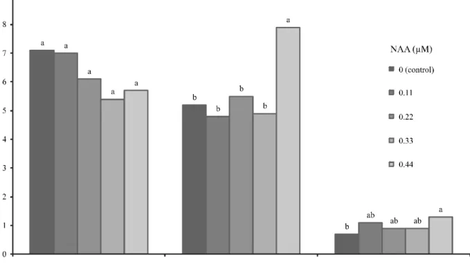
In vitrorooting: The supply of NAA increased the number of roots in the microcuttings. In the presence of 0.44 µM NAA, nearly to eight adventitious roots have been produced, an increase of 70% compared to the control which, in turn, does not differ from the other NAA concentrations (Figure 5).
Figure 5 - Effects of NAA onin vitro rooting of microcuttings ofL. rotundifolia, 40 days afterin vitro inoculation. Means followed by the same letters do not differ by Tukey’s test at 5% probability
and Bouchea fluminensis (RESENDE et al., 2014), another Verbenaceae. The supply of NAA, at all the concentrations evaluated, did not result in increasing in the shoots number and also was not effective in stimulating the shoots elongation (Figure 5).
Figure 4 - Averages of root number in cultures of L. rotundifolia in response to BAP and NAA, 40 days afterin vitroinoculation. Means followed by the same letters do not differ by Tukey’s test at 5% probability
ForLippia filifolia, thein vitrorooting was also promoted by NAA in the same concentration range
(PEIXOTO et al., 2006). The rhizogenesis occurs
through induction, initiation and expression phases. In every one of these developmental stages, auxins are critically and fundamentally important (SAUER; ROBERT; KLEINE-VEHN, 2013). However, the auxins should be supplied in suitable concentration, since excessive levels of these growth regulators inhibit the adventitious rooting, which occurs in response to ethylene production (GÜREL; WREN, 1995).
In the presence of 0.44 µM NAA, the roots showed a higher elongation (Figure 5) and improved morphological development, with more ramifications and greater thickness. In response to NAA, the cultures also showed no hyperhydricity symptoms nor the callus formation observed in multiplication phase in response to BAP (Figure 2b). Morphological and
physiological abnormalities are reported during in
vitro rooting, especially necrosis in shoots and callus development at the base of the explants (PARVEEN; SHAHZAD, 2011). However, in the present study,
microcuttings of L. rotundifolia showed no problems,
which was also no found toLippia filifolia (PEIXOTO
3. The NAA at 0.44 M provides a rooting 70% higher than in the control;
4. The acclimatization process was very successful, with more than 80% of plantlets survived;
5. The acclimatized plants bloomed one year after the transference to field conditions, showing that the in vitro culture did not affect the vegetative and reproductive development, which endorses the potential of micropropagation to reduce the extinction
risk ofL. rotundifolia.
ACKNOWLEDGEMENTS
We thanks the Fundação de Amparo à Pesquisa do Estado de Minas Gerais (FAPEMIG) and Programa de Apoio à Publicação da Universidade Federal de Juiz de Fora (PROPESQ-UFJF) for the financial support.
REFERENCES
AMIN, M. N.; JAISWAL, V. S. Micropropagation as an aid to rapid cloning of a guava cultivar.Scientia Horticulturae, v. 36, n. 1-2, p. 89-95, 1988.
ARRIGONI-BLANK, M. de F. et al. Micropropagação, aclimatização, teor e composição química do óleo essencial de genótipos de hortelã japonesa.Revista Ciência Agronômica, v. 42, n. 1, p. 175-184, 2011.
BABU, L. H.; PERUMAL, S.; BALASUBRAMANIAN, M. P. Myrtenal, a nature monoterpene, down-regulates TNF- expression and suppresses carcinogen-induced hepatocellular carcinoma in rats. Molecular and Cellular Biochemistry, v. 369, n. 1/2, p. 183-193, 2012.
BASHA, M. A. F. Spectroscopic, magnetic, and optical characterization of nanocomposite films of polyvinylpyrrolidone doped with cerium disulphate. Journal of Applied Polymer Science, v. 122, n. 3, p. 2121-2129, 2011.
BRAGA, V. F.et al. Micropropagation, antinociceptive and antioxidant activities of extracts of Verbena litoralis Kunth (Verbenaceae).Anais da Academia Brasileira de Ciências, v. 84, n. 1, p. 139-147, 2012.
BUSSELEZ, R. et al. Component dynamics in polyvinylpyrrolidone concentrated aqueous solutions. The Journal of Chemical Physics, v. 137, n. 8, 084902, 2012. COSTA, A. S. et al. Estabelecimento de alecrim-pimentain vitro.Horticultura Brasileira, v. 25, n. 1, p. 68-72, 2007. FOLCH-CANO, C.; OLEA-AZAR, C.; SPEISKY, H. Structural and thermodynamic factors on the adsorption process of phenolic compounds onto polyvinylpolypyrrolidone. Colloids and Surfaces A: Physicochemical and Engineering Aspects, v. 418, p. 105-111, 2013.
Ex vitroacclimatization: Rooted seedlings from culture media containing 0.44 M NAA were acclimatized
ex vitro. More than 80% of the plantlets removed from the test tubes survived to acclimatization. In this process, the plantlets were initially maintained in greenhouse under shade. After an initial period of two weeks, the plantlets were transferred to the greenhouse and kept under a micro-sprinkler, twice a day, for five minutes. After 45 days, the acclimatized plants were transferred to beds exposed to direct sunlight and watered weekly. In this condition, the plants showed faster vegetative growth reaching, after 12 months, about 1-1.5 m in height and exhibited a typical morphology of the species (Figure 2c), with leathery leaves strongly aromatic. The acclimatized plants from in vitro rooting phase showed normal vegetative development and underground rooting stem system like xylopodium, characteristic of the species, which allow the plants regrowth after the wildfires (SALIMENA-PIRES, 1991). The plants under field conditions also showed typical reproductive development. The flowering
occurred in spring (Figure 2d), showing that in vitro
protocols and culture conditions did not affect the life cycle of the species. Additionally, the literature showed
that the in vitro culture may increase the content of
terpenoids, revealing that the medicinal properties might
be improved in response toin vitro culture
(ARRIGONI-BLANKet al., 2011; SILVAet al., 2013).
In this work, the large-scale micropropagation of L. rotundifolia protocols were accomplished with absolute success considering the high plant survival observed in acclimatization phase. After the restoration of plants under field conditions were observed no
change in the phenotypic characteristics amongin vitro
propagated individuals and the donor plants, showing that
the protocols used are efficient forin vitro propagation
and conservation ofL. rotundifolia. Nowadays, several
plants of L. rotundifolia derived fromin vitro cultures
are completely adapted to field conditions, serving as sources of new propagules for different studies, allowing
the maintenance ofex vitro germplasm of this important
species inCampos Rupestres.
CONCLUSIONS
1. The field and laboratories disinfestation procedures and the supply of PVPP on culture media resulted in both reduced microbiological infection and phenol oxidation rates, with more than 90% of viable aseptic cultures;
Verbenaceae. Acta Botanica Brasilica, v. 28, n. 2, p. 184-189, 2014.
ROLLI, E.et al. Structure-activity relationships ofN -phenyl-N’-benzothiazol-6-ylurea synthetic derivatives: Cytokinin-like activity and adventitious rooting enhancement.
Phytochemistry, v. 74, p. 159-165, 2012.
SALIMENA-PIRES, F. R.Verbenaceae da Serra do Cipó, Minas Gerais, Brasil. 1991. 302 f. Dissertação (Mestrado em Botânica) - Instituto de Biociências, Universidade de São Paulo, São Paulo, 1991.
SANTOS, T. C.; ARRIGONI-BLANK, M. DE F.; BLANK, A. F. Propagação e conservaçãoin vitro de vetiver.Horticultura Brasileira, v. 30, n. 3, p. 507-513, 2012.
SAUER, M.; ROBERT, S.; KLEINE-VEHN, J. Auxin: simply complicated.Journal of Experimental Botany, v. 64, n. 9, p. 2565-2577, 2013.
SHIVA PRAKASH, N.; PENTAL, D.; BHALLA-SARIN, N. Regeneration of pigeonpea (Cajanus cajan) from cotyledonary nodevia multiple shoot formation.Plant Cell Reports, v. 13, n. 11, p. 623-627, 1994.
SHUKLA, M. R.et al.In vitro conservation of American elm (Ulmus americana): potential role of auxin metabolism in sustained plant proliferation. Canadian Journal of Forest Research, v. 42, n. 4, p. 686-697, 2012.
SILVA, B. O. et al. Micropropagation and in vitro production of secondary metabolites ofCroton floribundus Spreng. In vitro Cellular & Developmental Biology -Plant, v. 49, n. 3, p. 366-372, 2013.
THIYAGARAJAN, M.; VENKATACHALAM, P. Large scale in vitro propagation of Stevia rebaudiana (bert) for commercial application: Pharmaceutically important and antidiabetic medicinal herb.Industrial Crops and Products, v. 37, n. 1, p. 111-117, 2012.
VARSHNEY, A.; ANIS, M. Improvement of shoot morphogenesis in vitro and assessment of changes of the activity of antioxidant enzymes during acclimation of micropropagated plants of Desert Teak.Acta Physiologiae Plantarum, v. 34, n. 3, p. 859-867, 2012.
VIDYA, S. M.et al. Micropropagation ofClerodendrum serratum L. through direct and indirect organogenesis. Plant Tissue Culture and Biotechnology, v. 22, n. 2, p. 179-185, 2012. VITTA, F. A. Diversidade e conservação da flora nos Campos Rupestres da Cadeia do Espinhaço em Minas Gerais. In: Araújo, E. L.et al.Biodiversidade, conservação e uso sustentável da flora do Brasil. Recife: Sociedade Botânica do Brasil, 2002. p. 90-94.
GIULIETTI, A. M. et al. Caracterização e endemismo nos Campos Rupestres da Cadeia do Espinhaço.In: CAVALCANTI, T. B.; WALTER, B. M. T.Tópicos Atuais de Botânica. Brasília: EMBRAPA Recursos Genéticos, 2000. p. 311-318.
GUPTA, S. K.; KHANUJA, S. P. S.; SUSHIL, K. In vitro micropropagation ofLippia alba.Current Science, v. 81, n. 02, p. 206-210, 2001.
GÜREL, E.; WREN, M. J. In vitro development from leaf explants of sugar beet (Beta vulgaris L.) rhizogenesis and the effect of sequential exposure to auxin and cytokinin.Annals of Botany, v. 75, n. 1, p. 31-38, 1995.
HUETTEMAN, C. A.; PREECE, J. E. Thidiazuron: a potent cytokinin for woody plant tissue culture.Plant Cell, Tissue and Organ Culture, v. 33, n. 2, p. 105-119, 1993.
JULIANI JR, H. R. et al. Micropropagation of Lippia junelliana (Mold.) Trone. Plant Cell, Tissue and Organ Culture, v. 59, n. 3, p. 175-179, 1999.
KEVERS, C. et al. Hyperhydricity of micropropagated shoots: a typically stress-induced change of physiological state. Plant Cell, Tissue and Organ Culture, v. 77, n. 2, p. 181-191, 2004.
LEITÃO, S. G.et al. Analysis of the chemical composition of the essential oil extracted from Lippia lacunosa Mart. & Schauer and Lippia rotundifolia Cham. (Verbenaceae) by gas chromatography and gas chromatography-mass spectrometry.Journal of the Brazilian Chemical Society, v. 19, n. 7, p. 1388-1393, 2008.
MALIK, S.et al. Direct shoot regeneration from intact leaves of Arnebia euchroma (Royle) Johnston using thidiazuron.
Cell Biology International, v. 34, n. 5, p. 537-542, 2010. MURASHIGE, T.; SKOOG, F. A revised medium for rapid growth and bioassay with tobacco tissue culture.Physiologia Plantarum, v. 15, n. 3, p. 473-497, 1962.
PARVEEN, S.; SHAHZAD, A. A micropropagation protocol for Cassia angustifolia Vahl. from root explants. Acta Physiologiae Plantarum, v. 33, n. 3, p. 789-796, 2011. PASCUAL, M. E.et al.Lippia: traditional uses, chemistry and pharmacology: a review. Journal of Ethnopharmacology, v. 76, n. 3, p. 211-214, 2001.
PEIXOTO, P. H. P.et al. In vitro propagation of endangered Lippia filifolia Mart. and Schauer Ex Schauer.In vitro Cellular & Developmental Biology - Plant, v. 42, n. 6, p. 558-561, 2006.