Invited Review
Use of dual-energy x-ray absorptiometry in non-ruminant nutrition research
Candido Pomar1*, Marcos Kipper1,2, Marcel Marcoux1 1 Agriculture and Agri-Food Canada, Sherbrooke Research and Development Centre, Sherbrooke, Quebec, Canada. 2 Universidade Federal do Rio Grande do Sul, Faculdade de Agronomia, Departamento de Zootecnia, Porto Alegre, RS, Brazil.
ABSTRACT - Precise body composition measurements are essential in animal nutrition studies because the impact of treatments is evaluated based on changes in body weight and composition. Various indirect techniques for animal compositional evaluation have been developed and evaluated for applicability in animal nutrition studies. A fast, accurate, minimally invasive method that requires little input is considered the ideal for providing information about the animal. Measurements obtained by dual-energy x-ray absorptiometry (DXA) are highly correlated with those obtained by chemical analysis and dissection. The algorithms of DXA software partition the six chemical components of the body (lipids, water, proteins, carbohydrates, non-bone mineral, and bone mineral) into three compartments (total body mineral content, fat mass, and lean mass). Questions have been raised about how this partitioning affects the precision of the DXA method. In addition, the relationship between the DXA measurements and dissected carcass tissues is nonrepresentational of the relationship between DXA and chemical analysis. Furthermore, since DXA devices and their software were developed primarily for human medicine, they may not be fully adequate for animal evaluation. Calibration is required to obtain true values. The DXA method has some advantages and disadvantages that should be identified and controlled before calibration. Nonetheless, DXA is a valuable tool that provides precise, repeatable body composition measurements of live monogastric animals and their carcasses.
Key Words: alternative method, birds, body composition, DXA, pig
© 2017 Sociedade Brasileira de Zootecnia ISSN 1806-9290
www.sbz.org.br
R. Bras. Zootec., 46(7):621-629, 2017
Received: December 16, 2016
Accepted: March 11, 2017
*Corresponding author: candido.pomar@agr.gc.ca http://dx.doi.org/10.1590/S1806-92902017000700010
How to cite: Pomar, C.; Kipper, M. and Marcoux, M. 2017. Use of dual-energy x-ray absorptiometry in non-ruminant nutrition research. Revista Brasileira de Zootecnia 46(7):621-629.
Copyright © 2017 Sociedade Brasileira de Zootecnia. This is an Open Access article distributed under the terms of the Creative Commons Attribution License (http://creativecommons.org/licenses/by/4.0/), which permits unrestricted use, distribution, and reproduction in any medium, provided the original work is properly cited.
Introduction
Animals of all species can vary considerably in terms of weight and body composition depending on their growth stage, nutritional background, genetic potential, and other factors. Precise body composition measurements of experimental animals are essential in nutrition studies because the effect of treatments is evaluated based on changes in body weight and composition (Mitchell et al., 1997a; Pomar et al., 2001; Ryan et al., 2011). Nutrition research traditionally uses slaughter techniques to measure body composition, but this destructive method is expensive and precludes the possibility of taking repeated measurements of the same animal. Methods based on
indirect measurements allow repeated measurements of the same experimental animal, thus increasing accuracy and the ability to understand the growth and physiological processes involved in animal responses (Suster et al., 2006; Ryan et al., 2011). Indirect methods are preferred for determining animal nutritional requirements or for studying the effect of nutrition on animal growth. However, indirect methods require calibration to accurately estimate body composition (Szabo et al., 1999; Mitchell et al., 2003; Mercier et al., 2006).
Mercier et al., 2006), computed tomography (CT; Font-i-Furnols et al., 2013; Milisits et al., 2013), and magnetic resonance imaging (MRI; Davenel et al., 2000; Kremer et al., 2012b). The advantages and disadvantages of these methods are inherent to the technology and the principles used. For example, linear measurements are taken at specific locations on the body (body length, circumference, etc.). Many of these measurements are obviously related to total body weight or carcass wholesale cut weight, but they are of limited value for determining compositional patterns (Pomar et al., 2001; Pomar et al., 2009). Ultrasonic measurements of backfat and loin depth are taken on the assumption that these depths are closely related to body (or carcass) lean and fat masses (McLaren et al., 1991). These ultrasonic measurements have to be taken at specific body locations by experienced operators (McLaren et al., 1991; Oviedo-Rondón et al., 2007; Case et al., 2012). Linear body and ultrasonic measurements can, however, lose their accuracy when applied to animals of different weights, shapes, genetic backgrounds, etc. (Houghton and Turlington, 1992; Hassen et al., 1999). These accuracy losses are especially important when changes occur at body locations not being evaluated by these methods. In addition, ultrasonography has the disadvantage of not generating a complete body image due to insufficient penetration power (Liu and Stouffer, 1995). Other indirect methods, such as total body electrical conductivity, are not addressed in this paper because they may not be precise, since they are affected by the measuring environment and other limiting factors (Berg et al., 1994).
The DXA devices are more flexible than previous methods for estimating body composition in animals from different genetic lines (Mitchell et al., 1997b; Marcoux et al., 2005) or from different sexual categories because DXA evaluates the whole animal, not just parts of the body. Specific calibrations are, however, required for the evaluation of each animal type (i.e., pigs, sows, hens, poultry, etc.) or body parts (i.e., whole animal body, carcass, etc.). Other methods such as CT and MRI can also be used for whole-animal evaluation (Font i Furnols and Gispert, 2009; Barchia et al., 2010; Clarys et al., 2010; Kovner et al., 2010), but these methods are slow, expensive, and complex. However, the latter two technologies can be used to create methodologies for evaluating parts of the body to obtain an estimate for the whole animal (Kremer et al., 2012a; Kremer et al., 2012b). The purpose of this paper was to describe the use of DXA in non-ruminant nutrition research, highlighting some differences between DXA and the reference methods and identifying the advantages and disadvantages of this technology.
DXA as an alternative to in vivo body composition measurements
The value of DXA as a tool for evaluating the body chemical composition of live animals or the tissue composition of dissected carcasses has increased in recent years (Marcoux et al., 2005) because of its low instrumental and operating cost, high resolution, low radiation exposure, and rapid scan speed. Because this technique is non-invasive, live animals can be scanned at different production stages, thus improving research efficiency and reducing the number of animals required.
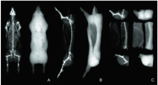
The DXA scanners were developed primarily for humans but are also used in animal studies. Several types of DXA devices are available, but they all operate in the same manner. An x-ray radiation source is aimed at a radiation detector placed directly opposite the site to be measured. The subject is placed on the table in the path of the radiation beam. The computer software records and analyzes the collected data (Blake and Fogelman, 1997). As the radiation source and detectors move over the subject, the DXA scanner generates a projected two-dimensional image, similar to the traditional x-ray (Figure 1). This two-dimensional image is made up of hundreds of pixels, which are the smallest units of an image (Tothill, 1995). The pixel size depends on the software configuration set prior to the scan.
bone mineral content (BMC) and bone mineral density, the latter of which is estimated as the ratio between BMC and the total area of bone pixels. Total DXA fat and lean masses are estimated by adding the masses of these tissues calculated in bone and non-bone soft tissues.
Measurements provided by DXA are highly correlated with total ash, protein, and lipid chemical composition, as well as with dissected adipose and muscle tissues. However, DXA tissue compositional data may not be the same across DXA devices or correspond to the chemical or dissected composition of the animal body or carcass. In fact, DXA tissue estimates normally have to be calibrated using acrylic materials rather than human or animal tissues (Scholz et al., 2007). Consequently, dissected bone does not correlate well with DXA BMC or DXA lean mass, since bones contain significant amounts of fat, protein, and water (Nielsen, 1973). Nonetheless, linear regression can be used to convert DXA measurements to body or carcass compositional measurements, but these regressions are specific to each DXA device, animal type, and body part (Mitchell et al., 1997b). It is important to be aware of differences between the principles of the methodologies when using one procedure to calibrate another (Marcoux et al., 2003). Most of the lack of agreement or adjustment between two methods can be attributed to what the results actually mean. In the following sections, we will discuss the main differences between measurements obtained by chemical analysis or dissection and those obtained by DXA.
Body chemical components vs. DXA measurements
The algorithms of DXA software partition the six chemical components of the body (lipids, water, proteins, carbohydrates, non-bone mineral, and bone mineral) into three DXA compartments (BMC, fat mass, and lean mass) (Pietrobelli et al., 1996; St-Onge et al., 2004) (Table 1). In animal bodies, carbohydrates and soft-tissue minerals are generally very small (Nielsen, 1973) and can be assumed to be negligible. In pigs, for example, DXA lean and fat values obtained with a GE Lunar Prodigy (Lunar Corp., Madison, WI, USA) device can be converted into the chemical equivalents of protein and lipid masses, as suggested by Pomar and Rivest (1996) in the following manner:
Total body protein (g) = −1384 + 0.216 × DXA lean (g) Total body fat (g) = 2825 + 1.009 × DXA fat (g) Body phosphorus (P) and calcium (Ca) can be estimated as the sum of the P and Ca present in the bones, lean, and
Main chemical
component of the body Chemical analysis compartment DXA compartment
Lipids Ether extract Fat mass
Water Water Lean mass
Proteins Nitrogen Lean mass
Carbohydrates -
-Non-bone minerals -
-Bone minerals Ash Bone mineral content
Table 1 - Partitioning of the six basic components into the chemical analysis and DXA compartments
DXA - dual-energy x-ray absorptiometry.
Adapted from Pietrobelli et al. (1996) and St-Onge et al. (2004). A: Image of a 90-kg live pig scanned in prone position. B: Image of a pig half-carcass. C: Image of pig primal cuts.
fat tissues of the body based on the DXA body measurements, assuming that P and Ca in bone accounts for 18% and 36% of BMC, respectively, that body proteins are associated with lean tissue containing 1.04% P and 0.042% Ca, and that body lipids are associated with fat tissue containing 0.05% P and 0.005% Ca (Nielsen, 1973; Letourneau-Montminy et al., 2015).
Some assumptions used by DXA in relation to bones should also be emphasized, since the technology is not designed to evaluate internal bone composition. Bone marrow fat is assumed to be constant between 4 and 5% (Mitchell et al., 1997a; Mercier et al., 2006), except in the skull, where the composition is assumed to be 17% due to the high lipid content of the brain (Hologic, 1996). However, these values depend on the brand of commercial DXA device used. The assumed values may constitute an important bias when comparing methods, since chemical analysis considers the actual lipid content, whereas the DXA method merely extrapolates the content to humans.
Lastly, although the dimensions of DXA equipment allow for pigs, dogs, and cats, among other non-ruminant animals, to be assessed, DXA software was developed solely to evaluate humans (except for some modes that allow small animals, such as rats, to be scanned). Consequently, any adjustments made to the software to better estimate body composition in humans (usually information considered confidential) may add a new source of bias in animal evaluation (Kipper et al., 2015).
Dissected carcass tissues vs. DXA measurements
The relationship between DXA measurements and dissected carcass tissues is somewhat nonrepresentational, as tissues obtained by dissection include several chemical components (Marcoux et al., 2003). The DXA method also identifies the various components within each dissected tissue (Table 2). Adipose and muscle tissues correspond to the respective content of lean and fat masses, while bone tissue corresponds to these two components plus BMC (Pietrobelli et al., 1996). The reason dissected tissues can be related to DXA measurements is that dissected adipose tissue has a strong correlation with DXA fat mass, dissected muscle tissues are correlated with DXA lean mass, and dissected bone tissues are correlated with BMC and lean mass (Marcoux et al., 2005). This is because fat is rich in lipids, the reference material of DXA fat mass; similarly, dissected muscle tissues are correlated with DXA lean mass, because lean beef is used as the reference for DXA lean mass (Pietrobelli et al., 1996). Bone tissue is correlated with BMC because DXA calibration can be carried out with artificial parts that simulate bones and its correlation with
lean mass is due to the strong positive relationship between muscle and bone (Pietrobelli et al., 1996; Schoenau, 2005). Therefore, it is necessary to take into consideration the fact that the manner in which the dissection method classifies the tissues will affect the model used to estimate their values using DXA measurements (Marcoux et al., 2003).
In some cases, due to practical or commercial issues, artificial assumptions are made to relate dissected carcass tissues and DXA measurements. Examples include the cartilage and skin, which can be treated as bone and adipose tissues, respectively, during dissection (Marcoux, 2001). The treatment of cartilage represents one of the greatest contrasts: it is often treated as bone during dissection, while DXA treats it as soft tissue. As a result, the bias in the DXA algorithms used to estimate dissected bone increases as the ratio of cartilage in the sample increases, especially in ribs (Marcoux et al., 2003; Mercier et al., 2006). Another point worth noting is that the entire foot can be classified as a bone in dissection, whereas the skin and foot tendons are considered to be part of the soft tissue compartment in DXA, thus generating additional bias. The skin can be treated as adipose tissue during dissection; however, skin has a lower lipid content than adipose tissue does. This chemical difference is also detected by DXA: adipose tissue (without skin) exhibits a fat mass of approximately 58%, while skin exhibits only 28% (unpublished data). A similar issue occurs in jowl classification: this meat cut can be treated as adipose tissue in dissection, but a small portion of muscle tissue is present. Different methods for estimating dissected tissues in pig and lamb carcasses and cuts can be found in Marcoux et al. (2005) and Mercier et al. (2006).
Advantages of DXA
Non-destructive methods like DXA allow for accurate, repeated measurements of total body composition, resulting in accurate evaluations of the effect of the experimental treatments used in non-ruminant nutrition research. With
Dissection DXA
Adipose tissue Fat mass
Lean mass
Muscle Fat mass
Lean mass
Bone Fat mass
Lean mass Bone mineral content
Table 2 - Relationship of DXA measurements to dissection tissues
DXA - dual-energy x-ray absorptiometry.
this technology, mathematical growth models can be calibrated using data obtained repeatedly over time for a given individual. The DXA method is less expensive than comparable slaughter techniques and reduces the error due to individual variability (Suster et al., 2006). Furthermore, DXA provides high reproducibility and minimizes operator effect (Raffan et al., 2006; Suster et al., 2006; Kipper et al., 2015). In addition, operator bias is usually high in the dissection of small (young) pigs, but this effect is not observed with DXA, in which accuracy is independent of animal size (Mitchell et al., 1998).
The DXA method has other advantages that are not directly related to theoretical concepts. Since slaughter is not necessary, some logistical issues are easier to manage, especially given that the trial requires fewer animals. Furthermore, DXA technology makes it possible to test a larger number of treatments in an experiment without increasing the number of animals. This method is extremely fast and the results are available immediately after image acquisition. Lastly, unlike actual destructive methods, the use of DXA in animal genetic selection could save individuals that exhibit advantageous characteristics or high genetic and economic value from being slaughtered.
Disadvantages of DXA
One of the major difficulties in using this technology is related to the hydration of the sample (i.e. the level of water in fat-free tissues [FFT]) (Emmans and Kyriazakis, 1997; St-Onge et al., 2004). Water content varies from one animal to another, being high in young and low in mature individuals (Emmans and Kyriazakis, 1995). After maturity is reached, the water:FFT ratio remains constant, around 0.73 in mammals (Wang et al., 1999). For example, in pigs, the ratio is approximately 0.87 at 1.5 kg body weight (BW), but this value falls to 0.76 at 54 kg and to approximately 0.73 at 145 kg BW (Shields et al., 1983). The water:FFT ratio is considered to be constant in DXA and can be deemed a bias in some situations (Roubenoff et al., 1993). This issue is so important that it has been extensively studied in juvenile and adult humans with disorders/diseases that alter the water equilibrium in the body (Snead et al., 1993; Georgiou et al., 1997). The water:FFT ratio bias must be taken into account in studies in which changes in BW of animals are an important factor.
Since the invention of DXA, thickness has been acknowledged as one of its most limiting factors (Goodsitt, 1992). The deeper the tissue, the lower the precision of consecutive measurements and, consequently, the less true the value (Jebb et al., 1995). In thick samples, the
low-energy x-ray is more attenuated than the high-low-energy x-ray, an effect known as the beam hardening effect (Seibert and Boone, 2005). Although the data provided by DXA are related to the values of the chemical methods, they are not identical, which means that some adjustments are required. Another point to be taken into account when making adjustments is that DXA devices and software were developed for use in human medicine and may not always be suitable for animal studies.
Accuracy, repeatability and reproducibility of DXA devices
Evaluating the accuracy of an instrument means determining the closeness between its measurements and the accepted reference values in terms of trueness and precision. The trueness of a measurement refers to the closeness of the agreement between the average value obtained from a large series of test results and an accepted reference value, while precision refers to the degree of internal agreement between independent measurements taken under specific conditions. A device is said to be accurate when it is true, i.e., when its measurements are close to the true values, and precise when there is no spread around the true value (International Organization for Standardization, 1993).
The accuracy, trueness, and precision of measurements obtained by DXA in pig half-carcasses and primal cuts were recently studied by our group (Kipper et al., 2015). Briefly, a trial was conducted to investigate the repeatability and reproducibility of DXA measurements taken in pig half-carcasses. The repeatability measures the error inherent to DXA readings. The repeatability conditions were created by scanning each carcass ten times in the same position. The reproducibility includes sources of variation in repeatability. The reproducibility was therefore related to the variation inherent to the positioning of the carcass on the DXA table. The reproducibility conditions were created by scanning each carcass once in each of ten different positions. After the scans, all images obtained under both repeatability and reproducibility conditions were analyzed either by using a custom rectangular region of interest (ROI) or by placing the carcass within the head, trunk, leg, and arm regions of interest of the standard grid for the human body. Coefficients of variation (CV) were computed for each carcass individually and then combined, assuming a normal variance distribution (Glüer et al., 1995).
technology was well calibrated. Methods with inadequate reproducibility can be easily adjusted and improved, but methods with inadequate repeatability require major structural changes (Burdick et al., 2005). The repeatability was generally lower than 1% and reproducibility was lower than 3%. Soft tissue was predicted with greater precision among the ROI. The custom ROI did not provide precise estimates of BMC, but it produced acceptable values for bone mineral density. The trunk ROI did not provide precise estimates of fat. This suggests that appropriate positioning of the half-carcass on the DXA table is required to avoid variability in the results.
The repeatability and the reproducibility of DXA devices have been studied in limited number of research projects. For example, Nielsen et al. (2004) described a measurement protocol suitable for use in postmortem specimens of low economic value (tarsus and carpus distally) from a large number of commercial pigs to quantify the precision of that protocol and to estimate accuracy of the technique for measuring BMC in this region. To achieve these objectives, these authors used the feet and tails of the pigs to evaluate bone mineralization. Scanning precision (i.e., repeatability) for each ROI was determined with data derived from ten separate scans from one pig. The reproducibility was evaluated with ten repeated scans performed as above, except that the specimen was removed from the table and repositioned before each scan. As in our study, reproducibility was always greater than repeatability, but an interesting finding was that small ROI presented higher CV and, therefore, ROI size needs to be established in accordance with the coveted precision of the measurement. Raffan et al. (2006) scanned ten dogs six times each, alternating between dorsal and lateral recumbency to determine the precision of body composition measurements in dogs. This study demonstrated that DXA is a precise method of body composition analysis in dogs.
Nevertheless, because differences were found between body positions and between operators, the protocols need to be properly standardized to ensure proper measurement repeatability.
Repeatability is related to the technology and cannot be easily corrected, while reproducibility is related to the measurement protocol and can therefore be adjusted to the required precision of the measurements.
Our team has recently published a study (Kipper et al., 2015) about how the thickness of meat samples can affect DXA measurements. Briefly, belly samples with a constant mass (and constant composition) were scanned, but between consecutive scans, the bellies were cut in half and stacked to increase the thickness. The samples were scanned using three software configurations. The scanning mode used was a total body, with one scan for each of the following options: thin (for samples less than 16 cm thick), standard (for samples of 16 to 25 cm thick), and thick (for samples more than 25 cm thick).
Neither the scanning mode options nor their interaction with the thickness had an effect on the DXA measurements. Thickness affected the percentage of fat, soft tissue, and lean mass, but did not affect the fat mass (Figure 2). The DXA measurements exhibited several different distribution patterns in relation to the thickness. The soft tissue and lean mass exhibited a quadratic pattern, while the percentage of fat was sigmoidal and the fat mass was not affected. There was a 43% variation between the lean masses estimated for the smallest and largest thicknesses. The percentage of fat increased up to a thickness of approximately 10 cm was similar between 10 and 20 cm and increased again beyond 20 cm. Despite their effect on the fat percentage, the fat mass values did not change (Figure 2). Thus, the variation in the fat percentage may be due to the soft tissue and its relation to fat mass. The results suggest that the most stable range of thicknesses is approximately 17 to 23 cm.
Table 3 - Coefficient of variation of DXA measurements obtained in pig half-carcasses using different regions of interest (ROI) under
repeatability and reproducibility conditions1
Custom ROI Head ROI Trunk ROI Arm ROI Leg ROI
Repeatability condition
BMD (g/cm2) 0.61 0.52 0.57 0.53 0.56
BMC (g) 0.68 0.56 0.68 0.56 0.55
Percentage of fat (%) 0.81 0.65 4.20 0.78 0.81
Soft tissue (kg) 0.07 0.04 0.10 0.04 0.04
Reproducibility condition
BMD (g/cm2) 0.64 0.64 0.67 0.67 0.62
BMC (g) 3.59 0.59 2.20 0.52 0.60
Percentage of fat (%) 2.30 1.71 21.16 2.64 2.69
Soft tissue (kg) 0.24 0.32 0.78 0.32 0.22
The differences observed in the DXA measurements may be due to a physical phenomenon called beam hardening, which is characterized by more pronounced attenuation of the low-energy x-ray (Seibert and Boone, 2005). This effect reduces R-values and consequently increases estimates of fat content. In addition, the effect of beam hardening increases in substances with higher R-values (Goodsitt, 1992). Thus, the greater the lean percentage, the more it will be underestimated due to this effect (Jebb et al., 1995). However, there were no differences in the magnitude of the effect when different scanning modes were compared. Since the configurations were designed to account for this effect, it could be deduced that these configurations do not work properly under the study conditions. However, more knowledge is required on this subject and this effect should be further examined in future research.
Conclusions
The DXA method is a valuable tool for providing accurate, repeatable, and reproducible body composition measurements of live monogastric animals and their carcasses. This non-destructive method is less expensive than the traditional slaughter techniques, allows repeated measures, reduces the errors due to individual weight or compositional variation, and removes operator biases. Subjects can be scanned quickly and compositional measurements are available soon after the scan. However, calibration is required to convert DXA values to the true chemical or dissected values. The factors that can affect the
precision of measurements and the disadvantages of this technology must be known and controlled to avoid potential bias. The subject hydration and thickness are between the most important ones.
References
Barchia, I. M.; Arthur, P. F.; Giles, L. R. and Eamens, G. J. 2010. Temporal growth and development of body tissues of pigs as assessed by X-ray computed tomography. Animal Production Science 50:322-328.
Berg, E. P.; Forrest, J. C. and Fisher, J. E. 1994. Electromagnetic scanning of pork carcasses in an on-line industrial configuration. Journal of Animal Science 72:2642-2652.
Blake, G. M., and Fogelman, I. 1997. Technical principles of dual energy X-ray absorptiometry. Seminars in Nuclear Medicine 27:210-228.
Burdick, R. K.; Borror, C. M. and Montgomery, D. C. 2005. Design and analysis of gouge R&R studies: making decisions with confidence intervals in random and mixed ANOVA models (ASA-SIAM Series on Statistics and Applied Probability). American Statistical Association, Alexandria, Virginia and Society for Industrial and Applied Mathematics, Philadelphia, Pennsylvania, USA.
Case, L. A.; Wood, B. J. and Miller, S. P. 2012. The investigation of ultrasound technology to measure breast muscle depth as a correlated trait to breast meat yield in turkey (Meleagris gallopavo). Journal of Animal Science 90:3410-3417.
Clarys, J. P.; Scafoglieri, A.; Provyn, S.; Louis, O.; Wallace, J. A. and De Mey, J. 2010. A macro-quality evaluation of DXA variables using whole dissection, ashing, and computer tomography in pigs. Obesity 18:1477-1485.
Craigie, C. R.; Navajas, E. A.; Purchas, R. W.; Maltin, C. A.; Bünger, L.; Hoskin, S. O.; Ross, D. W.; Morris, S. T. and Roehe, R. 2012. A review of the development and use of video image analysis (VIA) for beef carcass evaluation as an alternative to the current EUROP system and other subjective systems. Meat Science 92:307-318. Dänicke, S.; Halle, I.; Strobel, E.; Franke, E. and Jeroch, H. 2001. Effect
of energy source and xylanase addition on energy metabolism, performance, chemical body composition and total body electrical conductivity (TOBEC) of broilers. Journal of Animal Physiology and Animal Nutrition 85:301-313.
Davenel, A.; Seigneurin, F.; Collewet, G. and Rémignon, H. 2000. Estimation of poultry breastmeat yield: magnetic resonance imaging as a tool to improve the positioning of ultrasonic scanners. Meat Science 56:153-158.
Emmans, G. C. and Kyriazakis, I. 1995. A general method for predicting the weight of water in the empty bodies of pigs. Animal Science 61:103-108.
Emmans, G. C. and Kyriazakis, I. 1997. Models of pig growth: problems and proposed solutions. Livestock Production Science 51:119-129.
Font-i-Furnols, M.; Brun, A.; Tous, N. and Gispert, M. 2013. Use of linear regression and partial least square regression to predict intramuscular fat of pig loin computed tomography images. Chemometrics and Intelligent Laboratory Systems 122:58-64. Font i Furnols, M. and Gispert, M. 2009. Comparison of different
devices for predicting the lean meat percentage of pig carcasses. Meat Science 83:443-446.
Fortun-Lamothe, L.; Lamboley-Gaüzère, B. and Bannelier, C. 2002. Prediction of body composition in rabbit females using total body electrical conductivity (TOBEC). Livestock Production Science 78:133-142.
Georgiou, E.; Virvidakis, K.; Douskas, G.; Lambrinoudaki, I.; Voudiklari, S.; Katsoudas, S.; Mountokalakis, T. and Proukakis, C. 1997. Body composition changes in chronic hemodialysis patients before and after hemodialysis as assessed by dual-energy x-ray absorptiometry. Metabolism 46:1059-1062.
Glüer, C. C.; Blake, G.; Lu, Y.; Blunt, B. A.; Jergas, M. and Genant, H. K. 1995. Accurate assessment of precision errors: How to measure the reproducibility of bone densitometry techniques. Osteoporosis International 5:262-270.
Goodsitt, M. M. 1992. Evaluation of a new set of calibration standards for the measurement of fat content via DPA and DXA. Medical Physics 19:35-44.
Gotfredsen, A.; Bæksgaard, L. and Hilsted, J. 1997. Body composition analysis by DEXA by using dynamically changing samarium filtration. Journal of Applied Physiology 82:1200-1209.
Hassen, A.; Wilson, D. E. and Rouse, G. H. 1999. Evaluation of carcass, live, and real-time ultrasound measures in feedlot cattle: II. Effects of different age end points on the accuracy of predicting the percentage of retail product, retail product weight, and hot carcass weight. Journal of Animal Science 77:273-282.
Hologic. 1996. QDR 4500 fan beam X-ray densitometer, user’s guide. Hologic, Waltham, MA, USA.
Houghton, P. L. and Turlington, L. M. 1992. Application of ultrasound for feeding and finishing animals: a review. Journal of Animal Science 70:930-941.
International Organization for Standardization. 1993. Statistics - Vocabulary and symbols. Part l: Probability and general statistical terms. International Organization for Standardization, Geneva, Switzerland.
Jebb, S. A.; Goldberg, G. R.; Jennings, G. and Elia, M. 1995. Dual-energy X-ray absorptiometry measurements of body composition: effects of depth and tissue thickness, including corn parisons with direct analysis. Clinical Science 88:319-324.
Kim, K. H. 2010. X-ray filter design and its evaluation in dual-energy X-ray absorptiometry (DXA). IEEE Transactions on Nuclear Science 57:2155-2158.
Kipper, M.; Pomar, C.; Marcoux, M. and Radünz Neto, J. 2015. Évaluation de la technologie DXA pour étudier la composition des carcasses de porc et de ses coupes principales. Journées Recherche Porcine 47:31-36.
Kovner, I.; Taicher, G. Z. and Mitchell, A. D. 2010. Calibration and validation of EchoMRI™ whole body composition analysis based on chemical analysis of piglets, in comparison with the same for DXA. International Journal of Body Composition Research 8:17-29.
Kremer, P. V.; Fernández-Fígares, I.; Förster, M. and Scholz, A. M. 2012a. In vivo body composition in autochthonous and conventional pig breeding groups by dual-energy X-ray absorptiometry and magnetic resonance imaging under special consideration of Cerdo Ibérico. Animal 6:2041-2047.
Kremer, P. V.; Förster, M. and Scholz, A. M. 2012b. Use of magnetic resonance imaging to predict the body composition of pigs in vivo. Animal 7:879-884.
Letourneau-Montminy, M. P.; Narcy, A.; Dourmad, J. Y.; Crenshaw, T. D. and Pomar, C. 2015. Modeling the metabolic fate of dietary phosphorus and calcium and the dynamics of body ash content in growing pigs. Journal of Animal Science 93:1200-1217.
Liu, Y. and Stouffer, J. R. 1995. Pork carcass evaluation with an automated and computerized ultrasonic system. Journal of Animal Science 73:29-38.
Maiwashe, A. N.; Bradfield, M. J.; Theron, H. E. and van Wyk, J. B. 2002. Genetic parameter estimates for body measurements and growth traits in South African Bonsmara cattle. Livestock Production Science 75:293-300.
Marcoux, M. 2001. Estimation par absorptiométrie aux rayons X du poids total, de la quantité de muscle, de gras et d’os ainsi que le rendement de carcasses de porc. Thesis (M.Sc.). Universite´ Laval, Québec, Canada.
Marcoux, M.; Bernier, J. F. and Pomar, C. 2003. Estimation of Canadian and European lean yields and composition of pig carcasses by dual-energy X-ray absorptiometry. Meat Science 63:359-365.
Marcoux, M.; Faucitano, L. and Pomar, C. 2005. The accuracy of predicting carcass composition of three different pig genetic lines by dual-energy X-ray absorptiometry. Meat Science 70:655-663. Mazess, R.; Collick, B.; Trempe, J.; Barden, H. and Hanson, J. 1989.
Performance evaluation of a dual-energy X-ray bone densitometer. Calcified Tissue International 44:228-232.
McClure, E. K.; Scanga, J. A.; Belk, K. E. and Smith, G. C. 2003. Evaluation of the E+V video image analysis system as a predictor of pork carcass meat yield. Journal of Animal Science 81:1193-1201. McLaren, D. G.; Novakofski, J.; Parrett, D. F.; Lo, L. L.; Singh, S.
D.; Neumann, K. R. and McKeith, F. K. 1991. A study of operator effects on ultrasonic measures of fat depth and longissimus muscle area in cattle, sheep and pigs. Journal of Animal Science 69:54-66.
Mercier, J.; Pomar, C.; Marcoux, M.; Goulet, F.; Theriault, M. and Castonguay, F. W. 2006. The use of dual-energy X-ray absorptiometry to estimate the dissected composition of lamb carcasses. Meat Science 73:249-257.
Milisits, G.; Donkó, T.; Dalle Zotte, A.; Sartori, A.; Szentirmai, E.; Emri, M.; Opposits, G.; Orbán, A.; Pőcze, O.; Repa, I. and Sütő, Z. 2013. Application of computed tomography to assess the effect of egg yolk ratio on body composition in chickens of different genotype and gender at hatch and during the rearing period. British Poultry Science 54:611-619.
Mitchell, A. D.; Rosebrough, R. W. and Conway. J. M. 1997a. Body composition analysis of chickens by dual energy X-ray absorptiometry. Poultry Science 76:1746-1752.
Mitchell, A. D.; Solomon, M. B. and Rumsey, T. S. 1997b. Composition analysis of beef rib sections by dual-energy X-ray absorptiometry. Meat Science 47:115-124.
Mitchell, A. D.; Scholz, A. M. and Conway, J. M. 1998. Body composition analysis of pigs from 5 to 97 kg by dual energy X-ray absorptiometry. Applied Radiation and Isotopes 49:521-523. Mitchell, A. D.; Scholz, A. M. and Pursel, V. G. 2003. Prediction of
pork carcass composition based on cross-sectional region analysis of dual energy X-ray absorptiometry (DXA) scans. Meat Science 63:265-271.
Mollah, M. B. R.; Hasan, M. A.; Salam, M. A. and Ali, M. A. 2010. Digital image analysis to estimate the live weight of broiler. Computers and Electronics in Agriculture 72:48-52.
Nielsen, A. J. 1973. Anatomical and chemical composition of Danish Landrace pigs slaughtered at 90 kilograms live weight in relation to litter, sex and feed composition. Journal of Animal Science 36:476-483.
Nielsen, D. H.; McEvoy, F. J.; Poulsen, H. L.; Madsen, M. T.; Buelund, L. E. and Svalastoga, E. 2004. Dual-energy X-ray absorptiometry of the pig: Protocol development and evaluation. Meat Science 68:235-241.
Oviedo-Rondón, E. O.; Parker, J. and Clemente-Hernández, S. 2007. Application of real-time ultrasound technology to estimate in vivo breast muscle weight of broiler chickens. British Poultry Science 48:154-161.
Pomar, C.; Marcoux, M.; Gispert, M.; Furnols, M. F. i. and Daumas, G. 2009. Determining the lean content of pork carcasses. p.493-518. In: Improving the sensory and nutritional quality of fresh meat. Kerry, J. P. and Ledward, D. A., eds. Woodhead Publishing Limited, Cambridge, UK.
Pomar, C. and Rivest, J. 1996. The effect of body position and data analysis on the estimation of body composition of pigs by dual energy x-ray absorptiometry (DEXA). p.26. In: Proceedings of the 46th Annual conference of the Canadian Society of Animal Science (Abstr.), Lethbridge, Alberta, Canada.
Pomar, C.; Rivest, J.; Bailleul, P. J. D. and Marcoux, M. 2001. Predicting loin-eye area from ultrasound and grading probe measurements of fat and muscle depths in pork carcasses. Canadian Journal of Animal Science 81:429-434.
Raffan, E.; Holden, S. L.; Cullingham, F.; Hackett, R. M.; Rawlings, J. M. and German, A. J. 2006. Standardized positioning is essential for precise determination of body composition using dual-energy x-ray absorptiometry in dogs. Journal of Nutrition 136:1976S-1978S.
Riva, J.; Rizzi, R.; Marelli, S. and Cavalchini, L. G. 2004. Body measurements in Bergamasca sheep. Small Ruminant Research 55:221-227.
Roubenoff, R.; Kehayias, J. J.; Dawson-Hughes, B. and Heymsfield, S. B. 1993. Use of dual-energy x-ray absorptiometry in body-composition studies: not yet a “gold standard”. The American Journal of Clinical Nutrition 58:589-591.
Ryan, W. F.; Lynch, P. B. and O’Doherty, J. V. 2011. Compensatory effect of dietary phosphorus on performance of growing pigs and development of bone mineral density assessed using dual energy X-ray absorptiometry. Livestock Science 138:89-95.
Schoenau, E. 2005. From mechanostat theory to development of the “functional muscle-bone-unit”. Journal of Musculoskeletal and Neuronal Interactions 5:232-238.
Scholz, A. M.; Mitchell, A. D.; Förster, M. and Pursel, V. G. 2007. Two-site evaluation of the relationship between in vivo and carcass
dual energy X-ray absorptiometry (DXA) in pigs. Livestock Science 110:1-11.
Seibert, J. A. and Boone, J. M. 2005. X-ray imaging physics for nuclear medicine technologists. Part 2: X-ray interactions and image formation. Journal of Nuclear Medicine Technology 33:3-18. Shields, R. G.; Mahan, D. C. and Graham, P. L. 1983. Changes in
swine body composition from birth to 145 kg. Journal of Animal Science 57:43-54.
Snead, D. B.; Birge, S. J. and Kohrt, W. M. 1993. Age-related differences in body composition by hydrodensitometry and dual-energy X-ray absorptiometry. Journal of Applied Physiology 74:770-775.
St-Onge, M.-P., Z. M. Wang, M. N. Horlick, J. Wang, and S. B. Heymsfield. 2004. Dual-energy X-ray absorptiometry lean soft tissue hydration: independent contributions of intra- and extracellular water. American Journal of Physiology - Endocrinology and Metabolism 287:E842-E847.
Suster, D.; Leury, B. J.; Kerton, D. J. and Dunshea, F. R. 2006. Repeatability of pig body composition measurements using dual energy X-ray absorptiometry and influence of animal size and subregional analyses. Australian Journal of Experimental Agriculture 46:1447-1454.
Szabo, C.; Babinszky, L.; Verstegen, M. W. A.; Vangen, O.; Jansman, A. J. M. and Kanis, E. 1999. The application of digital imaging techniques in the in vivo estimation of the body composition of pigs: a review. Livestock Production Science 60:1-11.
Tothill, P. 1995. Dual-energy X-ray absorptiometry for the measurement of bone and soft tissue composition. Clinical Nutrition 14:263-268.
Tothill, P. and Hannan, W. J. 2002. Bone mineral and soft tissue measurements by dual-energy X-ray absorptiometry during growth. Bone 31:492-496.