Microanatomy of the lateral femoral cutaneous nerve in
relation to inguinal ligament and its clinical importance
Sadacharan Chakravarthy MarxI
DOI: 10.5935/MedicalExpress.2016.01.06
I American University of Antigua, School of Medicine, Department of Anatomy, University Park, Antigua, West Indies
BACKGROUND: A better knowledge of the composition and properties of connective tissue related to the Lateral Femoral Cutaneous Nerve (LFCN) and to the Inguinal Ligament may be important to understand the diagnosis and treatment applicable to injuries such as meralgia paresthetica.
OBJECTIVE: To determine the relative amounts of the non-fascicular components in the following areas: (i) proximal to the inguinal ligament [LFCN-1], (ii) deep to the inguinal ligament [LFCN-2], or (iii) distal to LFCN-2 [LFCN-3]. These amounts were discriminated as adipose [FAT] and non-adipose (connective) [NON-FAT] tissues.
METHOD: Samples of LFCN-1, LFCN-2 and LFCN-3 from 21 human cadaveric samples were used. Parain sections of these structures were processed by Masson’s trichrome stain for connective tissue. The number of fascicles was counted in each of these structures; FAT and NON-FAT areas were determined in the non-fascicular areas of the structures.
RESULTS: There were more fascicles in LFCN-3 vs. LFCN-1 or LFCN-2; there was more NON-FAT vs. FAT in LFCN-2 vs. LFCN-1 and LFCN-3; inversely, there was more FAT vs. NON-FAT in LFCN-3 vs. LFCN-1 and LFCN-2. All of these comparisons were statistically signiicant.
CONCLUSION: The presence of a higher content of NON-FAT in LFCN-2 and FAT in LFCN-3 may help to explain meralgia paresthetica resulting from compression or focal entrapment of the Lateral Femoral Cutaneous Nerve as it passes deep relative to the inguinal ligament.
KEYWORDS: Lateral femoral cutaneous nerve; Collagen ibers; Adipose tissue; Inguinal ligament.
Marx SC. Microanatomy of the lateral femoral cutaneous nerve in relation to inguinal ligament and its clinical importance. MedicalExpress (São Paulo, online). 2016;3(1)M160106
Received for Publication on October 17, 2015; First review on November 09 2015; Accepted for publication on November 30, 2015; Online on January 8, 2016
E-mail: [email protected]
■
INTRODUCTIONThe Lateral Femoral Cutaneous Nerve (LFCN) is formed by the dorsal branches of the ventral rami of L2 and L3 spinal nerves. After emerging from the lateral border of the psoas major, the nerve reaches the notch, on the medial side of the anterior superior iliac spine. It passes deep to the Inguinal Ligament (IL) and enters the thigh.1 Compression or damage of the nerve in this
osseofibrous or fibromuscular tunnel may cause pain
or anesthesia or both on the lateral side of the thigh.2 In the thigh, the nerve divides into anterior and posterior branches, innervates the skin on the lateral surface of
the thigh from the level of the greater trochanter to the mid-thigh and may also supply the gluteal region.1
Meralgia paresthetica is a painful mononeuropathy resulting from compression or focal entrapment of the lateral femoral cutaneous nerve as it passes deep to the IL. Meralgia paresthetica is characterized by numbness, burning, tingling, or pricking sensation over the anterolateral side of thigh3.
The factors protecting the peripheral nerves against stretching can be attributed to their anatomical structure and biomechanical characteristics4-7. In the general population, an incidence of 43 per 100,000 people has been reported. In people with diabetes mellitus, a
five times higher incidence has been reported, at 247
age, but it has been reported in all age groups. Meralgia paresthetica is usually unilateral but may be bilateral in as many as 50% of cases.8 Various studies have shown that the amount of connective tissue varies in different peripheral nerves, even in different parts of the same nerve.4,9 In the available literature, there are only data for the changes in the non-fascicular area of the following nervous structures: (A) human: Sural, Facial, Sciatic and
Radial nerves; Superficial Branch of the Radial Nerve,
Lateral and Medial Antebrachial Cutaneous Nerves of
the forearm; (B) rat: Tibial and Sciatic Nerves; (C) dog:
Trochlear Nerve.7,10-20 Knowledge of the crosssectional microanatomy and of the arrangement of connective tissue pattern of the Lateral Femoral Cutaneous Nerve in relation to Inguinal Ligament may be important to understand the diagnosis and treatment following injury.
Ray et al.21 reported the fascicular area and total cross sectional area of the LFCN as it passes deep to the inguinal ligament in 12 nerves using hematoxylin and eosin stain. There are no studies on the cross sectional microanatomy nor to changes in the non-fascicular components (adipose (FAT) and non-adipose (NON-FAT) tissues) of LFCN in relation to the IL.
The aim of the present study was to find out and
compare the cross sectional microanatomy and to determine the amounts of the non-fascicular components (adipose (FAT) and non-adipose (NON-FAT) tissues) of the Lateral Femoral Cutaneous Nerve proximal to IL (LFCN-1), deep to IL (LFCN-2) and distal to LFCN-2 (LFCN-3) which may help to explain meralgia paresthetica resulting from compression or focal entrapment of the LFCN as it passes deep to the inguinal ligament.
■
METHODSAnatomical dissection and materials
Twenty one samples of LFCN-1(proximal to IL), LFCN-2 (deep to IL) and LFCN-3 (distal to LFCN-2) were collected from 13 (ten male and three female) formalin embalmed cadavers for histological study shown in Figure 1.
These cadavers were not affected by meralgia paresthetica. These LFCN was observed to pass superomedial to the anterior superior iliac spine and deep to the IL. Out of 21 samples, 3 samples were collected from the right and two from the left side of 5 cadavers; the contralateral LFCNs of these cadavers were not used for histologic study because of damaged surroundings. Sixteen samples were collected from both sides of 8 cadavers.
The age of the 13 cadavers at the time of death ranged between 45 and 84 (mean ± std error of mean: 69.6 ± 3.1). These cadavers were donated to the Department of Anatomy, Kasturba Medical College, Manipal, India.
Tissue sampling and processing
Figure 1 - Anterior view of the right thigh shows the sites of LFCN-1, LFCN-2 and LFCN-3 sample collection. LFCN-1 – lateral femoral cutaneous nerve above inguinal ligament, LFCN-2 – lateral femoral cutaneous nerve at inguinal ligament, LFCN-3 – lateral femoral cutaneous nerve below inguinal ligament, ASIS – anterior superior iliac spine, IL – inguinal ligament.
Samples of the Lateral Femoral Cutaneous Nerve (LFCN-1, LFCN-2, and LFCN-3) measuring 1 cm each were collected, processed and embedded in paraffin for histological study. Serial 6-micron thick paraffin sections were made at approximately the mid region of each nerve by using a Rotary microtome (Leica RM2125RT,
Leica Biosystems Nussloch GmBH, Deutschland). In each
nerve sample, every 10th section was selected in such a way that the three consecutive sections were selected and stained using Masson’s trichrome. Morphometric analysis was performed under a light microscope on these stained sections and the obtained mean values were used.
Histological Masson’s trichrome staining for connective tissue (collagen ibers)22
Chemicals used were purchased from Sigma-Aldrich
Chemicals Private Limited, Bangalore, India. They
included 1% Ponceau-Fuchsin in 1% acetic acid, 1% Phosphomolybdic acid in distilled water and 2% light green in 2% acetic acid.
Staining protocol22
Six micron thick paraffin sections were placed on gelatin coated slides. These nerve sections were hydrated in a series of graded alcohol, brought to distilled water,
and stained in a Celestine blue Hemalum sequence
with Ponceau-Fuchsin solution. Sections were quickly
rinsed in distilled water and placed in phosphomolybdic acid. Sections were dried and stained with light green, and treated with 1% acetic acid to remove excess green covering. Sections were dehydrated in a graded series of alcohol, cleared with xylene and mounted with coverslips. These Masson’s trichrome stained sections were observed under the binocular light microscope and photographed with the Motic live image
programme (Version 2.0, Motic China Group Co., Ltd.) for
morphometric analysis.
Morphometric analysis
Morphometric analyses of the nerve sections were performed under a light microscope with a projection
screen at a magnification of 50x. The images were analyzed
using the in-house developed software named ‘‘Tissue Quant’’ (TQ, Version 1.0), which is designed for color
quantification. This software provides the facility to select
a color for selectively measuring the areas in the image containing that particular color.
For evaluating fascicle areas, circles were drawn manually around each of the fascicles in all the images. The circles were segmented out of the image by appropriately adjusting the color settings. The same setting was used for all the images. The area covered by the circle was then calculated by the software. In the same way, total cross section, Masson’s trichrome connective tissue (green and red color) areas were also selectively segmented out of the images by appropriately adjusting the color settings. The areas occupied by these tissues were then obtained in terms of number of pixels.
The captured (imaged) object was compared to a
micrometer in the microscope at a magnification of 50x
for calibration purpose. The number of pixels representing a length of 1 mm was calculated for both horizontal and vertical arrangements. This provides the calibration for
the number of pixels representing one square millimeter
of area. The non-fascicular area was obtained by difference between the total cross section area and fascicular area.
The first part of present study included the estimation
of the total number of fascicles (total Nf) present in the total LFCN-1, LFCN-2 and LFCN-3 cross sectional areas, the measurement of total LFCN-1, LFCN-2 and LFCN-3 cross sectional area (Asc), individual and total fascicular area (Af) and non-fascicular area (Anonf).
The second part of the study included the measurement of the non-adipose area (NON-FAT) in the non-fascicular areas of LFCN-1, LFCN-2 and LFCN-3 (green color) by Masson’s trichrome stain. The adipose tissue areas (FAT) in the LFCN-1, LFCN-2 and LFCN-3 non-fascicular areas were calculated by taking the difference between non-fascicular area and non-adipose area. (FAT = non-non-fascicular area - NON-FAT).
Statistical analysis
The data were analyzed using ‘‘SPSS’’ (Version 11.5, The Predictive Analytics Company) Statistical packages. Each set of data was analyzed for range, mean and standard error of mean (SEM).
For comparison of non-fascicular area, adipose tissue and non-adipose tissue at the three sites, One-Way Analysis of Variance (ANOVA) followed by Tukey’s ‘‘post hoc test’’ was used.
■
RESULTSThe study of the LFCN-1, LFCN-2 and LFCN-3 cross sections showed differences in number, size, shape, and distribution of the fascicles. The cross sections of the
LFCN-2 were flattened due to their deep course to the IL. The cross
sections of LFCN-1 and LFCN-3 were oval. In all cases, the fascicular patterns were of the polyfascicular type. The sites at which the 1 cm segments of nerve were collected had no
branches. We found no statistically significant differences
between right and left side nerve parameters. So, we treated them as independent samples.
In the cross sections of these nerves, non-fascicular and
fascicular areas were identified for morphometric analysis.
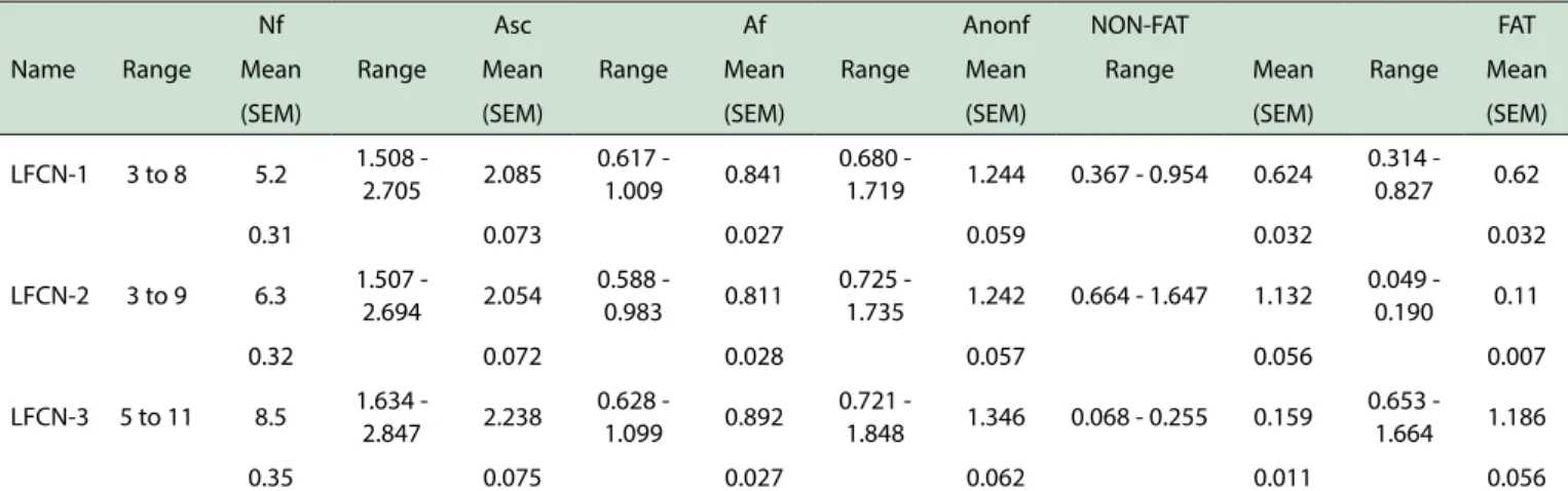
Crosssections of LFCN-1, LFCN-2 and LFCN-3 are shown in Figures 2, 3, 4, respectively. Measurements of cross sectional areas, nerve fascicles, fascicular and non-fascicular areas, NON-FAT and FAT areas for LFCN-1, LFCN-2 and LFCN-3 were obtained during morphometric analysis and range, mean, and SEM were calculated. Results are displayed in Table 1.
The LFCN-3 (range and mean; 5-11, 8.5) has a higher number of nerve fascicles as well as larger fascicular and cross sectional areas, when compared to LFCN-1 (range and mean; 3-8, 5.2), or to LFCN-2 (range and mean; 3-9, 6.3). This was due to division of the fascicle with inclusion of additional amounts of connective or adipose tissue for the newly divided fascicle. No morphometric differences were found between LFCN-1 and LFCN-2 number of fascicles (Nf), fascicular area (Af) and cross sectional area (Asc).
In the cross sections of LFCN-1, LFCN-2 and LFCN-3, the non fascicular areas were larger when compared to the fascicular area. The mean non fascicular area in LFCN-3 was larger when compared to LFCN-1 and LFCN-2.
There was no overall significant difference between
non-adipose vs. adipose tissue in the non-fascicular areas as shown in Figure 2. This similarity also applied to
LFCN-1. However, it did not hold for LFCN-2 and LFCN-3. In
LFCN-2 sections the non-fascicular areas consisted mainly of non-adipose tissue as seen in Figure 3. Concentrically arranged thick peri- and epineurium were observed around the LFCN-2 fascicles. This may have developed in order
to protect the LFCN fibers while passing deep to the IL.
Table 1 - Descriptive statistics of 21 evaluated cases LFCN-1, LFCN-2 and LFCN-3 mean morphometric parameters
Nf Asc Af Anonf NON-FAT FAT
Name Range Mean Range Mean Range Mean Range Mean Range Mean Range Mean (SEM) (SEM) (SEM) (SEM) (SEM) (SEM) LFCN-1 3 to 8 5.2 1.508 -
2.705 2.085
0.617 -
1.009 0.841
0.680 -
1.719 1.244 0.367 - 0.954 0.624
0.314 - 0.827 0.62 0.31 0.073 0.027 0.059 0.032 0.032 LFCN-2 3 to 9 6.3 1.507 -
2.694 2.054
0.588 -
0.983 0.811
0.725 -
1.735 1.242 0.664 - 1.647 1.132
0.049 - 0.190 0.11 0.32 0.072 0.028 0.057 0.056 0.007 LFCN-3 5 to 11 8.5 1.634 -
2.847 2.238
0.628 -
1.099 0.892
0.721 -
1.848 1.346 0.068 - 0.255 0.159
0.653 - 1.664 1.186 0.35 0.075 0.027 0.062 0.011 0.056
LFCN-1 - lateral femoral cutaneous nerve of thigh above inguinal ligament; LFCN-2 - lateral femoral cutaneous nerve of thigh at inguinal ligament; LFCN-3 - lateral femoral cuta-neous nerve of thigh below inguinal ligament; Nf - number of fascicles; ASc - cross sectional area; Af - total fascicular area; Anonf – non-fascicular area, NON-FAT – non-adipose (collagen) tissue area; FAT - adipose tissue area.
Figure 2 - Photomicrograph of the LFCN-1 above the inguinal ligament stained with Masson’s trichrome shows fascicular and non-fascicular tissue pattern. (A) LFCN-1 shows equal amount of adipose tissue (fat) and non-adipose tissue (collagen ibers) in the epifascicular and interfascicular connective tissue region (green area shows the more collagen ibers) (50 x); (B) The results of the automated measurement of the total cross section area (Asc) shown as white, individual fascicular area (Af ) shown as green and the non-fascicular area (Anonf ) (collagen ibers) shown blue of a single LFCN-1 were calculated by the image analysis software in (50 x). In (50 x) magniication, 1 mm2 fascicular or non-fascicular area = approx. 490 x 490 pixels. (C), (D) shows the area of igure (A) in (100 x) and (200 x) respectively. Figures 2(A)–(D) conirms clearly equal amount of adipose and non-adipose tissue in non-fascicular region. Arrows indicate the adipose tissue (fat) in non-fascicular area. Scale bar = 100 mm valid for all the images.
Figure 3 - Photomicrograph of the LFCN-2 at the inguinal ligament stained with Masson’s trichrome shows fascicular and non-fascicular tissue pattern. (A) LFCN-2 shows more amount of non-adipose tissue (collagen ibers) in the epifascicular and interfascicular connective tissue region (green area shows the collagen ibers) (50 x); (B) The results of the automated measurement of the total cross section area (Asc) shown as white, individual fascicular area (Af ) shown as green and the non-fascicular area (Anonf ) (collagen ibers) shown blue of a single LFCN-1 were calculated by the image analysis software in (50 x). In (50 x) magniication, 1 mm2 fascicular or non-fascicular area = approx. 490 x 490 pixels. (C), (D) shows the area of Figure (A) in (100 x) and (200 x) respectively. Figures 3(A)–(D) conirms clearly more amount of non-adipose tissue in non-fascicular region. Arrows indicate the non-adipose tissue (collagen ibers) in non-fascicular area. Scale bar = 100 mm valid for all the images.
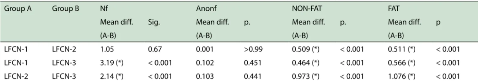
Therefore, the mean adipose tissue area in LFCN-3 was greater vs. LFCN-1 and LFCN-2; conversely, the mean non adipose tissue area in LFCN-2 was greater vs. LFCN-1 and LFCN-3. These data and their statistical analysis are displayed in Table 2.
■
DISCUSSIONThis study was undertaken to determine differences in the non-fascicular components (adipose and non-adipose
connective tissue) of the Lateral Femoral Cutaneous Nerve in areas (i) proximal to inguinal ligament (LFCN-1), (ii) deep to the inguinal ligament (LFCN-2) and distal to LFCN-2 (LFCN-3).
Neurosurgeons and orthopedicians, who usually perform microsurgical interventions on peripheral nerves,
are familiar with the nerve anatomy and its significance in
the diagnosis and management of nerve lesions.23
Figure 4 - Photomicrograph of the LFCN-3 below the inguinal ligament stained with Masson’s trichrome shows fascicular and non-fascicular tissue pattern. (A) LFCN-3 shows more amount of adipose tissue (fat) in the epifascicular and interfascicular connective tissue region (green area shows the less collagen ibers) (50 x); (B) The results of the automated measurement of the total cross section area (Asc) shown as white, individual fascicular area (Af ) shown as green and the non-fascicular area (Anonf ) (collagen ibers) shown blue of a single LFCN-1 were calculated by the image analysis software in (50 x). In (50 x) magniication, 1 mm2 fascicular or non-fascicular area = approx. 490 x 490 pixels. (C), (D) shows the area of Figure (A) in (100 x) and (200 x) respectively. Figures 4(A)–(D) conirms clearly more amount of adipose tissue in non-fascicular region. Arrows indicate the adipose tissue (fat) in non-fascicular area. Scale bar = 100 mm valid for all the images.
Table 2 - LFCN-1, LFCN-2 and LFCN-3 morphometric parameters comparison by using one way analysis of variance (ANOVA) - Tukey Test
Group A Group B Nf Anonf NON-FAT FAT
Mean dif. Sig. Mean dif. p. Mean dif. p. Mean dif. p
(A-B) (A-B) (A-B) (A-B)
LFCN-1 LFCN-2 1.05 0.67 0.001 >0.99 0.509 (*) < 0.001 0.511 (*) < 0.001 LFCN-1 LFCN-3 3.19 (*) < 0.001 0.102 0.451 0.464 (*) < 0.001 0.566 (*) < 0.001 LFCN-2 LFCN-3 2.14 (*) < 0.001 0.103 0.441 0.973 (*) < 0.001 1.076 (*) < 0.001
LFCN-1 - lateral femoral cutaneous nerve of thigh above inguinal ligament; LFCN-2 - lateral femoral cutaneous nerve of thigh at inguinal ligament; LFCN-3 - lateral femoral cutaneous nerve of thigh below inguinal ligament; Nf - number of fascicles; Anonf – non-fascicular area, NON-FAT - non-adipose (collagen) tissue area; FAT - adipose tissue area.
The factors protecting the peripheral nerves against stretching can be attributed to their anatomical structure and biomechanical characteristics. In the present study, LFCN-2 has a higher NON-FAT content, which may protect the LFCN as it passes deep to IL. The structural organization of peripheral nerves enables them to function while resisting and adapting to stresses placed on them by varying postures and movements. The nerve fascicles are loosely positioned within the connective tissue covering.3,13,24 The fascicles are exposed to combinations of tensile, shear, and compressive stresses that may result in nerve excursion, strain, and transverse contraction.6 In the present study, there may have been compressive stresses on the
LFCN-2 at IL which flattened the total nerve cross section and
compressed the fascicles in contrast to what was found in LFCN-1 and LFCN-3.
The LFCN-3 (range and mean; 5-11, 8.5) was observed to have a higher number of nerve fascicles as well as larger fascicular and cross sectional areas, when compared to LFCN-1 (range and mean; 3-8, 5.2) or LFCN-2 (range and mean; 3-9, 6.3) due to the branching of the fascicle with inclusion of additional amount of connective tissue in each newly formed branch. The number of reported fascicles in LFCN-2 ranges from 3 to 6 (mean, 4.5).21 The number of LFCN-2 (mean, 6.3) fascicles in the present study is higher when compared to previous work. As far as 1 & LFCN-3 are concerned, there is no available data in the literature. McCormick et al.25 compared the non-fascicular area of the antebrachial cutaneous nerve of the forearm with the sural nerve by using Masson’s trichrome method and concluded that the sural nerve has more adipose tissue than the antebrachial forearm nerve. Various studies have shown that the amount of connective tissue varies in different peripheral nerves, even in different parts of the same nerve.2, 24 In the present study also, the amount of connective tissue varied in different parts (LFCN-1, LFCN-2, and LFCN-3) of LFCN in relation to IL.
Ray et al.21 used the Hematoxylin-Eosin stain in 12
nerves and identified the larger non-fascicular relative
to the fascicular areas and stated that this is due to the
increased amount of collagen fibers covering the fascicles at
the inguinal ligament. The above study did not include the
identification of adipose vs. non-adipose tissue area of LFCN
in relation to IL. The present investigation used Masson’s trichrome method as described in by McCormick et al.25
The chances of injury to nerve are greater near the anterior superior iliac spine structure. Excessive traction or direct compression of the nerve is thought to be the
main causative factor for blood flow reduction. Mackinnon
et al.26,27 observed an increase in the thickness of the
epineurium and perineurium confined to the region of
nerve compression. Previous studies indicated that the
increased amount of collagen fibers is a consequence of
tissue. Consequently, the presence of increased amounts of collagen fibers in the LFCN at the level of IL could also
be related to aging.12,13,18,20 Our morphometric study on the adipose and non-adipose tissue was limited to older individuals because of unavailability of young cadaver specimens. The cadaveric specimens which had a history of meralgia paresthetica during their life were excluded from our study.
The results of the present study showed that the non-fascicular area of LFCN-2 sections was mainly occupied
by non-adipose tissue (collagen fibers) when compared
with LFCN-1 and LFCN-3. Concentrically arranged thick peri and epineurium was observed around the LFCN-2 fascicles. This may have evolved for the protection of the
LFCN nerve fibers while passing deep under the inguinal
ligament. As LFCN-2 with increased amount of collagen
fibers pass deep under the IL, the risk of compression or
focal entrapment may be higher when compared to LFCN-1
and LFCN-3. Therefore, our findings were consistent with
those of Mackinnon et al.26,27
It has been shown that in conditions such as
entrapment, hereditary neuropathies, acquired
neuropathies, trauma, partial or full nerve transactions, and nerve tumors, there is an increase in cross sectional, fascicular, and/or non-fascicular areas. Carai et al.30 suggested surgical decompression of the nerve as an option when the conservative treatments fail.30 Knowledge regarding the anatomy of the Lateral Femoral Cutaneous Nerve is essential for decompression and neurolysis of the nerve in the treatment of meralgia paresthetica.31 Variable courses and relationships of the LFCN to surface landmarks became significant in
pulse radiofrequency neuro-modulation or ultrasound
guided LFCN blockade.32,33 There are no available values for cross sectional, fascicular, and/or non fascicular areas even though they have been studied using various methods. This study includes only samples collected from normal individuals, but fails to compare with samples affected by meralgia paresthetica. This study is an attempt to build a normal database for the Lateral Femoral Cutaneous Nerve in relation to IL (LFCN-1, LFCN-2, and LFCN-3). Such data might be relevant for clinical use. Any deviation from this reference value captured on image analysis may help the clinician to diagnose problems related to LFCN.
■
CONCLUSIONMore non adipose tissue as compared to adipose tissue was found in LFCN-2. The presence of higher NON-FAT in LFCN-2 and FAT in LFCN-3 might help to understand meralgia paresthetica resulting from compression or focal entrapment of the Lateral Femoral Cutaneous nerve as it passes deep under the inguinal ligament.
■
AKNOWLEDGEMENTSSincere thanks to Professor K. Ramachandra Bhat,
Department of Anatomy, Kasturba Medical College, Manipal for his guidance and help.
■
CONFLICT OF INTERESTThe author declares no conflict of interest.
MICROANATOMIA DO NERVO CUTÂNEO
FEMORAL LATERAL EM RELAÇÃO AO LIGAMENTO INGUINAL E SUA IMPORTÂNCIA CLÍNICA
TEMA: Um melhor conhecimento da composição e
propriedades do tecido conjuntivo relacionadas ao Nervo Cutâneo Femoral Lateral (NCFL) e ao Ligamento Inguinal pode ser importante para compreender o diagnóstico e o tratamento aplicável a lesões como a meralgia parestética.
OBJETIVO: Determinar as quantidades relativas
dos componentes não-fasciculares nas seguintes áreas: (i) proximal ao ligamento inguinal [NCFL-1], (ii) em profundidade ao ligamento inguinal [NCFL-2], ou (iii) distal a NCFL-2 [NCFL-3]. Esses valores foram discriminados como tecido conjuntivo adiposo [FAT] ou não-adiposo [NON_FAT].
MÉTODO: Foram utilizadas amostras de NCFL-1,
NCFL-2 e NCFL-3 a partir de 21 amostras de cadáveres
humanos. As secções em parafina destas estruturas foram
processadas por coloração Masson para tecido conjuntivo. O número de fascículos foi contado em cada uma destas estruturas; áreas de gordura e sem gordura foram determinadas nas áreas não-fasciculares das estruturas.
RESULTADOS: Foram contados mais fascículos em
NCFL-3 vs. NCFL-1 ou NCFL-2; havia mais NON-FAT vs. FAT em NCFL-2 vs. NCFL-1 e NCFL-3; inversamente, houve mais FAT vs. NON-FAT em NCFL-3 vs. NCFL-1 e NCFL-2. Todas
estas comparações foram estatisticamente significativas.
CONCLUSÃO: A presença de um maior teor de
NON-FAT em NCFL-2 e NON-FAT em NCFL-3 pode ajudar a explicar o aparecimento de paresthetica meralgia resultante da compressão ou encarceramento focal do Nervo Cutâneo
Femoral Lateral que passa profundamente ao ligamento
inguinal.
PALAVRAS-CHAVE: Nervo cutâneo Femoral Lateral,
fibras colagenas, tecido adipose, ligamento inguinal
■
REFERENCES1. Williams , A. Pelvic girdle and lower limb. In: Standring , S (ed.) Gray’s Anatomy, The Anatomical Basis of Clinical Practice. Philadelphia:
Churchill Livingstone; 2005. p. 1399-1547.
3. Sunderland, S. Nerves and nerve injuries. New York: Churchill Livin-gstone; 1964.
4. Barkmeier JM, Luschei ES. Quantitative analysis of the anatomy
of the epineurium of the canine recurrent laryngeal nerve. J Anat. 2000;196(Pt 1):85–101. http://dx.doi.org/10.1046/j.1469-7580.2000.19610085.x
5. Phillips J, Smit X, De Zoysa N, Afoke A, Brown RA. Peripheral nerves
in the rat exhibit localized heterogeneity of tensile properties during limb movement. J Physiol. 2004; 557(Pt 3):879-87. http://dx.doi. org/10.1113/jphysiol.2004.061804
6. Topp KS, Boyd BS. Structure and biomechanics of peripheral ner -ves, Nerve responses to physical therapist practice. Phys Ther. 2006;86(1):92-109.
7. Vivo J, Morales JL, Diz A, Diz A, Galisteo AM, Monterde JG, et al. In
-tracranial portion of the trochlear nerve and dorsal oblique muscle
composition in dog: a structural and ultrastructural study. J Morphol. 2004;262(3):708-13. http://dx.doi.org/10.1002/jmor.10271 8. Parisi TJ, Mandrekar J, Dyck PJ, Klein CJ. Meralgia
paresthe-tica: relation to obesity, advanced age, and diabetes mellitus.
Neurology. 2011;77(16):1538-42. http://dx.doi.org/10.1212/
WNL.0b013e318233b356.
9. Tillett RL, Afoke A, Hall SM, Brown RA, Phillips JB. Investigating
mechanical behaviour at a coresheath interface in peripheral nerve. J Peripher Nerv Syst. 2004;9(4):255-62. http://dx.doi.org/10.1111/ j.1085-9489.2004.09411.x
10. Azcoitia I, Leonem E, Magnaghi V, Veiga S, Garcia Segura LM, Mel -cangi RC. Progesterone and its derivates dihydroprogesterone
and tetrahydroprogesterone reduce myelin fiber morphological abnormalities and myelin fiber loss in the sciatic nerve of aged rats.
Neurobiol Aging. 2003;24(6):853-60. http://dx.doi.org/10.1016/ S0197-4580(02)00234-8
11. Captier G, Canovas F, Bonnel F, Seignarbieux F. Organization and micros -copic anatomy of the Adult human Facial nerve: Anatomical and histo-logical basis for the surgery. Plas Reconstr Surg. 2005;115(6):1457-65. http://dx.doi.org/10.1097/01.PRS.0000160264.42201.F5
12. Ceballos D, Cuadras J, Verdu E, Navarro X. Morphometric and ul-trastructural changes with ageing in mouse peripheral nerve. J Anat. 1999;195(Pt 4):563–76. http://dx.doi.org/10.1046/j.1469-7580.1999.19540563.x
13. Chakravarthy Marx S, Kumar P, Dhalapathy S, Prasad K, Anitha Marx C. Microanatomical and immunohistochemical study of the human radial nerve at the antecubital fossa. Ann Anat. 2009 Oct;191(4):389-98. http://dx.doi.org/10.1016/j.aanat.2009.04.005.
14. Chakravarthy Marx S, Kumar P, Dhalapathy S, Marx AC. Microanato-mical and immunohistocheMicroanato-mical study of the human anterior branch of the medial antebrachial cutaneous nerve of forearm at the ante-cubital fossa and its clinical implications. Rom J Morphol Embryol. 2010;51(2):337–46.
15. Marx SC, Kumar P, Dhalapathy S, Prasad K, Marx CA. Microanatomical and immunohistochemical study of the human lateral antebrachial cutaneous nerve of forearm at the antecubital fossa and its clinical im-plications. Clin Anat. 2010;23(6):693-701. http://dx.doi.org/10.1002/ ca.20985
16. Chakravarthy Marx S, Kumar P, Dhalapathy S, Marx A. A comparative
microanatomical study on crosssections of superficial branch of radial
nerve in proximal and distal parts of the forearm: A cadaveric study. Morphologie. 2010;94(307):98-106.
17. Marx SC, Kumar PSD, Marx CA, Babu MS, Bhat KM. Histological and ultrasonographical study of the human superficial branch of the radial
nerve at distal forearm and its clinical implications. Rom J Morphol Embryol. 2010;51(4):751-8.
18. Jacobs JM, Love S. Qualitative and quantitative morphology of human sural nerve at different ages. Brain. 1985;108(Pt 4):897-924. http://
dx.doi.org/10.1093/brain/108.4.897 897-924
19. Marx SC, Kumar P, Dhalapathy S, Anitha Marx C. A comparative micro-anatomical study on cross sections of medial and lateral cutaneous nerves of forearm at the antecubital fossa: A cadaveric study. Ann Anat. 2010;192(2):107-15. http://dx.doi.org/10.1016/j.aanat.2009.12.004
20. Sladjana UZ, Ivan JD, Bratislav SD. Microanatomical structure of the
human sciatic nerve. Surg Radiol Anat. 2008;30(8):619-26. http:// dx.doi.org/10.1007/s00276-008-0386-6
21. Ray B, D’Souza A, Kumar B, Marx C, Ghosh B, Gupta N, et al. Variations
in the course and microanatomical study of the lateral femoral cuta-neous nerve and its clinical importance. Clin Anat. 2010;23(8):978-84. http://dx.doi.org/10.1002/ca.21043
22. Jones, M. Connective tissues and stains. In: Bancroft , J.D, Gamble , M (eds.) Theory and practice of histological techniques. New York:
Churchill Livingstone; 2002.p. 149-153.
23. Goldberg SH, Jobin CM, Hayes AG, Gardner T, Rosenwasser MP, Strauch RJ. Biomechanics and histology of intact and repaired digital nerves: an in vitro study. J Hand Surg Am. 2007;32(4):474-82. http://dx.doi.
org/10.1016/j.jhsa.2006.12.008
24. Dyck, P.J, Thomas , P.K, Lambert , E.H, Bunge, R.Peripheral Neuropathy. London: WB Saunders Company;1984.
25. McCormick SU, Buchbinder D, McCormick SA, Stark M. Microanatomic
analysis of the medial antebrachial nerve as a potential donor nerve in maxillofacial grafting. J Oral Maxillofac Surg. 1994;52(10):1022-5. http://dx.doi.org/10.1016/0278-2391(94)90166-X
26. Mackinnon SE, Dellon AL, Hudson AR, Hunter DA. Histopathology of compression of the superficial radial nerve in the forearm. J Hand
Surg Am. 1986;11(2):206-10. http://dx.doi.org/10.1016/S0363-5023(86)80052-1
27. Mackinnon SE, Dellon AL, Hudson AR, Hunter DA. Chronic
human nerve compression - A histological assessment. Neu-ropathol Appl Neurobiolm. 1986;12(6):547-65. http://dx.doi. org/10.1111/j.1365-2990.1986.tb00159.x
28. Sunderland S. The intraneural topography of the radial, median and
ulnar nerves. Brain. 1945;68:243-98. http://dx.doi.org/10.1093/
brain/68.4.243
29. Lundborg, G. Nerve injury and repair . New York: Churchill Livingstone;
1988.
30. Carai A, Fenu G, Sechi E, Crotti FM, Montella A. Anatomical variability
of the lateral femoral cutaneous nerve: Findings from a surgical series. Clin Anat. 2009;22(3):365-70. http://dx.doi.org/10.1002/ca.20766 31. Ducic I, Dellon AL, Taylor NS. Decompression of the lateral femoral
cuta-neous nerve in the treatment of meralgia paresthetica. J Reconstr Micro-surg. 2006;22(2):113-8. http://dx.doi.org/10.1055/s-2006-932505 32. Dalmau-Carolà J. Treatment of meralgia paresthetica with pulsed
radiofrequency of the lateral femoral cutaneous nerve. Pain Physician.
2009;12(6):1025-6.