ANATOMICAL STUDIES OF IN VITRO ORGANOGENESIS INDUCED
IN LEAF-DERIVED EXPLANTS OF PASSIONFRUIT1
BEATRIZ APPEZZATO DA GLORIA2, MARIA LUCIA CARNEIRO VIEIRA3 and MARCELO CARNIER DORNELAS4
ABSTRACT - With the aim of studying the organogenesis in vitro in Passiflora edulis Sims f. flavicarpa Deg., the passionfruit, leaf-derived explants were cultured on media containing NAA or BAP and incubated either in continuous darkness or in light. The histological events leading to de novo organ formation were evaluated. Darkness induces rhizogenesis in the presence of NAA, whereas direct shoot regeneration is stimulated by light and BAP. This latter condition is recommended for passionfruit micropropagation as several adventitious shoot buds were formed from meristemoids of parenchymal origin.
Index terms:Passiflora, tissue culture, micropropagation, histology.
ESTUDOS ANATÔMICOS DA ORGANOGÊNESE IN VITRO INDUZIDA EM EXPLANTES DE FOLHA DE MARACUJÁ
RESUMO - Com o objetivo de estudar a organogênese in vitro em Passiflora edulis Sims f. flavicarpa Deg., o maracujá-amarelo, explantes derivados de folha foram cultivados em meio contendo NAA ou BAP, no escuro e na presença de luz. Foram descritos os eventos histológicos que levam à formação de novo de órgãos. Concluiu-se que o escuro induz a rizogênese, na presença de NAA, enquanto a regeneração de brotos é estimulada pela luz e BAP. Esta condição é recomendada para micropropagar o maracujá uma vez que vários brotos adventícios são formados a partir de meristemóides de origem parenquimática. Termos para indexação: Passiflora, cultura de tecidos, micropropagação, histologia.
INTRODUCTION
Passionfruit (Passiflora edulis Sims f. flavicarpa
Deg.) is one of the most economically important fruit crop used in Brazil for juice processing. Although passionfruit is cultivated in a relatively large area (30,000 ha), little production is in organized orchards. Flowers are hermaphrodites and insect-pollinated, although a simple hand pollination
1Accepted for publication on November 16, 1998.
2Agronomist, Dra, Dep. de Botânica, Escola Superior
de Agricultura Luiz de Queiroz (ESALQ), USP, Caixa Postal 9, CEP 13418-900 Piracicaba, SP. E-mail: bagloria@carpa.ciagri.usp.br
3Biologist, Livre Docente, Dep. de Genética, ESALQ, USP.
E-mail: mlcvieir@carpa.ciagri.usp.br
4Agronomist, M.Sc., Instituto de Biotecnologia de Plantas,
Universidade de Paris XI, 91405 Orsay, França. CAPES scholar.
technique is often used to enhance productivity.
Passiflora species are normally propagated from
seeds and cuttings.
Passiflora micropropagation has been described by Moran Robles (1978) and recently by Kantharajah & Dodd (1990) and Kawata et al. (1995). Plant regeneration from tissue culture has also been reported from a diversity of Passiflora germplasm, including the cultivated species (Dornelas & Vieira, 1994). Regeneration occurred by organogenesis on the surface of the explants.
numbers, micropropagation by cuttings has a relatively low multiplication rate. For commercial ap-plication, high rates of shoot development are required. Regeneration rates (percentage of explants that produced shoots) varied from 70% in the wild species up to 96% in Passiflora edulis f. flavicarpa
(Dornelas & Vieira, 1994).
Meristem-derived cultures without a significant dedifferentiation step give rise to normal plants. Among those types of cultures that regenerate after a dedifferentiated (callus) phase, one can distinguish between regeneration by organogenesis or somatic embryogenesis. It is clear from histological studies that organogenesis often involves more than one cell that act in a coordinate manner (Brown & Thorpe, 1986). In the case of somatic embryogenesis, the em-bryoid is often derived from a single cell, although evidence for a multicellular origin has also been obtained (Dornelas et al., 1992).
Some authors have argued that plants regenerated from direct somatic embryogenesis even by direct organogenesis ought to contain fewer mutations than those regenerated via callus phase. Also, many investigators have noted an increase in variability with an increase in culture age (Lee & Phillips, 1987). Our studies on species of Passiflora showed that plant regeneration occurs via direct organ differen-tiation after a short culture period of 28 days. These cultures have been able to produce viable plants by subsequent rooting (Dornelas & Vieira, 1994). However, the occurrence of direct organogenesis has to be clearly demonstrated by anatomical evidence. Thus, the present work focuses on the sequences of events, specially at the anatomical level, leading to the process ofpassionfruit plant regeneration. This investigation also deals with the effects of two light regimes and different hormone treatments on this process.
MATERIAL AND METHODS
The experiments were carried out at Escola Superior de Agricultura Luiz de Queiroz, at Piracicaba, Brazil, dur-ing the year of 1994. The protocol for obtaindur-ing de novo organ formation consisted of culturing leaf explants on MS basal medium (Murashige & Skoog, 1962) containing 0.3% (w/v) sucrose and supplemented with 1.0 mg L-1 BAP (6-benzyl aminopurine) or 1.0 mg L-1 NAA
(1-naphthaleneacetic acid). As reported previously, these growth regulator concentrations lead to in vitro regenera-tion of shoots and roots, respectively (Dornelas & Vieira, 1994). The media were solidified with 0.15% Phytagel (w/v, Sigma) and distributed (10 mL per flask) into 25 x 85 mm flasks, which were autoclaved for 20 minutes. The pH was adjusted to 5.8 prior to adding the gelling agent.
Leaf explants of P. edulis f. flavicarpa were excised from 60-day-old plants as described by Dornelas & Vieira (1994). The explants were taken from the central part of the leaves, thus containing the midrib. Using a cork-borer (11 mm in diameter) 48 discs (6 treatments x 4 culture conditions x 2 replicates) were halved and placed individually into the flasks that were sealed with plastic film.
In order to study the influence of light regime in the process of in vitro regeneration the cultures were grown either under 23 µmol m-2 second-1 light radiation
(16 hours photoperiod) provided by a cold-white bulb (80 W), or darkness. The temperature of the culture room was kept at 25±2°C.
Leaf explants samples were collected after 0, 3, 7, 14, 21 and 28 days of culture. Samples were collected from the four culture conditions, raised from the combinations of MS + 1.0 mg L-1 of BAP or NAA and the two light
regimes.
Samples were fixed in FA A 50, dehydrated in an ethanol series, embedded in paraffin, serially cut into 8 µm sections (Sass, 1951) and stained in basic fuchsin-astra blue combination (Roeser, 1972).
RESULTS AND DISCUSSION
Explant characterization
Analysis of cross sections of P. edulis f.
flavicarpa collected at the beginning of in vitro
presents the vascular system with four collateral bundles arranged in a circle and enclosed by the fundamental parenchyma. This characterization confirms the data presented by Abanto & Müller (1972) for P. edulis leaf anatomy.
The explants did not present any anatomical changes after three days. In contrast, Moran Robles (1979) and Scorza & Janick (1980) reported changes in the internal structures of the stem segments of
P. edulis and P. suberosa L., cultivated during the same period on Nitsch medium (Nitsch et al., 1968) supplemented with 2.0 mg L-1 kinetin and on MS
containing 0.1 mg L-1 6-benzylaminopurine,
respectively. According to the investigators, proliferation of large cells with a prominent nucleus was observed in the sectioned areas. Usually, these cells originated from cortical tissue. This pattern was not observed in this study.
Explants cultivated on MS + 1.0 mg L-1 BAP, under
23 µmol m-2 sec-1 light radiation
Plant tissue culture systems offer the possibility of analyzing the point of determination of cytodifferentiation (Fukuda & Komamine, 1985). Under these conditions (light and BAP stimulus), the explants presented alterations in the leaf border region after the 7th day of culture. The mesophyll
displayed small cells with a dense cytoplasm and conspicuous nucleus originated from the chlorophyll parenchyma, associated or not with the explant vascularisation. The first divisions in the fundamen-tal parenchyma of the midrib were also observed during this phase.
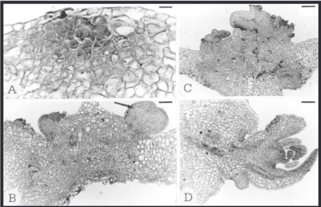
On the 14th day of culture, the palisade
parenchyma presented more elongated cells in some of the mesophyll sectors. Predominantly periclinal divisions in the mesophyll close or not to the vascularisation were observed in other sectors. The divisions in the more peripheral layers led to the formation of meristematic areas (Fig. 1A) similar to those described by Saravitz et al. (1993) as meristemoids. These consisted of small cells with a dense cytoplasm and prominent nucleus. At the same time, divisions on several planes were observed in the fundamental parenchyma on the adaxial surface of the midrib, especially in the subepidermal layers,
which led to the formation of meristematic structures similar to those described for the mesophyll.
On the 21st day of culture, these structures were
more developed and were found to be lined with the protodermis of epidermal origin (Fig. 1B, arrow), as also occurs in Pinus radiata (Villalobos et al., 1985) and Picea abies (Von Arnold et al., 1988). During this phase, there was an increased activity of the fundamental parenchyma leading to disorganization of the vascular system of the midrib, which progressed with culture (Fig. 1C). The presence of several shoot buds in different developmental phases (Fig. 1C and 1D) was observed on the 28th day,
confirming that these buds originated from the meristematic structures described. The absence of synchrony in bud formation was also noted by Von Arnold et al. (1988) and Saravitz et al. (1993).
Thus, the adventitious shoot buds of P. edulis f.
flavicarpa were developed from meristemoid areas after the second week of culture, as the result of the mitotic activity of parenchymatous cells, given support to the results reported by Scorza & Janick (1980) for P. suberosa. The predominance of cell divisions in the explant epidermis and subjacent layers in response to BAP has been observed in conifers (Villalobos et al., 1985; Von Arnold & Gronroos, 1986; Saravitz et al. 1993). In Begonia x
erythrophyla petiole sections both shoots and roots are formed directly from cells of epidermal origin (Burritt & Leung, 1996). Yeung et al. (1981) suggested that the epidermal and subjacent layers are not completely determined at the time of excision, leading these cells to respond to cytokinin stimulus.
Explants cultivated on MS + 1.0 mg L-1 BAP, in the
dark
meristematic activity under the stomata, which persisted during the course of culture. In embryos of
Picea abies treated with BAP the surrounding cells of stomata started to proliferate and meristemoid cells developed during the second week of culture (Von Arnold et al., 1988). Barciela & Vieitez (1993) also reported the morphogenetic potential of the adaxial epidermis of Camellia cotyledons. These data support the results of Kaneko & Matsushima (1984) in stem segments of Nicotiana tabacum
although no development of shoot buds from the cell proliferation was observed in this material.
Divisions in several planes of the fundamental parenchyma were observed and became more intense on the 21st and 28th day of culture.
However, the development of meristemoid areas was not observed.
The relationship between morphogenetic ability and light regime has been reported in the literature. The exhaustive study carried out by Seibert et al. (1975) showed the effects of light intensity and spectral quality on callus formation and bud regeneration in tobacco explants. These investiga-tors observed that light contributed to an increase in the callus fresh matter weight and in the number of regenerated buds, comparing to cultures maintained in the dark. Previously, Weiss & Jaffe (1969) had de-scribed the stimulating effects of blue light on the process of organogenesis in tobacco apex cultures. Moran Robles (1979) also reported the influence of
FIG. 1. Transections of a leaf explant of Passiflora edulis f. flavicarpa placed on MS medium supplemented with 1.0 mg L-1 BAP under 23 µmol m-2 second-1 light radiation, after 14 (A), 21 (B) and 28 (C, D) days
luminosity in the number of growing shoots in
Passiflora cultures comparing to explants kept in the dark.
Explants cultivated on MS + 1.0 mg L-1 NAA
The first explant alterations promoted by the NAA treatment in cultures maintained under light started on the 14th day. Divisions on several planes were
observed in the fundamental parenchyma of the midrib and leaf border. However, neither the increase in the meristematic activity nor the root meristemoid formation usually stimulated by an auxin were observed during the course of this type of culture.
In the dark, the first anatomical changes relating to organogenesis were observed on the 21st day of
culture. In the mesophyll, the cells of the palisade parenchyma divided periclinally and the spongy
parenchyma cells divided along several planes. In the midrib, root meristemoids developed from meristematic activity initiating near the procambium of the midvein vascular system (Fig. 2A and 2B, arrows). Moran Robles (1979) also observed rhizogenesis in internodal explants of P. edulis var.
flavicarpa Deg. and P. mollissima Bailey, after the 21st day of culture.
By the 28th day of culture, in addition to the
adventitious roots developed in the midrib, several roots were observed along the mesophyll. The formation of these structures was endogenous and always associated with the explant vascularisation (Fig. 2C and 2D, arrows). As proposed before, the sites of origin of adventitious roots have been located in cells of the procambium or close to it, in the phloem (Hicks, 1987; Ranjit et al., 1988).
FIG. 2. Transections of a leaf explant of Passiflora edulis f. flavicarpa placed on MS medium supplemented with 1.0 mg L-1 NAA cultured in the dark, after 21 (A, B) and 28 (C, D) days. A and B: The arrows
Harbageet al. (1993) by inducing rhizogenesis in
Malus observed that cells of several tissues
divided in response to IBA treatment but, only divisions in the phloem parenchyma led to the development of adventitious roots. Similarly, adventitious root formation observed in the present study occurred near the explant vascularisation although the chlorophyll parenchyma showed divisions on several planes.
CONCLUSIONS
1. Leaf explants of P. edulis f. flavicarpa exhibit distinct morphogenic reactions as a function of the hormone added to the culture medium and are highly responsive and become strongly determined by the 28th day of culture.
2. Rhizogenesis occurs in the presence of NAA whereas direct shoot regeneration is stimulated by BAP.
3. The white light is essential for bud formation in leaf-derived explants cultured on medium supplemented with BAP.
4. The dark condition acts synergistically with the effect of auxin leading to adventitious root formation.
ACKNOWLEDGEMENTS
To the Conselho de Desenvolvimento Científico e Tecnológico (CNPq) and the Fundação de Amparo à Pesquisa do Estado de São Paulo (FAPESP) for financial support; to Mr. C. A. de Oliveira and Mrs. M. A. R. Machado for excellent technical assistance.
REFERENCES
ABANTO, A.M.; MÜLLER, L. Algunos aspectos morfológicos del maracuyá, Passiflora edulis. Turrialba, v.22, p.268-274, 1972.
BARCIELA, J.; VIEITEZ, A.M. Anatomical sequence and morphometric analysis during somatic embryo-genesis on cultured cotyledon explants of Camellia japonica L. Annals of Botany, Oxford, v.71, p.395-404, 1993.
BROWN, D.C.W.; THORPE, T.A. Plant regeneration by organogenesis. In: VASIL, I. K. (Ed.). Cell culture
and somatic cell genetics of plants. Orlando: Academic, 1986. v.3, p.49-73.
BURRITT, D.; LEUNG, D.W.M. Organogenesis in cultured petiole explants of Begonia x erythrophylla: the timing and specificity of the inductive stimuli. Journal of Experimental Botany, Oxford, v.47, p.557-567, 1996.
DORNELAS, M.C.; VIEIRA, M.L.C.; GLÓRIA, B.A. da. Histological analysis of organogenesis and somatic embryogenesis induced in immature tissues of Stylosanthes scabra Vog. Annals of Botany, Oxford, v.70, p.477-482, 1992.
DORNELAS, M.C.; VIEIRA, M.L.C. Tissue culture studies on species of Passiflora. Plant Cell, Tissue and Organ Culture, Dordrecht, v.36, p.211-217, 1994.
FUKUDA, H.; KOMAMINE, A. Cytodifferentiation. In: VASIL, I.K. (Ed.). Cell culture and somatic cell genetics of plants. Orlando: Academic, 1985. v.2, p.149-212.
HARBAGE, J.F.; SIMART, D.P.; EVERT, R.F. Anatomy of adventitious root formation in microcuttings of Malus domestica Borkh. Gala. Journal of the American Society for Horticultural Science, Geneva, v.118, p.680-688, 1993.
HICKS, G.S. Adventitious rooting of apple microcuttings in vitro: an anatomical study. Canadian Journal of Botany, Ottawa, v.65, p.1913-1920, 1987. KANEKO, Y.; MATSUSHIMA, H. Direct observation
by low-temperature scanning electron microscopy of fresh-shoot apices and floral buds induced to form on Nicotiana tabacum stem cultures. Journal of Electron Microscopy, Tokyo, v.33, p.248-251, 1984. KANTHARAJAH, A.S.; DODD, W.A. In vitro micropropagation of Passiflora edulis (Purple passionfruit). Annals of Botany, Oxford, v.65, p.337-339, 1990.
LEE, M.; PHILLIPS, R. L. Genomic rearrangements in maize induced by tissue culture. Genome, Ottawa, v.29, p.122-128, 1987.
MORAN ROBLES, M.J. Multiplication végétative, in vitro, des bourgeons axilaires de Passiflora edulis var. flavicarpa Deg. et de P. mollissima Bailey. Fruits, Paris, v.33, p.693-699, 1978.
MORAN ROBLES, M.J. Potentiel morphogénétique des entrenoeuds de Passiflora edulis var. flavicarpa Deg. et P. mollissima Bailey en culture in vitro. Turrialba, v.29, p.224-228, 1979.
MURASHIGE, T.; SKOOG, F. A revised medium for rapid growth and bioassays with tobacco tissue cultures. Physiologia Plantarum, Kobenhavn, v.15, p.473-497, 1962.
NITSCH, J.; NITSCH, C; HAMON, S. Réalisation expérimentale de landrogénèse chez divers Nicotiana. Comptes Rendus de la Societé de Biologie, Paris, v.162, p.369-372, 1968.
RANJIT, M.; KESTER, D.E.; POLITO, V.S. Micro-propagation of cherry rootstocks: III. Correlations between anatomical and physiological parameters and root initiation. Journal of the American Society for Horticultural Science, Geneva, v.113, p.155-159, 1988.
ROESER, K.R. Die Nadel der Schwarzkiefer - Massen Produkt und Kunstwert der Natur. Mikrokosmos, Stuttgart, v.61, p.33-36, 1972.
SARAVITZ, C.H.; BLAZICH, F.A.; AMERSON, H.V. Histology of in vitro adventitious bud development on cotyledons and hypocotyls of Fraser fir. Journal of the American Society for Horticultural Science, Geneva, v.118, p.163-167, 1993.
SASS, J.E. Botanical microtechnique. Ames: Iowa University Press, 1951. 228p.
SCORZA, R.; JANICK, J. In vitro flowering of Passiflora suberosa L. Journal of the American Society for Horticultural Science, Geneva, v.105, p.892-897, 1980.
SEIBERT, M.; WETHERBEE, P.J.; JOB, D.D. The effects of light intensity and spectral quality on growth and shoot initiation in tobacco callus. Plant Physiology, Rockville, v.56, p.130-139, 1975. VILLALOBOS, V.M.; YEUNG, E.G.; THORPE, T.A.
Origin of adventitious shoots in excised radiata pine cotyledons cultured in vitro. Canadian Journal of Botany, Ottawa, v.63, p.2172-2176, 1985. VON ARNOLD., S.; GRONROOS, R. Anatomical
changes and peroxidase activity after cytokinin treatments inducing adventitious bud formation on embryos of Picea abies. Botanical Gazette, Chicago, v.147, p.425-431, 1986.
VON ARNOLD, S.; ALSTERBORG, E.; WALLES, B. Micromorphological studies of adventitious bud formation on Picea abies embryos treated with cytokinin. Physiologia Plantarum, Kobenhavn, v.72, p.248-256, 1988.
WEISS, J.F.; JAFFE, M.J. Photoenhancement by blue light of organogenesis in tobacco pith cultures. Physiologia Plantarum, Kobenhavn, v.22, p.171-176, 1969.