Vol.56, n.3: pp. 421-430, May-June 2013
ISSN 1516-8913 Printed in Brazil BRAZILIAN ARCHIVES OF
BIOLOGY AND TECHNOLOGY
A N I N T E R N A T I O N A L J O U R N A L
Organogenesis from in vitro-derived Leaf and Internode
Explants of Hoya wightii ssp. palniensis - a Vulnerable
Species of Western Ghats
Subbaiah Revathi Lakshmi
1, Jambarapu Herald Franklin Benjamin
1, Tirupathi Senthil
Kumar
2, Garimella Venkata Suryanarayana Murthy
3and Mandali Venkateswara Rao
1*1Department of Plant Science; 2Department of Industry University Collaboration; Bharathidasan University;
Tiruchirappalli 620 024; Tamil Nadu. 3Botanical Survey of India; Southern Circle; Coimbatore- 641 003; Tamil Nadu
ABSTRACT
An efficient system was developed for indirect plant regeneration from in vitro-derived leaf and internode explants of Hoya wightii ssp. palniensis. Maximum percentage of the organogenic callus was obtained on MS medium supplemented with NAA (1.0 mg/l) and 2,4-D (2.0 mg/l). The best shoot bud induction was observed on MS medium with BA (1.0 mg/l) +IBA (0.5 mg/l). The coconut water (15%) was better, resulting in a differentiation of the shoot initials in to well-developed shoots. The elongated shoots (› 3cm long) were rooted on a full strength MS basal medium, supplemented with 0.2 mg/l of IBA. Finally, the rooted plants were transferred to the soil with 80% success rate. This protocol was utilized for the in vitro propagation of this endangered plant species.
Key words:In vitro explants, organogenesis, Hoya wightii ssp. Palniensis, vulnerable plant, growth regulators
*
Author for correspondence: mvrao_456@yahoo.co.in
INTRODUCTION
Hoya wightii ssp. palniensis, commonly known as wax plant due to its waxy nature, belongs to the family Asclepiadaceae. It is a woody trailing herb, endemic to Pambar Shola of Western Ghats of Tamil Nadu, India. Mathew (1992) and Matthew
(1996) reported an individual population of H.
wightii ssp. palniensis in the ravine of Pambar Shola, Tamil Nadu, India. The genus Hoya is of horticultural importance in Europe, Australia and
America. H. wightii ssp. palniensis possesses
white flowers with red coronas which are borne in an umbel that can last for a week. Seed setting is very rare. Single follicle setting was observed during 2007 at Vattakanal Conservation Trust by the authors. These features limit its population to
Western Ghats of Tamil Nadu, India. Imminent extinction threat of this species necessitates
developing the conservation strategies and in vitro
tissue culture techniques appear as a promising alternative to ensure its regeneration and
conservation. Plant regeneration through
organogenesis and somatic embryogenesis has
been reported in other species of Hoya such as H.
kerrii (Tube et al. 2007) and H. carnosa (Maraffa et al. 1981) and micropropagation of this species (Lakshmi et al. 2010).
The present study describes a protocol for the
organogenesis of this species from in vitro derived
leaf and internode explants since the use of in vitro
derived explants will eliminate the need for
disinfection. Shoot regeneration from in vitro
such as Decalepis hamiltonii by Giridhar et al.
(2004); Vaccinium corymbosum (Billings et al.
1988); Pyrus communis (Leblay et al. 1991);
Elaegnus angustifolia (Economou and Maloupa,
1995); Pistachio vera L. cv. Siirt (Tilkat and
Onay, 2009); Morus alba (Chitra and Padmaja,
2005); Phellodendron amurense (Azad et al.
2005). This is the first report on indirect organogenesis and the establishment of complete plantlets from the regenerated leaf and internode explants of H. wightii ssp. palniensis.
MATERIALS AND METHODS
Establishment of In Vitro Shoot Cultures
Single long apical bud of 0.5 – 1.0 cm was aseptically cultured in the conical flasks, each containing 50 ml of MS medium. To establish the shoot cultures, the apical shoot tips were cultured on the MS medium containing KN (1.0 mg/l) + IBA (0.3 mg/l), in addition to 30 g/l sucrose and 8 g/l agar. A detailed method for the optimized surface sterilization and culture initiation from
mature H. wightii spp. palniensis has been
reported by Lakshmi et al. (2010). Adventitious shoot buds from the initiation medium were sub-cultured on the fresh initiation medium every 2-3 weeks. The regenerated adventitious shoot buds
from the in vitro cultures were maintained and
proliferated on the initiation medium for about six months. The pH of the medium was adjusted to 5.7 using 0.1 N NaOH or 0.1 N HCl, prior to autoclaving at 1210C for 20 min. All the cultures were placed and maintained in a growth room under a 16-h photoperiod (60 µ mol m-2s-1) with
day and night temperatures of 25±20C.
Callus Induction from Axenic Leaf and Internode Explants
To initiate the callus induction and shoot regeneration from the axenic leaf and internode tissues, in vitro-regenerated shoots maintained for nearly six months on the culture initiation medium were used as source material. Semi mature leaf and internode were excised and abaxial sides in contact with the medium. The MS callus induction medium supplemented with 3% (w/v) sucrose at different concentrations of auxins 2,4 – D or NAA or IBA or Pic at 0.5, 1.0, 1.5, 2.0, 3.0 and 4.0 mg/l were tested in the callus induction experiment. These cultures were incubated at 25±20C in dark for seven days and then they were transferred to
16/8h light incubation. The cultures were observed constantly for any morphological response and after four weeks in culture, the percentage of explants showing callus induction was recorded. Each treatment contained at least 20 explants and the experiment was repeated two times.
Shoot Regeneration from Callus
The primary callus of green compact nature was then transferred to the culture bottles containing shoot regeneration medium. The MS medium supplemented with 3% sucrose and different concentrations and combinations of BA, KN or TDZ at 0.5, 1.0, 1.5 and 2.0 mg/l and auxins such as IAA, IBA and NAA at the concentrations of 0.1, 0.2, 0.5 and 1.0 mg/l were tested in the shoot regeneration experiments. The cultures were observed constantly for the percentage of explants regenerating shoots via adventitious shoot buds induction and proliferation.
Shoot Multiplication and Elongation
The adventitious shoots regenerated from the
adventitious buds were used for further
multiplication. GA3 (0.5 mg/l) was added to the
shoot regeneration medium for shoot elongation. The shoot regeneration medium used was MS medium supplemented with BA (1.0 mg/l) + IBA (0.5 mg/l). After elongation, the clusters of apical shoots (5-10mm long) were segmented and used for proliferation and multiplication studies. Four different concentrations each of AA (25, 50, 100 and 150 mg/l), CA (5, 10, 25 and 50 mg/l), PVP (10, 25, 50 and 100 mg/l), CE (25, 50, 100 and 150 mg/l), YE (25, 50, 100 and 150 mg/l), ME (25, 50, 100 and 150 mg/l) and CW (5, 10, 15 and 25%) were added separately to shoot regeneration medium, i.e., MS+BA (1.0 mg/l)+ IBA (0.5 mg/l)
and GA3 (0.5 mg/l) were added based on the
response observed in previous two experiments, and they were added in this assessment along with the additives to enhance its shoot multiplication rate. For shoot proliferation and multiplication, each treatment was replicated twice and each replicate consisted of at least 20 explants. Four weeks after the culturing, the percentage of number of shoots and shoot length of newly formed shoots were recorded.
In Vitro Rooting of Shoots
rooting medium containing the MS medium with 3% sucrose and 8 g/l agar. The effects of the different concentrations of IBA, IAA and NAA (0.1, 0.2 and 0.3 mg/l) on rooting were evaluated. For rooting studies, each treatment was replicated three times, and each experiment consisted of at least 10 explants. After four weeks, the percentage of rooted shoots and the number of roots per shoot were recorded.
Acclimatization
The rooted shoots were carefully taken out of the medium and washed thoroughly in the running tap water, being careful not to damage the roots, to remove the traces of the medium attached to the roots. Then, they were planted in 3-cm paper cups containing a sterile 1:1:1 (w/w/w) red soil to sand and coconut coir mixture, enriched with a ¼ strength MS salt solution, covered with a polythene bag to maintain the high humidity, and
placed in a culture room at 25±20C (day and
night). The polythene bags were opened for 5 mins each the first two days, the length of the opening time doubled each subsequent day. Then, following four week period of acclimatization by the progressive reduction of the humidity level from 90 to 60%, the established plants were transferred to earthen pots and kept in a shade house for further growth and observation at Vattakanal Conservatory Trust, Kodaikanal as a part of reintroduction.
Experimental Design and Data Collection
All the experiments were conducted using the completely randomized block design. Significance was determined by the analysis of variance, and the least significant (P≤ 0.05) differences among the mean values were estimated using Duncan’s new Multiple Range Test.
RESULTS AND DISCUSSION
A number of preliminary experiments revealed that the phytohormones were essential for the induction of callus from the leaf and internode explants and no callus was induced by the basal MS medium only. A series of attempts were made to initiate the shoots from the primary callus using the individual cytokinins. It was noticed that the KN, BA and TDZ failed to initiate the shoot buds from the callus and the callus explant died within a short period. In order to obtain the alternate source
of explant for indirect shoot regeneration, axenic cultures derived by the protocol of Lakshmi et al. (2010) was used as explant. This is the first report on organogenesis of H. wightii ssp. palniensis. In
general, the explants derived from the
micropropagated shoots have an early and greater capacity for morphogenesis than the tissue excised from the field plants (George 1996), which is attributed to the absence of lag period between the explanting and adaptation to in vitro conditions. The presence of smaller yet active meristematic centers of the microplants compared to relatively larger but quiescent meristems of the shoots of mature plants might be the reason for the successful regeneration of the explants from the micropropagated shoots in the present study (Amin and Jaiswal 1987).
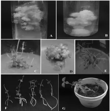
The leaf and internode explants from the axenic shoots served as the explants for the induction of organogenic callus. The callus initiation was observed on the cut edges of leaf and internodes 15 days after the culture incubation (including initial seven days dark incubation). The induction of callus was mainly influenced by the auxin type and concentration used (Table 1). Initially (15 days), all the calli were yellow to white in color (Fig.1) with soft, friable and unorganized morphology. This callus was sub-cultured every two weeks (15d) for 60 days on to fresh medium of the same composition after which the callus showed varied nature on the different types of auxins used (Table 1). Most of the calli became yellow to green compact organogenic in nature after a total of 60 days (Fig. 1 A and B). Only green compact callus was investigated further and reported in this study.
The mean percentage of callus induction of in vitro leaf ranged from 20.0-83.6% while the internode ranged from 20 – 100% based on the concentration of auxins used (Table 1). Maximum percentage of the organogenic callus was obtained on MS medium supplemented with NAA (1.0 mg/l) and 2,4-D (2.0 mg/l). NAA supplementation on callus induction has been reported on Saintpanlia ionantha (Khan et al. 2007).
Effect of Cytokinins on Shoot Regeneration
when the culture medium was supplemented with BA 1.0 mg/l (78%) with 8.4 shoots/explant (Table 2 and Fig. 1 C). For the internode-derived callus, significant difference was detected among the cytokinin media supplementation treatments, and the explant responsiveness ranged from 56.0-87.2% and maximum number of shoots/explant was obtained on MS medium fortified with BA 1.0 mg/l. BA has long been used most frequently for
the in vitro shoot regeneration and multiplication studies of many plants. It showed high cytokinin activity by promoting the shoot multiplication and
growth of leaf explant of Momordica cymbalaria
(Nikam et al. 2009); Elaeagnus angustifolia
(Karami and Piri 2009) and from internode of
Withania somnifera (Kulkarni et al. 2000) and
Piper colubrinum (Kelkar and Krishnamurthy 1998). This was reflected in the present study too.
Table 1 - Callus induction of Hoya wightii ssp. palniensis, from leaf and internode explant on MS medium
supplemented with different concentrations of auxins, after 60 days of culture.
PGRs (mg/l)
Percentage of response Color and nature of the callus
Leaf Internode Leaf Internode
0 Pic 0.5 1.0 1.5 2.0 3.0 2,4-D 0.5 1.0 1.5 2.0 3.0 NAA 0.5 1.0 1.5 2.0 3.0 IBA 0.5 1.0 1.5 2.0 3.0 0 75.4±0.2c 83.6±0.5a 83.5±0.1a 80±0.3b 25±0.7g 33.3±0.6f 50.0±0.8d 50.0±0.2d 40.0±0.4e 20.0±0.2h 75.0±0.1c 80.0±0.7b 40.0±0.6e 33.0±0.3f 20.0±0.5h 40.0±0.3e 40.0±0.8e 20.0±0.4h - - 0 83.5±0.5b 100.0±0a 100.0±0a 25.0±0.4i - 33.3±0.2h 57.1±0.6f 75.0±0.5d 50.0±0.8g 50.0±0.1g 50.0±0.4g 100.0±0a 80.0±0.1c 60.0±0.5a 50.0±0.3g 80.0±0.6c 60.0±0.2e 20.0±0.3j - - - WFr WFr WFr WFr WFr YC YC YC YC YC GC GC GC GC GC Brfr Brfr Brfr - - - GC GC GC GC GC Wfr GC Wfr GC GC Brfr GC YC Wfr Wfr Brfr Brfr Brfr - -
Values followed by the same letter within the columns are not significantly different at the 5% level of significance according to Duncan’s Multiple Range Test.
Table 2 - Shoot regeneration from callus of Hoya wightii ssp. palniensis on MS medium supplemented with
different concentrations of cytokinins, after 60 days of culture.
Plant growth regulators (mg/l) Percentage of explants forming
shoots
No. of shoots/explants Shoot length (cm)
BA KN TDZ Leaf Internode Leaf Internode Leaf Internode
0.0 0.5 1.0 1.5 2.0 0.0 0.5 1.0 1.5 2.0 0.0 0.5 1.0 1.5 2.0 - 75±0.3b 78±0.5a 70±0.7d 66±0.8f 68±0.3e 72±0.2c 65±0.8g 57±0.7i 60±0.6h 65±0.1g 58±0.5i 55±0.4j - 78±0.2b 82±0.6a 75±0.4c 70±0.3e 72±0.6d 75±0.8c 68±0.7f 60±0.2i 63±0.1h 70±0.6e 65±0.4g 56±0.6j - 2.9±0.5h 8.4±0.3a 8.1±0.4ab 7.8±0.3bc 3.0±0.5h 6.3±0.7de 6.0±0.8e 5.2±0.9f 4.0±0.2g 7.2±0.3c 6.8±0.5d 5.6±0.8f - 4.0±0.2g 11.2±0.4b 11.8±0.9a 11.0±0.4b 4.2±0.3g 10.4±0.8c 9.8±0.9d 9.4±0.5e 6.0±0.6f 11.5±0.2a 10.3±0.1c 9.6±0.6de - 1.2±0.4h 3.4±0.5a 2.5±0.7c 2.0±0.2e 2.0±0.5e 2.8±0.7b 2.6±0.5bc 2.1±0.3d 1.4±0.7g 1.6±0.8f 2.0±0.2e 2.0±0.4e - 1.8±0.2g 4.0±0.4a 3.4±0.5b 2.6±0.1d 2.3±0.4e 3.2±0.2c 2.6±0.5d 2.0±0.6f 1.2±0.4i 1.7±0.6h 2.5±0.3d 2.5±0.8d
Figure 1 - Organogenesis of Hoya wightii ssp. Palniensis. A – Leaf callus; B – Internode callus; C – Shoot bud induction from leaf callus; D – Shoot bud induction from internode callus; E – Shoot multiplication; F – Rooting; G –Hardening of in vitro raised plantlets.
The better effect of BA over other cytokinins for shoot bud induction has been attributed to the abilities of plant synthetic growth regulators or to the ability of BA to induce the endogenous
production of Zeatin. The MS medium
supplemented with KN also showed marked influence on shoot formation but the number of shoots formed was lower than on BA amended medium. This could be due to the fact that the KN was less effective than the BA in the formation of multiple shoot buds (Murashige 1974). TDZ showed least percentage of response on both the explants used (Table 2). The difference in the number of shoots formed in the leaf and internode explant could be a result of difference in the regeneration potential of different explants, which was attributed by the physiological state, age and cellular differentiation among the constituent cells (Murashige 1974). Among the explants tested, the internode explant showed high morphogenic efficiency. The efficiency of the internode callus over the leaf callus might be due to the passage of some internode components from the pre-existing
axillary buds that were essential to evoke the
caulogenesis. Moreover, stem internodes
contained sufficient cytokinins at the time of excision for the adventitious shoot production (Douglas 1984).
Effect of Auxin on Shoot Multiplication
The effect of three different auxins (IAA, IBA and NAA) was tested on shoot multiplication in the MS basal medium containing BA (1.0 mg/l). The MS basal medium amended with BA 1.0 mg/l and IBA 0.5 mg/l showed high percentage of response- 82.5 and 84% on leaf and internode explants, respectively. A superior response for the number of shoots 12.5 and 19.5 from the leaf and internode explants was observed on the MS medium incorporated with BA 1.0 mg/l + IBA (0.5 mg/l) (Fig. 1 E).
The superiority of the IBA over NAA and IAA on shoot regeneration through organogenesis has also
been reported in other plants, including
Ophiorrhiza prostrata (Beegum et al. 2007);
candelabrum (Beena et al. 2003) and Curcuma zedoaria (Loc et al. 2005). Next to IBA, NAA showed maximum numbers but the quality of shoots was not desirable. The NAA supplemented shoot multiplication medium resulted in basal callus formation at the concentrations used. The IAA supplementation on MS+BA 1.0 mg/l produced the least number of shoots and lower percentage for shoot multiplication. For the shoot
elongation, the shoots regenerated on MS+BA 1.0
mg/l+ IBA 0.5 mg/l was transferred to GA3
supplementation at 0.5 mg/l on shoot regeneration medium (MS+BA 1.0 mg/l +IBA 0.5 mg/l). The
GA3 supplementation not only increased the shoot
length, but also increased the shoot number. GA3
stimulates the elongation by inhibiting the action of auxins in meristematic regions (Taiz and Zeiger 1998).
Table 3 - Shoot regeneration from callus of Hoya wightii ssp. palniensis on MS medium supplemented MS+BA (1.0
mg/l) in combination with auxins and GA3, after 60 days of culture. Plant growth regulators
(mg/l)
Percentage of explants
forming shoots No. of shoots/explants Shoot length (cm)
IAA NAA IBA GA3 Leaf Internode Leaf Internode Leaf Internode
0.0 0.1 0.5 1.0
0.0
0.1 0.5 1.0
0.0
0.1 0.5 1.0 0.5 0.5
55±0.4i 65.0±0.4f 62.3±0.4g 60.0±0.8h 70.0±0.9e 78.0±0.7c 74.0±0.2d 75.0±0.2d 82.5±0.3b 76.4±0.6c 85.6±0.4a
56±0.6j 67.4±0.3g 64.5±0.4h 63.6±0.2i 72.5±0.4f 76.4±0.3d 76.2±0.8e 78.0±0.5d 84.0±0.4b 80.0±0.6c 87.2±0.5a
5.6±0.8j 8.9±0.7i 9.2±0.8g 9.0±0.4h 9.2±0.6g 10.4±0.6e 10.0±0.4f 10.6±0.3d 12.5±0.5b 11.8±0.7c 15.2±0.3a
9.6±0.6j 12.3±0.3i 13.6±0.5g 13.0±0.6h 16.2±0.7f 18.5±0.3c 17.9±0.8e 18.2±0.3d 19.5±0.4b 18.5±0.7c 20.2±0.8a
2.0±0.4j 3.6±0.2i 4.7±0.4e 5.6±0.5b 4.2±0.7g 5.6±0.6b 4.8±0.5d 3.8±0.4h 4.3±0.7f 5.2±0.6c 6.4±0.3a
2.5±0.8j 3.4±0.2h 3.8±0.3f 2.8±0.5i 3.9±0.4f 4.6±0.7d 4.2±0.6e 3.6±0.8g 4.8±0.3c 5.6±0.4b 6.2±0.7a
Values followed by the same letter within the columns are not significantly different at the 5% level of significance according to Duncan’s Multiple Range Test.
Effect of Antioxidants on Shoot Multiplication
The effect of three different antioxidants (AA, CA and PVP) was tested on shoot multiplication in the MS medium containing BA 1.0 mg/l+IBA 0.5
mg/l+GA3 0.5 mg/l. The shoot multiplication
obtained from the regeneration medium containing the antioxidants showed maximum number of shoots than that on regeneration medium containing auxins and cytokinins, i.e., MS+BA 1.0
mg/l+IBA 0.5 mg/l+GA3 0.5 mg/l. MS medium
incorporated with BA 1.0 mg/l+IBA 0.5
mg/l+GA3 0.5 mg/l+AA 150 mg/l was higher to
that containing the CA or PVP on shoot
multiplication (Table 4). The optimum
concentration of the AA, CA and PVP on shoot multiplication was 150, 50 and 25 mg/l,
respectively. The maximum number of
shoots/explant (31.4) was obtained at 150 mg/l AA supplementation.
AA has also been shown to possess a stimulatory effect during the organogenesis by increasing the
shoot multiplication rate of Salvia broussanetii
(Mederos-Molina 2006), Lathyrus cicera
(Sahin-Demirbag et al. 2008) and Sterculia urens
(Hussain et al. 2008). From this study, it was
found that the addition of ascorbic acid in the regeneration medium was beneficial for shoot
multiplication. CA supplementation showed
moderate and PVP supplemented medium showed least response for shoot multiplication (Table 4).
Effect of Organic Supplements on Shoot Multiplication
The role of organic supplements on shoot multiplication was examined after optimizing the synergistic effect of auxin and cytokinin. The influence of different organic supplements (CH, YE, ME and CW) was tested in MS basal medium
containing 1.0 mg/l BA+ 0.5 mg/l IBA and GA3
0.5 mg/l for shoot multiplication. The addition of ME and CW increased the number of shoots, whereas CH and YE showed least influence over shoot multiplication (Table 4). Shoots regenerated
on MS+BA 1.0 mg/l+ IBA 0.5 mg/l+GA3 0.5
mg/l+ CW or ME produced more number of shoots than shoot regeneration obtained on
MS+BA 1.0 mg/l+ IBA 0.5 mg/l+GA3 0.5 mg/l+
1.0 mg/l+ IBA 0.5 mg/l+GA3 0.5 mg/l+ CW 15%
was superior to that containing malt extract or yeast extract or casein hydrolysate on shoot regeneration and multiplication (Table 4). The higher efficiency of CW over other supplements on organogenesis has also been proved in other
plants, including Ananas comosus by Atique
Akbar et al. (2003), Curcuma zedoaria by Loc et
al. (2005) and Sterculia urens (Hussain et al.
2008). This study showed that CW
supplementation could improve the shoot
multiplication in H. wightii ssp. palniensis as it was composed of many amino acids, nitrogenous compounds, inorganic supplements, organic acids,
sugars and their alcohols, vitamins, growth substances (Cytokinins and auxins) and many other unknown components (George 1993). This could make CW a unique natural supplement in plant tissue culture studies. The optimum concentration of CW, ME, CH and YE on shoot multiplication was 15%, 25 mg/l, 25 mg/l and 50 mg/l respectively. The maximum number of shoots/explant (31.6) was produced at 15% CW supplementation. Malt extract supplementation produced moderate response, whereas YE and CH amended medium showed least response for shoot multiplication (Table 4).
Table 4 - Shoot regeneration from callus of Hoya wightii ssp. palniensis on MS medium supplemented MS+BA (1.0
mg/l) +IBA (0.5 mg/l) +GA3 (0.5 mg/l) with additives after 3 weeks. Additives
(mg/l)
Percentage of response No. of Shoots /explant Shoot length (cm)
Leaf Callus Internode Callus Leaf callus Internode callus Leaf callus Internode callus
0 AA 25 50 100 150 CA 5 10 25 50 YE 25 50 100 150 CH 25 50 100 150 ME 25 50 75 100 CW (%) 5 10 15 25 PVP 10 25 50 100 12.3±0.5p 31.5±0.2gh 34.2±0.4g 39.3±0.3fg 42.0±0.5fg 20.2±0.2kl 21.1±0.9ijk 22.3±0.1ij 22.6±0.3ij 47.2±0.4d 50.5±0.5c 34.2±0.3g 30.3±0.4gh 19.5±0.2klm 16.2±0.4n 14.6±0.3no 15.3±0.7no 21.4±0.8ij 26.3±0.6hi 25.2±0.4hi 23.0±0.3ij 42.6±0.3f 62.1±0.5ab 53.0±1.2c 63.7±0.7a 20.5±0.6kl 19.7±0.7klm 19.5±0.3klm 19.0±0.2klm 11.6±0.2a 49.5±0.2e 70±0.3g 59±0.7ef 72.3±0.6g 23.8±0.3ef 23.4±0.4gf 24.1±0.4def 24.1±0.3def 49.1±0.6d 52.2±1.5e 36.7±1.3c 31.6±0.9c 19±0.8a 17±0.5a 15±0.4a 17±0.6a 23±0.7b 28±0.9bc 27±0.2bc 25±0.4bc 34±1.2c 36±1.5c 39±1.3cd 45±0.7d 23.2±0.6fg 22.3±0.8g 22.3±0.3g 22.3±0.2g 15.2±0.3m 24.8±0.3c 22.6±0.2fg 31.4±0.7a 29.8±0.5b 22.4±0.3gh 21.6±0.2jk 22.6±0.6fg 23.4±0.4e 21.4±0.7jkl 22.6±0.8fg 22.2±0.1hi 21.6±0.4jk 22.6±0.5fg 22.4±0.4gh 23.2±0.8e 23.2±0.9e 22.2±0.5hi 22.0±0.6f 22.0±0.7i 21.2±0.4jkl 22.4±0.4gh 31.4±0.3a 31.6±0.5a 23.8±0.3d 21.6±0.5j 21.0±0.6l 21.6±0.3jk 21.2±0.7jkl 20.2±0.8l 25.4±0.5e 24.0±0.3f 30.6±0.5c 30.2±0.4d 21.0±0.3o 23.0±0.5i 22.6±0.3i 23.4±0.2h 21.6±0.2m 22.0±0.7l 21.0±0.4o 22.0±0.6l 22.6±0.6j 22.4±0.7j 22.4±0.3j 23.0±0.5i 21.6±0.6m 22.2±0.3k 21.6±0.2m 23.8±0.1g 23.0±0.5i 30.8±0.6b 31.2±0.1a 22.2±0.5k 21.6±0.2m 21.6±0.3m 20.8±0.7o 21.2±0.2n 6.4±0.3q 9.66±0.3l 9.18±0.4p 9.82±0.7k 10.3±0.6i 9.26±0.2o 9.54±0.5m 10.3±0.6j 12.1±0.6b 10.3±0.1i 12.2±0.6a 10.2±0.4j 11.8±0.4c 10.8±0.5f 11.4±0.6d 9.68±0.2l 10.4±0.7h 11.1±0.6e 11.4±0.6d 11.4±0.9d 10.2±1.0j 9.3±0.4n 10.4±0.5h 9.3±0.7n 9.4±0.1n 10.2±0.2j 10.3±0.4i 10.7±0.7g 9.5±0.2m 6.2±0.7o 8.66±0.2n 9.12±0.5m 9.6±0.7j 9.2±0.3lm 9.0±0.5k1 9.5±0.7h 10.1±0.9h 12.7±0.3a 9.6±0.2j 12.1±0.5b 10.0±0.6h 12.1±0.3b 11.3±0.5c 11.3±0.7c 10.1±0.5h 10.0±0.3h 11.08±0.6d 10.8±0.4c 9.9±0.7i 9.9±0.8i 9.2±0.7l 9.9±0.9i 9.5±0.4k 9.6±0.5j 10.3±0.5f 10.2±0.7g 10.4±0.5f 9.9±0.3i
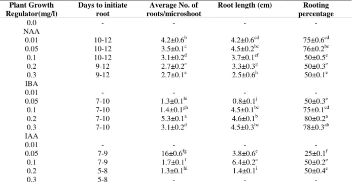
Rooting
The shoots after multiplication were isolated and transferred on to the MS medium containing different auxins. The mean percentage of rooting ranged from 50-76, 0- 50 and 0-80% were recorded on the rooting medium, supplemented with NAA, IAA and IBA, respectively. The highest rooting response (80%) (Table 5) was obtained on the MS medium supplemented with
IBA, with an average root length of 4.6cm. These observations were consistent with those reported by Salma et al. (2008), Raha and Roy (2003),
Neeta Mishraet al. (2003) and Atique Akbar et al.
(2003). The efficacy of the NAA at lower
concentrations in vitro rooting has been reported in
various medicinal plants, e.g., Verbascum thapsus
L. (Turker et al. 2001) and Santolina canescens
Lagasca (Casado et al. 2002).
Table 5 - In vitro rooting of Hoya wightii ssp. palniensis on MS medium supplemented with different
concentrations of auxins, after 30 days of culture period. Plant Growth
Regulator(mg/l)
Days to initiate root
Average No. of roots/microshoot
Root length (cm) Rooting
percentage 0.0
NAA 0.01 0.05 0.1 0.2 0.3 IBA 0.01 0.05 0.1 0.2 0.3 IAA 0.01 0.05 0.1 0.2 0.3
-
10-12 10-12 10-12 9-12 9-12
- 7-10 7-10 7-10 7-10
- 7-9 7-9 5-8 5-8
-
4.2±0.6b 3.5±0.1c 3.1±0.2d 2.7±0.2e 2.7±0.1e
- 1.3±0.1hi 1.4±0.1gh 5.3±0.1a 3.1±0.2d
- 16±0.6fg 1.7±0.1f 1.3±0.1hi
-
-
4.2±0.6cd 4.5±0.2bc 3.7±0.1ef 3.3±0.3g 2.5±0.6h
- 0.8±0.1j 4.5±0.1bc
4.6±0.1b 4.5±0.3bc
- 3.8±0.6e 6.4±0.2a 1.4±0.1i
-
-
75±0.6cd 76±0.2bc 50±0.5e 50±0.3e 50±0.1e
- 50±0.3e 75±0.1cd 80±0.2a 78±0.3ab
- 25±0.1f 50±0.2e 50±0.4e
-
Values followed by the same letter within the columns are not significantly different at the 5% level of significance according to Duncan’s Multiple Range Test.
Acclimatization of Regenerated Plants
The rooted plants with three-four fully expanded leaves and well-developed roots were transferred to the pots containing red soil, sand and coconut coir (1:1:1). Normal growth for the potted plants was observed after four weeks of transfer. After two months, they were transferred to larger pots containing the same ingredients and moved to a green house. After transplantation to soil, 70% of the rooted plantlets survived and grew to maturity when transferred to a green house (Fig. 1 G). The regenerated plants did not show visible varieties in the morphological or growth characteristics when compared to the donor plants.
CONCLUSION
This is the first report describing a protocol for organogenesis of H. wightii ssp. palniensis. The presence of the cytokinin BA in combination with IBA and GA3 was required for the efficient shoot
multiplication and proliferation. The addition of AA and CW was useful for the improvement of shoot regeneration. The IBA favored the root induction and development. The explants derived from the micropropagated shoots showed better organogenic potential than the explants from the mature plants.
The organogenesis and plant regeneration system developed in this study could be utilized in future
for the in vitro culture, transformation and
ACKNOWLEDGEMENTS
We acknowledge financial support from the University Grants Commission, New Delhi and Mr. R.W. Stewart and Mrs. Tanya Balcar and their
team of Vattakkanal Conservation Trust,
Kodaikanal for the help rendered during the reintroduction program.
REFERENCES
Amin MN, Jaiswal VS. Clonal propagation of guava through in vitro shoot proliferation on nodal experiments of mature trees. Plant Cell Tiss Org Cult. 1987;9; 235-244.
Atique Akbar M, Karmakar BK, Roy SK. Callus induction and high-frequency plant regeneration of Pineapple (Ananas comosus (L.) Merr.). Plant Tiss Cult. 2003;13(2): 109-116.
Azad MAK, Yokota S, Ohkubo T, Andoh Y, Yahara S, Yoshizawa N. In vitro regeneration of the medicinal woody plant Phellodendron amurense Rupr. through excised leaves. Plant Cell Tiss Org Cult. 2005; 80: 43-50.
Beegum AS, Martin KP, Zhang CL, Nishitha IK, Ligimol Slater, Madhusoodanan A. Organogenesis from leaf and internode explants of Ophiorrhiza prostrata, an anticancer drug (camptothecin) producing plant. Electronic J Biotech. 2007; 10(1): 114-123.
Beena MR, Martin KP, Kirti PB, Molly Hariharan. Rapid in vitro propagation of medicinally important Ceropegia candelabrum. Plant Cell Tiss Org Cult. 2003; 72:285-289.
Billings SG, Chin CK,Jelenkovic G. Regeneration of blue-berry plantlets from leaf segments. Hort Sci. 1988; 23: 763-766.
Casado JP, Navarro MC, Utrilla MP, Martinez A, Jimenez J. Micropropagation of Santolina canescens Lagasca and in vitro volatile production by shoot explants. Plant Cell Tiss Org Cult.2002; 69: 147-153. Chitra DS, Padmaja G. Shoot regeneration via direct
organogenesis from in vitro derived leaves of mulberry using thidiazuron and 6-benzylaminopurine. Sci Hort. 2005; 106: 593-602.
Douglas GC. Formation of adventitious buds in stem internodes of Populus species cultured on in vitro on basal medium: influence of endogenous properties of explants. J Plant Physiol.1984; 116: 313-321. Economou AS, Maloupa EM. Regeneration of
Elaeagnus angustifolia from leaf segments of in vitro-derived shoots. Plant Cell Tiss Org Cult. 1995; 40: 285-288.
George EF. Plant Propagation by Tissue Culture. Part II. Exegetics Ltd., Erdington, UK.1996.
Giridhar P, Vinod Kumar, Ravishankar GA. Somatic embryogenesis, organogenesis and regeneration from leaf callus culture of Decalepis hamiltonii Wight & Arn., an endangered shrub. In vitro Cell Dev Biol – Plant. 2004; 40: 567-571.
Hussain TM, Chandrasekhar T, Gopal GR. Micropropagation of Sterculia urens Roxb., an endangered tree species from intact seedlings. Afr J Biotechnol. 2008; 7(2): 95-101.
Karami O, Piri K. Shoot organogenesis in Oleaster (Elaeagnus angustifolia L.). Afr J Biotechnol. 2009; 8(3): 438-440.
Kelkar SM, Krishnamurthy KV. Adventitious shoot regeneration from root, internode, petiole and leaf explants of Piper colubrinum Link. Plant Cell Rep. 1998; 17: 721-725.
Khan S, Naseeb S, Ali K, Callus induction, plant regeneration and acclimatization of African violet (Saintpaulia ionantha) using leaves as explants. Pak J Bot. 2007; 39(4):1263-1268.
Kulkarni AA, Thengane SR, Krishnamurthi, KV. Direct shoot regeneration from node, internode, hypocotyl and embryo explants of Withania somnifera. Plant
Cell Tiss Org Cult. 2000;2
: 203-209.
Lakshmi SR, Franklin Benjamin JH, Senthil Kumar T, Murthy GVS, Rao MV. In vitro propagation of Hoya wightii ssp. palniensis K.T.Mathew, a highly vulnerable and endemic species of Western Ghats of Tamil Nadu. Afr J Biotech. 2010; 9(5): 620-627. Leblay C, Chevreau E, Raboin LM. Adventitious shoot
regeneration from in vitro leaves of several pear cultivars (Pyrus communis L.). Plant Cell Tiss Org Cult. 1991;25: 99-105.
Loc NH, Duc DT, Kwon TH, Yang MS. Micropropagation of zedoary (Curcuma zedoaria Roscoe) – a valuable medicinal plant. Plant Cell Tiss Org Cult. 2005; 81: 119-122.
Maraffa SB, Sharp WR, Tayama HK, Fretz TA. Apparent asexual embryogenesis in cultured leaf sections of Hoya carnosa. Z Pflanzen Physiol. 1981; 102: 45-56.
Mathew KT. The Hoyan. 1992; 14 (1): 3, t.1.
Matthew KM. Matthew. III. Fl. Palni Hills, 1996; t. 511.
Mederos-Molina S. Micropropagation of Salvia broussonetii Benth. – A medicinal Plant Species. Plant Tissue Cult Biotech. 2006; 16 (1): 19-23. MurashigeT. Plant propagation through tissue culture.
Annu Rev Plant Physiol. 1974; 25: 136-166.
Murashige T, Skoog F. A revised medium for rapid growth and bioassays for tobacco tissue cultures. Physiol Plant. 1962; 15: 473-497.
Nikam TD, Ghane SG, Nehul JN, Barmukh RB. Induction of morphogenic callus and multiple shoot regeneration in Momordica cymbalaria Fenzl. Indian J Biotechnol. 2009; 8: 442-447.
Raha S, Roy SC. Efficient plant regeneration in Holarrhena antidysentrica Wall, from shoot segment-derived callus. In Vitro Cell Dev Biol- Plant. 2003; 39: 151-155.
Sahin-Demirbag N, Kendir H, Khawar KM, Ciftci CY. In vitro regeneration of Tirkish dwarf chickling (Lathyrus cicera L.) using immature zygotic embryo explant. Afr J Biotechnol. 2008; 7(12): 2030-2033. Salma U, Rahman MSM, Islam S, Haque N, Jubair TA,
Haque AKMF, Mukti IJ. The influence of different hormone concentration and combination on callus induction and regeneration of Rauwolfia serpentina L. Benth. Pak J Biol Sci. 2008; 11(12): 1638-1641. Silva FAB, Pereira LAR, Silveira CES.
Micropropagation of Alibertia edulis Rich. Braz Arch Biol Technol. 2008; 51(6): 1103-1114.
Taiz L, Zeiger E. Plant Physiology. Massachusetts: Sinauer Associates, 1998; 792.
Tilkat E, Onay A. Direct shoot organogenesis from in vitro-derived mature leaf explants of pistachio. In vitro Cell Dev Biol Plant. 2009; 45: 92-98.
Tube Y, Xiao T, Leung K, Leung K-P, Yan G, Xiao S-Y, et al. Leaf ball tissue culture and rapid propagation test Hoya kerrii in vitro and its rapid propagation. Tropical Agric Sci Technol. 2007; 30(2) (Abstract only).
Turker AU,Camper ND, Gurel E. In vitro culture of common mullein (Verbascum thapsus L.). In Vitro Cell Dev Biol Plant. 2001; 37: 40-43.