*Corresponding author. E-mail: [email protected] (J. Walter).
aDivision of Allergy and Immunology, Children’s Research Institute, University of South Florida, St. Petersburg, FL, United States. bClinics Hospital, Universidade Federal do Paraná, Curitiba, PR, Brazil.
cDepartment of Biology, University of Tampa, Tampa, FL, United States.
dDivision of Allergy, Clinical Immunology and Rheumatology, Department of Pediatrics, Universidade Federal de São Paulo, São Paulo, SP, Brazil.
eDepartment of Immunology, Institute of Biomedical Sciences, Universidade de São Paulo, São Paulo, SP, Brazil. fDivision of Allergy and Immunology, Johns Hopkins All Children’s Hospital, St. Petersburg, FL, United States. gDivision of Pediatric Allergy and Immunology, Massachusetts General Hospital, Boston, MA, United States. Received on August 22, 2018.
In tIme: the value and global
ImplIcatIons of newborn screenIng
for severe combIned ImmunodefIcIency
In time: Importância e implicações globais da triagem
neonatal para a imunodeficiência grave combinada
Cristina Meehan
a,*
, Carmem Bonfim
b, Joseph F. Dasso
a,c,
Beatriz Tavares Costa-Carvalho
d, Antonio Condino-Neto
e, Jolan Walter
a,f,gSEVERE COMBINED
IMMUNODEFICIENCY (SCID)
S
evere combined immunodeficiency (SCID) is recognized as a global pediatric emergency that manifests early in infancy.1 In the absence of adaptive cellular and humoralimmune response, infants with SCID are prone to life threat-ening infections around 4–6 months of age, as they lose protec-tive maternal antibodies. Therefore, there is a narrow window of opportunity for early detection of infants with SCID during the asymptomatic period around birth. Newborn screening (NBS) is an essential solution for timely recognition and treatment of this otherwise fatal pediatric disease.
Specifically, infants with SCID are highly susceptible to a broad spectrum of bacterial, fungal and viral infec-tions. In addition to typical and opportunistic infections, live attenuated vaccine agents including Bacillus Calmette-Guérin (BCG) for tuberculosis, the oral poliovirus and rota-virus vaccines can result in severe complications including disseminated disease.2-4 Therefore, it is imperative to
per-form NBS for SCID before live vaccines are administered, so patients at risk can be identified and the potentially harmful routine live vaccinations can be avoided for this vulnerable patient population.
Since the discovery of SCID in the 1960s, two major breakthroughs in treatment have re-defined clinical outcomes (Figure 1).5-16
• bone marrow transplant (BMT) of healthy donor hema-topoietic stem cells to SCID patients was introduced in 1968 in the United States.17 If successful, this approach
can fully restore a normal immune system—T, B and natural killer (NK) cells;
• gene therapy was introduced in 1990.18 Through this
process, the abnormal gene can be corrected in the patient’s own hematopoietic stem cell by viral trans-fer of the normal gene and, therefore, donor cells are not needed. This therapy has been implemented for two variants of SCID: adenosine deaminase deficiency (ADA-SCID) and X-linked SCID with IL2RG mutation.
with NBS. Ideally, SCID patients identified by NBS receive treatment before infection occurs, which greatly increases sur-vival outcomes.19
Implementation of nbs for
scId in the united states
Most patients with SCID will present severe naïve T-cell lymphopenia secondary to impaired T-cell development in the thymus.20,21 The United States is pioneering in
imple-mentation of SCID NBS, with an assay based on the detec-tion of early abnormal T-cell development via T-cell recep-tor excision circles (TRECs). TRECs are generated during the process of T-cell receptor gene rearrangement in T-cell precursors in the thymus. Therefore, TRECs are enriched in the new immigrant naïve T-cells leaving the thymus. As T-cells get activated and proliferate, they will not prop-agate TRECs. Therefore, activated cells will have low lev-els TRECs. Thus, TRECs are an indirect measure of naïve T-cells and thymic function. The assay was originally designed to assess remnant thymic function in peripheral blood of patients infected by human immunodeficiency virus (HIV) with T-cell lymphopenia.22 Chan and Puck have applied this
assay first for evaluation of patients with SCID.23 For NBS
for SCID, the detection and quantification of TRECs are
Figure 1 Timeline of severe combined immunodeficiency therapy.
BMT: bone marrow transplant; SCID: severe combined immunodeficiency; ADA: adenosine deaminase deficiency; X-SCID: X-linked severe combined immunodeficiency; NBS: newborn screening; U.S.: United States; EU: Europe Union.
1968 First Bone
Marrow Transplant (BMT) for SCID
from a sibling donor by Robert A Good5
1977 First BMT in Latin America
performed Curituba, Brazil6
1990 First gene therapy clinical
trial for ADA-SCID8,9
1998 First BMT for SCID in Brazil10
2010 X-SCID self-inactiva-ting retroviral gene therapy
vector
2016 Brazil initiates
pilot NBS for SCID as the first country in
Latin America12-14 1988
First applica-tion of gene-marked cells in humans7
1992-2006 ADA-SCID, X-SCID, clinical
trials of retro-viral gene
therapy8,9
2009 Wisconsin as the first state initiates pilot NBS for SCID in
US11
2012 ADA-SCID with
self-inactiva-ting lentiviral
vector
2017 The first gene therapy as drug is approved for ADA-SCID in EU (Strimvelis)15,16
1968 1977 1988 1990 1992-2006 1998 2009 2010 2012 2016 2017
accomplished through extraction and amplification of deoxy-ribonucleic acid (DNA) from Guthrie cards obtained from infants around birth.
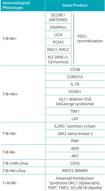
B-cell development can also be affected in several types of SCID. In addition to TRECs, a DNA-based assay has been developed to detect B-cell immunoglobulin light chain kappa receptor chain excision circle (KRECs). The absence of KRECs reflects abnormal B-cell development in the bone marrow and can accompany abnormal TRECs in forms of SCID that affect gene rearrangements, such as recombination activating gene (RAG) deficiency and components of the non-homologous end-joining complex (Table 1).24
Every country has different considerations regarding the inclusion of SCID on NBS panels. We believe that NBS for SCID should be implemented globally, which requires inter-national efforts due to disparities in healthcare. In the United States, a disease must meet the following criteria to be consid-ered for inclusion on the NBS panel:25,26
• minimum incidence of 1:100,000;
• fatality without treatment;
• improvement of outcomes with early treatment;
• development of a robust feasible test;
• a reasonable false positive rate;
T-B-NK+ Immunological Phenotype: DCLRE1: DNA cross-link repair 1C (artemis); DNA-PKcs: DNA-dependent protein kinase, catalytic subunit; LIG4: DNA ligase IV; XLF: XRCC4-like factor (Cernunnos) or NHEJ1: non-homologous end-joining factor; RAG1: recombination
activating gene 1; RAG2: recombination activating gene 2;
PMG3: phosphoglucomutase 3.
T-B+NK+ Immunological Phenotype: CD3δ: cluster of differentiation
3 delta chain; CORO1A: coronin-1A; IL-7R: interleukin-7 receptor;
FOXN1: forkhead box N1; 22q11.2 deletion (Full DiGeorge Syndrome); TBX1: T-box 1; LAT: linker for activation of T-cells; T-B+NK- Immunological
Phenotype.
IL2RG: interleukin 2 receptor subunit gamma (“common γ chain”);
JAK3: Janus kinase 3; PNP: purine nucleoside phosphorylase; ADA: adenosine deaminase deficiency; AK2: adenylate kinase 2; CD45: cluster of differentiation (leukocyte common antigen); RMRP: RNA component of mitochondrial RNA processing endoribonuclease; DKC1: dyskerin pseudouridine synthase 1; TERT: Telomerase reverse transcriptase; TINF2: TERF1-interacting nuclear factor 2; DCLRE1B: DNA cross-link repair 1B protein (apollo).
Table 1 Genetic background of severe combined immunodeficiency (SCID) listed by immunological phenotype.
Immunological
Phenotype Gene Product
T-B-NK+
DCLRE1 (ARTEMIS)
V(D)J
recombination DNAPKcs
LIG4
PGM3
RAG1, RAG2
XLF (NHEJ1, Cernunnos)
T-B+NK+
CD3δ
CORO1A
IL-7R
FOXN1 2q11 deletion (full DeGeorge syndrome)
TBX1 LAT
T-B+NK-IL2RG “common γ chain JAK3 Janus kinase 3
PNP
T-B-NK- ADA
AK2
T-B-/+NK+/low CD45
T-B+NK+/low RPP25 (RMRP)
T+B-NK-Hoyeraal-Hreidarsson Syndrome DKC1 (dyskeratin), TERT, TINF2, DCLRE1B (Apollo)
Fi g u r e 2 Severe combined immunodeficiency newborn screening implementation worldwide as of August 2018.30
Source: Immune Deficiency Foundation, available at: https:// primaryimmune.org/idf-scid-center
Screening
Pilots and Planning in 2018
The US Secretary’s Advisory Committee on Heritable Disorders in Newborns and Children (SACHDNC) recom-mends the list of disorders to be screened by NBS. To date, 34 congenital disorders have been added to the Recommended Uniform Screening Panel, and SCID was added in 2009.27,28
However, since the implementation of SCID NBS depends on state legislatures, the implementation time is variable across the United States. Since the first pilot program began in Wisconsin in 2009, 47 of the 50 states, the District of Columbia and Puerto Rico have sequentially implemented or have committed to implement SCID NBS (Jeffrey Modell Foundation; Figure 2).11,29,30
Apart from the United States, SCID NBS programs have implemented natiowide in Israel, Norway, and Taiwan, and in parts of Canada, and Spain (Figure 2), according to the Jeffrey Modell Foundation. In other countries, pilot screen-ing programs have been initiated in France (2006), Germany (2010), Sweden (2013), United Kingdom (2013), and Belgium (2012).13,31-37 Routine, nationwide implementation of pilot
or regional NBS programs have been limited by financial and legislative issues.
*alias CIITA, RFXANK, RFX5, RFXAP.
Table 2 Alphabetized list of conditions and/or genetic defects associated with T cell lymphopenia identified by newborn screening (NBS) for severe combined immunodeficiency (SCID).
ATM (ataxia
telangiectasia) DOCK8
Moesin
deficiency SMARCAL1
BCL10 IKBKB, MTHFD1 STAT5B
BLC11B IKBK2 NOLA2 STIM1
CARD11 IL-21R NOLA3 STK4 (MST1)
CD3e ITK ORAI1
(CRACM1)
TAP1/TAP2/ tapasin
CD3g Jakobsen PCFT TCN2
CD3z LCK/p56 PRKDC TCRa
CD8A
MAGT1 (X-MEN syndrome)
PTPRC
Trisomy 21 (Down syndrome
CHARGE
(CHD67) MALT-1 RAC2 TTC7A
DOCK2 MHCII* RHOH UNC119
ZAP70
confirmatory testing and treatment
following positive nbs for scId
Once a patient is screened positive by NBS for SCID, the diagnosis needs to be confirmed with laboratory testing. These tests assess the immune system of the patient, includ-ing the lymphocyte count with subset analysis of naïve and memory T-cells, B and NK cells and lymphocyte prolifer-ation studies. Low counts of autologous T-cells (<300 cells / µl) with low T-cell proliferation (<10% of lower level of normal) upon stimulation with phytohemagglutinin (PHA) are currently the diagnostic criteria of classical SCID.39
There are additional SCID variants (e.g., leaky SCID, Omenn syndrome and variant SCID) that present higher counts of autologous T-cells (300–1,000 cells / µl) with improved, but low T-cell proliferation (10–30% of lower level of nor-mal lymphocyte proliferation with PHA).39 In addition, it is
also recommended that naïve T-cell count and fraction are determined, as it reflects well the abnormal thymic activity and T-cell development.
While being prepared for hematopoietic stem cell trans-plantation (HSCT), the patient must be isolated at home or in the hospital to avoid exposure to infectious agents. Currently, there is no consensus on whether asymptomatic patients should be hospitalized. As patients may contract infections, strategies need to be developed for monitor-ing of infections and avoidmonitor-ing them by use of prophylac-tic antimicrobials and other interventions. About 42% of SCID infants identified by NBS develop infections prior to receiving definitive therapy.40 Cytomegalovirus (CMV)
is serious and life-threatening in SCID infants and is asso-ciated with increased risk for graft vs. host disease (GVHD) in patients receiving allogeneic transplantation. CMV is transmissible from a mother’s birth canal and/or breast milk. Therefore, infants with SCID whose mothers are seroposi-tive should not be breastfed. The indication for prophylac-tic treatment for CMV is debated as it can cause neutro-penia.41,42 While waiting for transplant, bridging therapies
include immunoglobulin replacement, antimicrobial (fungal, viral and bacterial) treatments and in specific cases enzyme replacement therapy for SCID with adenosine deaminase (ADA) deficiency (Table 3).43
During bridge therapy, the patient waits for the opti-mal setting of HSCT from a full human leukocyte antigens (HLA)-matched sibling or unrelated donor. If not available, most SCID patients receive haploidentical stem cells from par-ents (haploidentical transplant), especially if T-cells are absent, and, therefore, the likelihood of GVHD is lower. For patients without an optimal donor, autologous HSCT gene therapy (HSCT-GT) may be an option and has been highly successful.
In fact, HSCT-GT is recommended as an equal first line ther-apy for ADA-deficiency and is advantageous for avoiding risk of severe GVHD.44
Haploidentical donors increase the risk for GVHD. Therefore, SCID patients, especially those with T-cells, may require conditioning.40 With reduced intensity
con-ditioning, the bone marrow environment is optimized for engraftment of donor hematopoietic stem cells. There is a debate regarding the earliest time when conditioning can be safely used. Some centers have a long track record of no conditioning in infancy even at the expense of partial immune reconstitution with low B-cell function and the need for lifelong immunoglobulin replacement therapy. Depending on the underlying genetic defect, outcomes may be improved by using conditioning regardless of age, for example in SCID patients with hypomorphic RAG deficiency or DNA repair (non-homologous end joining) defects (Table 1).45
Prophylaxis in
Newborn Drug
Time of
initiation Alternatives Comments
PCP
TMP-SMX orally (5 mg TMP/kg once a day for 2 consecutive days weekly)
1 month old
Atovaquone orally (30 mg/kg
once a day)
Verify that bilirubin is <2X’s upper limit of normal before starting.
Monitor ALT, AST, and bilirubin every 2-4 weeks
HSV Acyclovir orally (20 mg/kg/
dose 3 times a day) At first visit
Follow BUN and creatinine every 2-4 weeks
Respiratory syncytial virus
Palivizumab (15 mg/kg/
I.M.) 1 month old
Given during peak RSV season, typically November-March in the
northern hemisphere
General (bacterial/ viral)
IVIG (0.4–0.5 g/kg every
month) or SCIG 1 month old
Monitor troughs monthly and maintain Ig>600 mg/dl; Based on subcutaneous fat and body surface
area to volume of medication administered, could consider SCIG
in select patients
Fungal Fluconazole (6 mg/kg once
daily) 1 month old
Follow AST, ALT, and bilirubin every 2–4 weeks
In family members
or close contacts
Influenza Inactivated influenza vaccine Seasonally
Pertussis Tdap vaccine
Per routine childhood vaccinations
One booster for adolescents (11–12 years age); adults 19–64 years age
and adults >65 years age
Table 3 Recommended infectious disease prophylaxis for newborns with suspected of severe combined immunodeficiency (SCID).
PCP: pneumocystis carinii pneumonia; HSV: herpes simplex virus; TMP-SMX: trimethoprim / sulfamethoxazole; IVIG: intravenous immunoglobulin; SCIG: subcutaneous immunoglobulin; ALT: alanine aminotransferase; AST: aspartate aminotransferase; BUN: blood urea nitrogen; RSV: respiratory
syncytial virus; SCIG: subcutaneous immunoglobulin.
Source: Thakar et al.23
Early live vaccinations and exposure to a wide variety of infectious agents may lead to clinical infections that worsen transplant outcomes and increase healthcare costs for man-agement of these patients.46 Therefore, the outcome in
countries without NBS for SCID remains sub-optimal with increased morbidity and mortality despite advances in ther-apy. The initiation of SCID NBS faces challenges in Brazil. National efforts for SCID NBS should be supported by sev-eral centers with high diagnostic and transplant expertise in SCID. These centers should be evenly dispersed across the country to ensure access and coverage. Ideally, these centers should also prioritize and allocate resources for the routine care of SCID patients, including beds, organization of an inpatient and outpatient clinical care team and development of hospital protocols.
The introduction of the Guthrie card in 1963 has resulted in the widespread use of this simple but universal NBS device that is available globally. Blood spots on the card, obtained from a heel prick, can be analyzed to detect rare genetic, metabolic, and endocrine diseases. DNA remains stable on this card and can be a reliable source of detection of TRECs. NBS began in Brazil in 1976, and, from 2001 to 2005, about 13 million newborns were screened, with coverage increasing from 55 (in 1976) to 80.2% (in 2005).12 Despite these advancements in
programs for NBS in Brazil. The first Brazilian SCID NBS pilot launched in 2016 and screened 8,715 newborns using the TRECs assay.13 The second pilot launched in 2017 and
screened 6,881 newborns using both the TRECs and KRECs assays, with sample collections in several metropolitan areas in the São Paulo region.14 Both of these pilot programs
con-firmed that SCID NBS assay is reliable and feasible for future implementation on a national scale in Brazil.
Without effective infrastructure for early HSCT, there is only partial value in NBS for SCID. Yet, many countries among Central and Latin America are leading efforts to improve treat-ment for SCID. In 1976, Colombia was the first country to conduct a HSCT. Similarly, since that time Brazil has estab-lished infrastructure to provide many key therapies for SCID. In 1979, the first organized Brazilian HSCT program was estab-lished in the city of Curitiba, in state of Paraná. To improve the HLA-matching for donor and recipient, HSCT program initially began with sibling matched donors and evolved to alternative donor transplantation in 1995. With the intro-duction of post-transplantation cyclophosphamide to prevent GVHD, haploidentical transplantation was initiated. The first HSCT for patients with SCID were conducted in Central and Latin America in 1985 in Costa Rica and in 1998 in Brazil, respectively.10 For a population of over 200 million inhabitants
in Brazil, there are close to a hundred BMT medical units. Of approximately 3,000 HSCT performed in the period of 1979–2018 for various health conditions in Curitiba, 90% of these were allogenic. This magnitude of population and grow-ing level of expertise underscores the importance of screengrow-ing program for SCID in Brazil.
Families are getting smaller in Brazil, as in most developed countries, thereby decreasing the chance of finding a sibling donor. Brazilian BMT units are unable to do haploidentical transplant with T-cell depletion, and thus use post-HSCT treat-ment with cyclophosphamide to remove donor T-cells is needed to reduce the risk of GVHD. Brazil has developed a donor regis-try entitled Registro Nacional de Doadores Voluntários de Medula Óssea (REDOME), that currently has more than four million donors registered. Therefore, it is the third largest bone marrow volunteer donor registry in the world. In addition, there are 11 public cord blood banks in Brazil, even though cord blood transplantation is decreasing after the emergence of HSCT treat-ment with post-transplant cyclophosphamide. Unfortunately, despite the ample infrastructure for HSCT technology, there are inadequate numbers of personnel trained in the specialized HSCT for SCID patients in Brazil and Latin America.
Since the initial pilot studies, Brazil has reached the fourth phase of implementation of SCID NBS within the coun-try. Experts in immunology advocate on all levels for the
implementation of NBS for SCID and other primary immu-nodeficiencies during the first year of life as it would decrease clinical costs and improve public health. In fact, the Brazilian Society of Allergy and Immunology is currently applying to incorporate the NBS for SCID and possibly other primary immune deficiencies (PIDs) in the national screening pro-gram together with other rare diseases. This request is pending approval and funding.13,14
To optimize the implementation of these advancements, it is essential to ensure that patients have access confirmatory services for the diagnosis of SCID after positive NBS. These diagnostic services include machinery to quantify lymphocytes subpopulations (T and naïve T-cells) and function (lympho-cyte proliferation assays). Unfortunately, these tests are not uni-versally available, but only in large academic research centers.
economic impact of nbs for scId
From a long-term economic perspective, screening programs and treatments for early diagnosis of asymptomatic SCID patients are less expensive than providing healthcare to a child that has a delayed diagnosis and complicating infections before defin-itive therapies are initiated.
Globally, short-term implementation costs may be a barrier to adding SCID to NBS panels, but it could be justified by the cost difference between transplanting a child above and below 3.5 months of age with or without infections. For example, in the United States in 2014, the mean total charges for late trans-plantation for SCID per patient were four times greater than early treatment ($ 1.43 million vs. $ 365,785 respectively) without consideration of the potential need for intensive care services.47
The cost-effectiveness of early treatment for SCID provided strong economic justification for the addition of SCID screening to NBS programs in all states in the United States by 2018. Brazil has not performed a thorough cost-benefit analysis of the cost of SCID NBS and treatment before or after the onset of infections.14
Making the cost of SCID NBS comparable to or less than that for treatment on a population level will facilitate government approval of nationwide SCID NBS programs. Healthcare cost for SCID treatment, including HSCT, are lower in Europe48 and in the developing world than in the
United States. Therefore it is less expensive for these countries to treat SCID once symptoms present. Thus, implementing countrywide SCID NBS programs may be a lesser health-care priority in most of Europe and in developing countries than in the US. However, not implementing these programs results in greater infant mortality and morbidity;32,47 failure
for SCID costs approximately $ 5 pe patient in the United States.47-49 A German study lowered the cost of SCID NBS to
€ 2 per sample ($ 2.33) by reducing the sample size used for testing, devising a more efficient DNA extraction technique and using internal controls selectively.50 The reduced cost of
the new SCID NBS method, the marked increase in cost of late versus early SCID treatment, and the long-term mone-tary value of saving lives with early screening and treatment51
are strong economic rationales, besides ethical justification, for considering SCID NBS throughout Brazil and the rest of the world where it is not performed.
Impact of nbs on scId
incidence and patient survival
SCID NBS saves lives. For example, a multi-site study conducted by the Primary Immune Deficiency Treatment Consortium found that infants not tested until symptoms presented had a 58% survival rate, compared to 85% survival for infants tested at birth.40 The implementation of SCID NBS on the Recommended
Uniform Screening Panel has dramatically changed the clinical presentation of SCID in the United States. Analysis of screen-ing of three million newborns for SCID after the initiation of SCID NBS confirmed a higher-than-expected prevalence of 1:58,000, increasing from 1:100,000 in 2009 prior to NBS. In the United States, X-linked SCID remains the most common variant among SCID patients. However, its relative frequency has decreased from 46 to 19% and recombinase activating gene (RAG1/2) deficiency is becoming dominant in leaky SCID vari-ants.41,52 Pathogenic variants are now the norm. Furthermore, the
frequency of SCID across racial and ethnic groups is increasing following implementation of SCID NBS. There is also founder mutation penetrance in communities with frequency up to 1:2,000, found in communities of Somali, Amish, Mennonite, Navajo Indians and Irish Traveler descent.53-56
To broaden newborn screening for immunodeficiency, a new program, “Following Infants with Low Lymphocytes” (FILL), has been organized by the Clinical Immunology Society (CIS)
and the United States Immunodeficiency Network (USIDNET). This program is designed to track the diagnoses and outcomes of non-SCID patients identified with T-lymphopenia in the NBS program57 (Table 2).
CONCLUSION
Early diagnosis of SCID is feasible by using a Guthrie screening card shortly after birth. Although the method is relatively inex-pensive, it requires centralized laboratory testing and a network of clinical immunologists to confirm the clinical and genetic diag-nosis, and a BMT team to perform HSCT with optimal timing and selection of donor and conditioning regimen. With inter-national effort addressing the challenges and solutions to man-aging SCID in newborns, the dire consequences of this disease can be thwarted, thus relieving the tremendous fiscal, social, and emotional burden of affected children and families worldwide.
ACKNOWLEDGEMENTS
We acknowledge the efforts of the government of Brazil and the Jeffrey Modell Foundation in the funding of pilot programs for newborn screening of SCID within Brazil.
We thank and acknowledge Dr. Jane Carver from the University of South Florida, for their assistance in the editing of this document.
funding
This work was partly supported by Jeffrey Modell Diagnostic and Research Center at John Hopkins All Children’s Hospital and the Robert A. Good Chair Endowment from the University of South Florida Foundation. Dr. Beatriz Tavares Costa-Carvalho and Dr. Antonio Condino-Neto have received Jeffrey Modell Foundation funding to implement newborn screening in Brazil.
conflict of interests
The authors declare no conflict of interests.
REFERENCES
1. Bonilla FA, Khan DA, Ballas ZK, Chinen J, Frank MM, Hsu JT, et al.
Practice parameter for the diagnosis and management of primary
immunodeficiency. J Allergy Clin Immunol.
2015;136(5):1186-205.e1-78. https://doi.org/10.1016/j.jaci.2015.04.049
2. Bakare N, Menschik D, Tiernan R, Hua W, Martin D. Severe combined immunodeficiency (SCID) and rotavirus vaccination: reports to the Vaccine Adverse Events Reporting System
(VAERS). Vaccine. 2010;28(40):6609-12. https://doi.
org/10.1016/j.vaccine.2010.07.039
3. Marciano BE, Huang CY, Joshi G, Rezaei N, Carvalho BC, Allwood Z, et al. BCG vaccination in patients with severe combined immunodeficiency: complications, risks, and vaccination
4. Shearer WT, Fleisher TA, Buckley RH, Ballas Z, Ballow
M, Blaese RM, et al. Recommendations for live viral and bacterial vaccines in immunodeficient patients and their
close contacts. J Allergy Clin Immunol. 2014;133(4):961-6.
https://dx.doi.org/10.1016%2Fj.jaci.2013.11.043
5. Thomas ED, Lochte H Jr. Studies on the biochemical defect
of pernicious anemia. I. In vitro observations on oxygen consumption, heme synthesis and deoxyribonucleic acid
synthesis by pernicious anemia bone marrow. J Clin Invest.
1958;37(2):166-71. https://dx.doi.org/10.1172/JCI103595
6. Kanegae MPP, Barreiros LA, Sousa JL, Brito MAS, Oliveira Junior EB, Soares LP, et al. Newborn Screening for Severe Combined Immunodeficiencies Using Trecs and Krecs: Second
Pilot Study in Brazil. Rev Paul Pediatr. 2017;35(1):25-32. https://doi.org/10.1590/1984-0462/;2017;35;1;00013
7. Rosenberg AS, Katz SI, Singer A. Rejection of skin allografts by CD4+ T cells is antigen-specific and requires expression
of target alloantigen on Ia- epidermal cells. J Immunol. 1989;143(8):2452-6.
8. Appelbaum FR. Hematopoietic-Cell Transplantation at 50. N
Engl J Med. 2007;357(15):1472-5. https://doi.org/10.1056/
NEJMp078166
9. Wirth T, Parker N, Yla-Herttuala S. History of gene therapy.
Gene 2013;525(2):162-9. https://doi.org/10.1016/j. gene.2013.03.137
10. Fasth A. Osteopetrosis--more than only a disease of the bone. Am J Hematol. 2009;84(8):469-70. https://doi.org/10.1002/ ajh.21454
11. Verbsky J, Thakar M, Routes J. The Wisconsin approach to newborn screening for severe combined immunodeficiency.
J Allergy Clin Immunol. 2012;129(3):622-7. https://doi. org/10.1016/j.jaci.2011.12.004
12. Carvalho TM, dos Santos HP, dos Santos IC, Vargas PR, Pedrosa J. Newborn screening: a national public health
programme in Brazil. J Inherit Metab Dis. 2007;30(4):615. https://doi.org/10.1007/s10545-007-0650-7
13. Kanegae MP, Barreiros LA, Mazzucchelli JT, Hadachi SM, Guilhoto LMFF, Acquesta AL, et al. Neonatal screening for
severe combined immunodeficiency in Brazil. J Pediatr (Rio J). 2016;92(4):374-80. https://doi.org/10.1016/j. jped.2015.10.006
14. Kanegae MPP, Barreiros LA, Sousa JL, Brito MAS, Oliveira Junior EB, Soares LP, et al. Newborn Screening for Severe Combined Immunodeficiencies Using Trecs and Krecs: Second
Pilot Study in Brazil. Rev Paul Pediatr. 2017;35(1):25-32. https://doi.org/10.1590/1984-0462/;2017;35;1;00013
15. Miller N. Glybera and the future of gene therapy in the European Union. Nat Rev Drug Discov. 2012;11(5):419.
16. Morrison C. $1-million price tag set for Glybera gene therapy.
Nat Biotechnol. 2015;33(3):217-8. https://doi.org/10.1038/
nbt0315-217
17. Gatti RA, Meuwissen HJ, Allen HD, Hong R, Good RA.
Immunological reconstitution of sex-linked lymphopenic immunological deficiency. Lancet. 1968;292(7583):1366-9.
https://doi.org/10.1016/S0140-6736(68)92673-1
18. Kaufmann KB, Büning H, Galy A, Schambach A, Grez M. Gene
therapy on the move. EMBO Mol Med. 2013;5(11):1642-61. https://doi.org/10.1002/emmm.201202287
19. Pai SY, Logan BR, Griffith LM, Buckley RH, Parrott RE,
Dvorak CC, et al. Transplantation outcomes for severe combined immunodeficiency, 2000-2009. N Engl J Med. 2014;371(5):434-46. https://doi.org/10.1056/NEJMoa1401177
20. Puck JM. Neonatal Screening for Severe Combined Immunodeficiency (SCID). Curr Opin Pediatr.
2011;23(6):667-73. https://doi.org/10.1097/MOP.0b013e32834cb9b0
21. Clinical and Laboratory Standards Institute. NBS06-A: Newborn blood spot screening for severe combined
immunodeficiency by measurement of T-cell receptor
excision circles; approved guideline [Internet]. 2013 [cited on Sept. 12, 2018]. Avaliable at: https://clsi.org/media/1488/
nbs06a_sample.pdf
22. Douek DC, McFarland RD, Keiser PH, Gage EA, Massey
JM, Haynes BF, et al. Changes in thymic function with age and during the treatment of HIV infection. Nature.
1998;396(6712):690-5. https://doi.org/10.1038/25374
23. Chan K, Puck JM. Development of population-based newborn screening for severe combined immunodeficiency. J Allergy
Clin Immunol. 2005;115(2):391-8. https://doi.org/10.1016/j. jaci.2004.10.012
24. Jyonouchi S , Jongco AM, Puck J, Sullivan KE . Immunodeficiencies Associated with Abnormal Newborn
Screening for T Cell and B Cell Lymphopenia. J Clin Immunol. 2017;37(4):363-74. https://doi.org/10.1007/ s10875-017-0388-4
25. Kwan A, Abraham RS, Currier R, Brower A, Andruszewski K, Abbott JK, et al. Newborn screening for severe combined immunodeficiency in 11 screening programs in the United
States. JAMA. 2014;312(7):729-38. https://doi.org/10.1001/ jama.2014.9132
26. Baker MW, Laessig RH, Katcher ML, Routes JM, Grossman WJ, Verbsky J, et al. Implementing routine testing for severe combined immunodeficiency within Wisconsin’s newborn
screening program. Public Health Rep. 2010;125(Suppl. 2):88-95. https://doi.org/10.1177/00333549101250S211
27. Watson MS, Mann MY, Lloyd-Puryear MA, Rinaldo P, Howell
RR. Newborn Screening: Towards a Uniform Screening Panel and System. Genetic Med [Internet]. 2006 [cited on Sept. 14, 2018];8(Suppl. 1):1S-252S. Available at: https://www.
ncbi.nlm.nih.gov/pmc/articles/PMC3111605/
28. Kwan A, Puck JM. History and current status of newborn screening for severe combined immunodeficiency. Semin
Perinatol. 2015;39(3):194-205. https://doi.org/10.1053/j. semperi.2015.03.004
29. Martínez-Morillo E, Prieto García B, Álvarez Menéndez
FV. Challenges for Worldwide Harmonization of Newborn
Screening Programs. Clin Chem. 2016;62(5):689-98. https:// doi.org/10.1373/clinchem.2015.240903
30. Jeffrey Modell Foundation. Newborn screening for SCID.
Update on the implementation of newborn screening for
SCID in the United States [Internet]. August 2018 [cited on Sept. 25, 2018]. Available at: http://www.info4pi.org/
town-hall/newborn-screening
31. Adams SP, Rashid S, Premachandra T, Harvey K, Ifederu A, Wilson MC, et al. Screening of neonatal UK dried blood spots using a duplex TREC screening assay. J Clin
32. Audrain M, Thomas C, Mirallie S, Bourgeois N, Sebille V, Rabetrano H, et al. Evaluation of the T-cell receptor excision circle assay performances for severe combined
immunodeficiency neonatal screening on Guthrie cards in a French single centre study. Clin Immunol. 2014;150(2):137-9. https://doi.org/10.1016/j.clim.2013.11.012
33. Barbaro M, Ohlsson A, Borte S, Jonsson S, Zetterström
RH, King J, et al. Newborn Screening for Severe Primary
Immunodeficiency Diseases in Sweden-a 2-Year Pilot
TREC and KREC Screening Study. J Clin Immunol.
2017;37(1):51-60. https://doi.org/10.1007/s10875-016-0347-5
34. Borte S, von Döbeln U, Fasth A, Wang N, Janzi M, Winiarski J, et al. Neonatal screening for severe primary immunodeficiency diseases using high-throughput triplex
real-time PCR. Blood. 2012;119(11):2552-5. https://doi. org/10.1182/blood-2011-08-371021
35. Chien YH, Chiang SC, Chang KL, Yu HH, Lee WI, Tsai LP, et al. Incidence of severe combined immunodeficiency
through newborn screening in a Chinese population. J Formos Med Assoc. 2015;114(1):12-6. https://doi. org/10.1016/j.jfma.2012.10.020
36. Cross C. Ontario newborns now screened for SCID. CMAJ. 2013;185(13):E616. https://doi.org/10.1503/ cmaj.109-4580
37. Olbrich P, de Felipe B, Delgado-Pecellin C, Rodero R, Rojas
P, Aguayo J, et al. [A first pilot study on the neonatal
screening of primary immunodeficiencies in Spain: TRECS
and KRECS identify severe T- and B-cell lymphopenia]. An
Pediatr (Barc). 2014;81(5):310-7. https://doi.org/10.1016/j. anpedi.2014.08.002
38. Wilson K, Duque DR, Murphy MSQ, Hawken S, Pham-Huy
A, Kwong J, et al. T-cell receptor excision circle levels
and safety of paediatric immunization: A
population-based self-controlled case series analysis. Hum Vaccin
Immunother. 2018;14(6):1378-91. https://doi.org/10.1 080/21645515.2018.1433971
39. Shearer WT, Dunn E, Notarangelo LD, Dvorak CC, Puck
JM, Logan BR, et al. Establishing diagnostic criteria for
severe combined immunodeficiency disease (SCID), leaky SCID, and Omenn syndrome: the Primary Immune
Deficiency Treatment Consortium experience. J Allergy Clin
Immunol. 2014;133(4):1092-8. https://doi.org/10.1016/j. jaci.2013.09.044
40. Heimall J, Logan BR, Cowan MJ, Notarangelo LD, Griffith LM, Puck JM, et al. Immune reconstitution and survival of
100 SCID patients post-hematopoietic cell transplant: a PIDTC natural history study. Blood. 2017;130(25):2718-27. https://doi.org/10.1182/blood-2017-05-781849
41. Dorsey MJ, Dvorak CC, Cowan MJ, Puck JM. Treatment of infants identified as having severe combined immunodeficiency by means of newborn screening. J Allergy Clin Immunol. 2017;139(3):733-42. https://doi. org/10.1016/j.jaci.2017.01.005
42. Griffith LM, Cowan MJ, Notarangelo LD, Kohn DB, Puck JM, Shearer WT, et al. Primary Immune Deficiency
Treatment Consortium (PIDTC) update. J Allergy Clin Immunol. 2016;138(2):375-85. https://doi.org/10.1016/j. jaci.2016.01.051
43. Thakar MS, Hintermeyer MK, Gries MG, Routes JM, Verbsky JW. A Practical Approach to Newborn Screening for Severe Combined Immunodeficiency Using the T Cell Receptor Excision Circle Assay. Front Immunol. 2017;8:1470. https:// doi.org/10.3389/fimmu.2017.01470
44. Kohn DB, Hershfield MS, Puck JM, Aiuti A, Blincoe A, Gaspar HB, et al. Consensus approach for the management
of severe combined immune deficiency caused by
adenosine deaminase deficiency. J Allergy Clin Immunol.
2018;S0091-6749(18):31268-5. https://doi.org/10.1016/j. jaci.2018.08.024
45. Schuetz C, Neven B, Dvorak CC, Leroy S, Ege MJ, Pannicke U, et al. SCID patients with ARTEMIS vs RAG deficiencies following HCT: increased risk of late toxicity in ARTEMIS-deficient SCID. 2014;123(2):281-9. https://doi.org/10.1182/
blood-2013-01-476432
46. Mazzucchelli JT, Bonfim C, Castro GG, Condino-Neto AA, Costa NM, Cunha L, et al. Severe combined immunodeficiency
in Brazil: management, prognosis, and BCG-associated complications. J Investig Allergol Clin Immunol. 2014;24(3):184-91.
47. Kubiak C, Jyonouchi S, Kuo C, Garcia-Lloret M, Dorsey MJ, Sleasman J, et al. Fiscal implications of newborn screening in the diagnosis of severe combined immunodeficiency. J
Allergy Clin Immunol Pract. 2014;2(6):697-702. https://doi. org/10.1016/j.jaip.2014.05.013
48. Clément MC, Mahlaoui N, Mignot C, Le Bihan C, Rabetrano H, Hoang L, et al. Systematic neonatal screening for severe combined immunodeficiency and severe T-cell lymphopenia: Analysis of cost-effectiveness based on French real field
data. J Allergy Clin Immunol. 2015;135(6):1589-93. https:// doi.org/10.1016/j.jaci.2015.02.004
49. Modell V, Knaus M, Modell F. An analysis and decision tool to measure cost benefit of newborn screening for severe
combined immunodeficiency (SCID) and related T-cell lymphopenia. Immunol Res. 2014;60(1):145-52. https:// doi.org/10.1007/s12026-014-8485-4
50. Tagliaferri L, Kunz JB, Happich M, Esposito S, Bruckner T, Hübschmann D, et al. Newborn screening for severe combined immunodeficiency using a novel and simplified method to measure T-cell excision circles (TREC). Clin Immunol.
2017;175:51-5. https://doi.org/10.1016/j.clim.2016.11.016
51. Brown L, Xu-Bayford J, Allwood Z, Slatter M, Cant A, Davies EG, et al. Neonatal diagnosis of severe combined
immunodeficiency leads to significantly improved survival outcome: the case for newborn screening. Blood. 2011;117(11):3243-6. https://doi.org/10.1182/blood-2010-08-300384
52. Fischer A, Notarangelo LD, Neven B, Cavazzana M, Puck
JM. Severe combined immunodeficiencies and related
disorders. Nat Rev Dis Primers. 2015;1:15061. https://doi.
org/10.1038/nrdp.2015.61
53. Sanchez JJ, Monaghan G, Børsting C, Norbury G, Morling N, Gaspar HB. Carrier frequency of a nonsense mutation in the
adenosine deaminase (ADA) gene implies a high incidence of
ADA-deficient severe combined immunodeficiency (SCID) in
Somalia and a single, common haplotype indicates common ancestry. Ann Hum Genet. 2007;71(3):336-47. https://doi.
54. Strauss KA, Puffenberger EG, Bunin N, Rider NL, Morton MC, Eastman JT 3rd, et al. Clinical application of DNA microarrays:
molecular diagnosis and HLA matching of an Amish child
with severe combined immune deficiency. Clin Immunol.
2008;128(1):31-8. https://doi.org/10.1016/j.clim.2008.02.016
55. Li L, Moshous D, Zhou Y, Wang J, Xie G, Salido E, et al. A founder mutation in Artemis, an SNM1-like protein, causes SCID in Athabascan-speaking Native Americans. J Immunol.
2002;168(12):6323-9.
56. Casey JP, Nobbs M, McGettigan P, Lynch S, Ennis S. Recessive mutations in MCM4/PRKDC cause a novel syndrome involving a primary immunodeficiency and a disorder of DNA repair.
J Med Genet. 2012;49(4):242-5. https://doi.org/10.1136/ jmedgenet-2012-100803
57. USIDNET CISC. Immune Deficiency Foundation, Jeffrey
Modell Foundation. About FILL Program [Internet] [cited in 2018]. Available at: www.usidnet.org/fill