Combating oncogene activation
associated with retrovirus-mediated
gene therapy of X-linked severe
combined immunodeficiency
1Setor de Vetores Virais, Laboratório de Genética e Cardiologia Molecular,
Faculdade de Medicina
2Departamento de Biologia Celular e do Desenvolvimento, Instituto de Ciências
Biomédicas, Universidade de São Paulo, São Paulo, SP, Brasil
3Instituto do Milênio: Rede de Terapia Gênica, MCT, Brasília, DF, Brasil
B.E. Strauss1,3
and E. Costanzi-Strauss2,3
Abstract
A successful gene therapy clinical trial that also encountered serious adverse effects has sparked extensive study and debate about the future directions for retrovirus-mediated interventions. Treatment of X-linked severe combined immunodeficiency with an oncoretrovirus harboring a normal copy of the γc gene was applied in two clinical trials, essentially curing 13 of 16 infants, restoring a normal immune system without the need for additional immune-related therapies. Approximately 3 years after their gene therapy, tragically, 3 of these children, all from the same trial, developed leukemia as a result of this experimental treatment. The current understanding of the mechanism behind this leukemogenesis involves three critical and cooperating factors, i.e., viral integration, oncogene activation, and the function of the therapeutic gene. In this review, we will explore the causes of this unwanted event and some of the possibilities for reducing the risk of its reoccurrence.
Correspondence
B.E. Strauss
Instituto do Coração, FM, USP Av. Dr. Enéas C. Aguiar, 44 Bloco 2, 10º andar 05403-000 São Paulo, SP Brasil
Fax: +55-11-3069-5022 E-mail: [email protected] Research supported by FAPESP (Nos. 00/12156-5 and 98/15120-0) and CNPq (Nos. 473587/04-1 and 420036/2005-9).
Received June 28, 2006 Accepted December 13, 2006
Key words
•Retrovirus
•Insertional mutagenesis •Severe combined
immune deficiency •Gene therapy •Adverse event •Clinical trial
Introduction
The clinical history of gene therapy is quite short, dating back only to 1990. The first gene therapy trial was the retrovirus-mediated gene transfer of a normal copy of the adenosine deaminase (ADA) gene for the treatment of immune deficiency caused by the loss of cellular ADA. This trial and others for ADA-X-linked severe combined immunodeficiency (ADA-X-SCID) have provided clinical benefit without side effects associated with the retroviral vector (1).
ge-nome seems to be less likely to cause an adverse effect, though clinical experience with these vectors is limited. In contrast, retroviral vectors have a long history in the laboratory and in the clinic, their popularity being due to their ease of use and stability in the transduced cell.
Although much has been learned about the choices of gene transfer vectors, disease targets and therapeutic genes, the relatively limited success and highly publicized risks indicate a clear need for further improve-ment before gene therapy will become widely accepted. The risk and benefits of gene thera-py are typified by the clinical trial for X-SCID conducted by Drs. Alain Fischer and Marina Cavazzana-Calvo and colleagues (Hôpital Necker Enfants Malade, Paris, France). In that trial, a normal copy of a defective gene was introduced via retroviral transduction into hematopoietic stem cells of 1- to 11-month-old infants. The trial was extremely successful, restoring normal im-mune function in 9 of the 10 children. How-ever, 3 of the patients developed T-cell leu-kemia due, in part, to the cooperation be-tween the viral insertion, activation of a cellular oncogene, and the growth-promot-ing characteristics of the therapeutic gene. Interestingly, a similar trial conducted in England, led by Dr. Adrian Thrasher (3), was also successful in treating 4 of 6 X-SCID patients with this strategy, and no adverse events have been observed. This
suggests that an unknown factor may have been present in the French trial that exacer-bated the onset of leukemia.
In the present article, we will summarize the key components of the French trial, ana-lyze the factors that contributed to the un-wanted leukemic transformation and explore some of the options for reducing this risk. As stated by the key administrators of these X-SCID clinical trials, “research must go on, particularly on vector design” (4). Among the molecular tools currently available, we propose that specialized control of viral ex-pression could help reduce the risk of un-wanted oncogene activity. Our work has involved retrovirus-mediated gene transfer for more than 12 years and we are currently developing retroviral vectors with novel tran-scriptional regulation. We describe here how exploration of vector design as well as trans-duction and transplantation procedures may improve retrovirus-mediated gene therapy.
Molecular basis of X-linked SCID
X-SCID is caused by a mutation in the γ
subunit of the common cytokine receptor (γc
or IL2RG), preventing T-cells and natural killer
cells from developing. Without functional T-cells, activation of B-cells is also limited, leav-ing the patient with a severely impaired im-mune system. The role of the wild-type γc
chain (encoded on chromosome Xq13) is to participate in the cytokine receptor complex
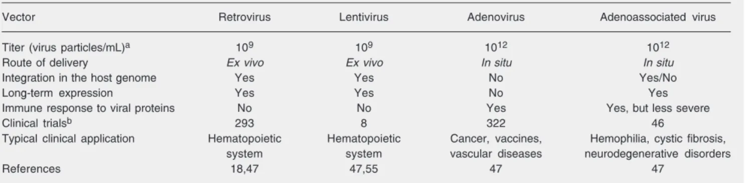
Table 1. Principal features of viral vectors.
Vector Retrovirus Lentivirus Adenovirus Adenoassociated virus
Titer (virus particles/mL)a 109 109 1012 1012
Route of delivery Ex vivo Ex vivo In situ In situ
Integration in the host genome Yes Yes No Yes/No
Long-term expression Yes Yes No Yes
Immune response to viral proteins No No Yes Yes, but less severe
Clinical trialsb 293 8 322 46
Typical clinical application Hematopoietic Hematopoietic Cancer, vaccines, Hemophilia, cystic fibrosis,
system system vascular diseases neurodegenerative disorders
References 18,47 47,55 47 47
that mediates cellular activation by IL-2, IL-4, IL-7, IL-9, IL-15, and IL-21 (5,6). Since cy-tokine activation is often associated with pro-liferation, the cytokine receptor complex, in-cluding the γc chain, may represent a growth-promoting assembly.
Without treatment, infants born with X-SCID would die within less than a year due to infection (7,8). Treatment requires a bone marrow transplant from a suitable donor, yet not all patients are able to find a match. Perfect matches may be found from siblings, with a 90% successful transplant rate, or up to about 70% when the donor is a haploidentical parent or unrelated individual (9,10).
Following transplant, these children are at risk for lymphoma if the donor had a history of infectious mononucleosis. In addition, some transplant recipients do not recover complete T-cell function and most patients have defi-cient B-cell function, requiring life-long im-mune-globulin-replacement therapy (9). Ef-forts to improve B-cell function include chemoablation prior to transplant so that he-matopoiesis is restored entirely from trans-planted cells, although this approach involves higher mortality (10). Clearly, X-SCID is a devastating disease and standard treatment modalities are inadequate.
Successful gene therapy for X-SCID is associated with three serious adverse events
A group of French researchers has con-ducted the most successful gene therapy trial to date, with considerable clinical benefit (though only time will tell if a true cure has been achieved) to 9 of 10 children with X-SCID (11-13). A second clinical trial for this gene therapy approach has been carried out in England and has also been successful in the treatment of 4 of 6 X-SICD patients (3). Until recently, these trials have served as the prime example of how to use molecular tools to treat and essentially cure a disease by the simple insertion of a functional gene into cells that,
due to an inherited mutation, would otherwise be lacking that gene’s function. The strategy used by Drs. Alain Fischer, Marina Cavazzana-Calvo, Adrian Thrasher, and colleagues was
ex vivo transduction of CD34+ stem cells,
derived from the patient’s own bone marrow, with a replication-defective Moloney murine leukemia virus (MLV) vector encoding a nor-mal copy of the γc gene. The transduced cells were then infused into the patients without preparative conditioning. Note that no per-fectly matching donor was available for any of the patients in this trial. The age at treatment ranged from 1-11 months (11,14). Of the 16 children treated in these two trials, 15 are still alive and 13 recovered normal T- and B-cell function, requiring no further treatment, such as immunoglobulin replacement. One infant from the French trial who did not recover immune cell function underwent allogeneic bone marrow transplant (14).
The successfully treated children are able to live in a normal environment and to re-ceive vaccines for common childhood dis-eases resulting in reactive T- and B-cells. Some of the patients were treated more than 6 years ago and are considered to be essen-tially cured, though long-term follow-up will be necessary to firmly establish this conclu-sion (11-13). In any case, these positive results are the most striking among those obtained in all gene therapy trials.
(13,15). However, the site of viral integra-tion was shown to be near a known onco-gene, LMO2 (15), as will be addressed in more detail below. The third child was shown to carry 4 viral insertions in the leukemic cells, including at the LMO2 locus (16,17). Much study and debate followed the an-nouncement of these adverse events and a clearer picture is beginning to emerge that depicts their cause, including retroviral inte-gration, oncogene activation and growth-promoting properties of the transgene.
The vector and risk of insertional mutagenesis
Retroviral vectors based on the Moloney MLV have long been used as the workhorse of gene transfer studies. These vectors are easily manipulated in the laboratory and have several inherent safety features. To date, retroviral vectors have been applied in 293 (representing 23% of all) clinical trials, mak-ing this one of the most popular gene trans-fer vehicles historically used in the clinical setting which has been applied for the treat-ment of a variety of diseases, including can-cer and hematopoietic deficiencies (http:// www.wiley.co.uk/genmed/clinical/). Except for the French X-SCID trial, no serious ad-verse events were reported to be associated with the retroviral vector.
Since the virus integrates in the host ge-nome, daughter cells will inherit a copy of the virus. This strategy is beneficial when long-term expression is desired, as in the case of X-SCID (18). Without integration, the viral sequence would eventually be lost after cell division since the virus cannot replicate as an episome. Until recently, the only integrating virus approved for clinical trials was the retrovirus, though eight clini-cal trials utilizing lentivirus have now been listed for the treatment of HIV (http:// www.wiley. co.uk/genmed/clinical/ and Ref. 19). Therefore, the retrovirus is the most popular vector currently available for
treat-ments that require prolonged virus expres-sion, such as the correction of immune defi-ciencies.
The insertion of a recombinant retroviral vector has long been theorized to cause al-teration in host-gene expression that could lead to cellular changes, a process known as insertional mutagenesis, but was thought to be too rare an event to be of clinical signifi-cance. Mutagenesis, for example, may occur if the virus disrupts an essential gene, thus killing the cell. This is not a threat to the organism or to the gene therapy strategy since this would be a very rare event in a large population of independently transduced cells. Thus, the vast majority of cells would survive the insertion of the viral genome. Moreover, insertion occurs in only one chro-mosome, leaving the second allele intact, rendering the problem of disabling an essen-tial gene or tumor suppressor less critical (20). In another example, the virus may in-sert itself near a proto-oncogene and activate its expression. In this case, the presence of the virus would give a proliferative advan-tage to the cell, a critical first step towards transformation.
In fact, retroviral insertion has been used in order to induce experimental tumors and identify oncogenes involved in the process of transformation, leading to the creation of the Retroviral Tagged Cancer Gene Data-base (http://RTCGD.ncifcrf.gov). The fact that retroviruses can and do promote tumori-genesis is not new, but was thought to be unrelated to the clinical situation. Experi-mentally induced tumors are generally cre-ated using replication-competent viruses (21,22), whereas the clinical application uti-lizes viruses that are not able to replicate. Until recently, tumors induced by non-repli-cating retroviruses had not been observed in pre-clinical gene therapy animal models.
pro-moter, called the long-terminal repeat (LTR) or an internal promoter (such as CMV, SV40, ubiquitin, etc.) or by both, permitting a single virus to carry two independent expression cassettes. These promoters encode binding sites for the transcriptional machinery and may act as enhancers which, by definition, may exert an influence over a large distance. In addition, the 3' LTR is a functional pro-moter that could drive expression of down-stream coding regions (20).
When driven by the LTR, retroviral ex-pression is quite strong and promiscuous, especially in cultured cells. However, retro-viral expression is often lost in transduced hematopoietic stem cells upon transplanta-tion since differentiatransplanta-tion of these cells may lead to chromatin remodeling and methyla-tion of the viral promoter, mechanisms that silence the LTR (23). Expression may be sustained under two conditions: 1) modifi-cations are made in the LTR in order to protect it from methylation (24), and 2) posi-tive selection of transduced cells occurs. In the latter case, the presence of the virus and transgene is beneficial to cell survival, se-lecting cells that harbor an expressed provi-rus. The Moloney MLV LTR can drive trans-gene expression to achieve clinical benefit, though not in all circumstances.
The theoretical risk of activating an on-cogene upon recombinant virus insertion in vivo was experimentally proven only re-cently. Li et al. (25) transduced mouse bone marrow cells with a retroviral vector ex-pressing the dLNGFR marker gene (26) and then transplanted these cells in isogenic re-cipients. These animals showed no sign of hematologic disturbances when the bone marrow of these primary recipients was pooled and used to transplant secondary re-cipients. All 10 of the secondary recipients, but only the secondary recipients, developed hematologic disorders, including acute my-eloid leukemia in 6 mice (probably due to a clonal population in the pooled cells used for the second transplantation). The virus was
shown to insert near the Evi1 oncogene, activating its expression, and presumably contributing to proliferation. Interestingly, other marker genes did not lead to leukemic transformation in control animals and the authors commented that the transgene, meant to serve as a marker, may have protected the cells against apoptosis, suggesting that the over-abundance of the oncogene combined
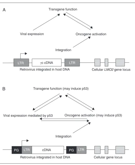
Figure 1. Overview of the influence of the virus on cellular and therapeutic gene expression. Integration of retrovirus in the host genome has been shown to favor regions proximal to the promoter of cellular genes. Viral expression is controlled by the long-terminal repeat (LTR) (viral promoter) which may also influence expression of neighboring cellular genes. A, In the cases of leukemia noted in the French X-SCID gene therapy trial, viral integration occurred near the LMO2 oncogene. In addition, γc gene activity may have cooperated with
with the resistance to programmed cell death brought these cells two steps closer to trans-formation. This study was the first to conclu-sively prove that insertional mutagenesis by a non-replicating, recombinant retroviral vec-tor could contribute to neoplastic transfor-mation in an animal model (25). Moreover, this study followed the model that viral in-sertion, oncogene action and transgene func-tion may cooperate and lead to transforma-tion.
Retroviruses were thought to integrate ran-domly within the host genome. However, this has been proven to be an overly simplistic view. Genomic-style strategies to identify in-sertion sites and their proximity to candidate genes and regulatory elements (27,28) have shown that MLV retroviruses, such as that used in the X-SCID trial, tend to insert near the promoter region of genes (28). Wu et al. (28) have shown that integration occurs at the locus of a confirmed gene (listed in the RefSeq database) in 34% of MLV insertion events, whereas HIV or recombinant lentivirus inser-tions land in RefSeq genes at a frequency of 62 or 50%, respectively. Even more striking was the observation that MLV vectors show a preference for insertion near (± 1 kb) CpG islands associated with transcriptional start sites, yet HIV integrates along the length of the gene locus downstream of the promoter. This may imply that the influence of viral elements on host-gene expression may be stronger for MLV than HIV or lentiviral vectors (28).
These recent observations have helped define the risk associated with retroviral inser-tion. Especially for MLV, the integration of the virus near gene promoters may represent an increased risk of subsequent mutagenesis.
Cooperation between retroviral insertion, oncogene activity and therapeutic gene function
A recent study suggests that at least three distinct paths merged and directed the leuke-mogenic transformation observed in these
patients of the French X-SCID gene therapy trial. As described above, insertion near the promoter of a cellular oncogene may bring about activation of its expression. By ana-lyzing a database of insertion sites from cases of mouse hematopoietic tumors caused by wild-type leukemia viruses, Davé et al. (29) have found examples of viral insertion at either the LMO2 gene or the γc chain gene, or both. First, this suggests, as expected, that oncogene activation will contribute to trans-formation. Second, and unexpectedly, acti-vation of γc could also promote leukemo-genesis. Finally, a specific cooperation may exist between LMO2 and γc chain functions, in this instance upon increased expression after viral insertion, that contributes to pro-liferation and transformation. This study points out that viral integration alone was not responsible for leukemic events, but re-quired other cooperating factors.
Understanding LMO2 may shed some light on its role in the adverse events associated with gene therapy of X-SCID. The LMO2
oncogene encodes a nuclear LIM-only protein that acts as a transcription factor necessary for erythroid development (16,30). LMO2-/- mice die at embryonic day 10.5 due, in part, to a lack of hematopoiesis (31); yet transgenic mice overexpressing LMO2 in the thymus devel-oped T-cell leukemia (32,33). Normal LMO2
expression occurs in a variety of tissue types, such as kidney, liver, lung, spleen, and the central nervous system (34), in addition to myeloid and erythroid cells (31). LMO2 has also been implicated in promoting angiogene-sis and capillary remodeling, processes clearly impeded in LMO2-/- mice (35). Therefore,
LMO2 is necessary, but uncontrolled expres-sion can contribute to leukemia. In the X-SCID patients who developed leukemia, LMO2
activity is thought to have played a major part in promoting cellular transformation.
that each patient received, it has been esti-mated that each patient would have been injected with 10-100 cells harboring inser-tion at a given expressed gene locus, includ-ing the LMO2 site (30). Since this integra-tion site was identified in relatively few patients, it may not represent a favored site,
but rather an available one. Subsequently, cooperation between LMO2 and γc may have provided an advantage for proliferation, se-lectively expanding this population of cells (Figures 1 and 2). Therefore, the insertion event, activation of LMO2 and its potential cooperation with γc chain functions all
tributed to the leukemogenesis. This under-standing may then serve as a starting point for improving future gene therapy applica-tions (36).
A clear and proportional relationship be-tween the transduction efficiency, number of proviral integration sites, and tendency to clonal expansion (including leukemogenesis) has been observed when oncoretroviral par-ticles are introduced into hematopoietic pro-genitor cells. Transduction, the process of introducing a recombinant virus into a target cell, can be characterized by the number of virus particles applied to the cells. This rela-tionship, called multiplicity of infection (MOI), reveals the number of virus particles/ target cell present during transduction. For example, 4000 virus particles applied to 2000 cells would have an MOI of 2. In this ex-ample we may be tempted to expect that all of the cells would be transduced, but the actual transduction efficiency is much lower, since some particles may never enter a cell and some cells may receive more than one particle.
At low virus concentrations (MOI ≤1), transduction of a target cell with a retroviral vector typically results in a single particle entering the cell. However, at higher MOI several virus particles may enter a single target cell, as was recently shown by quanti-tative methods (37). Therefore, seeking higher transduction efficiencies by increas-ing the MOI may lead to increased risk of insertional mutagenesis, though single in-sertion sites were observed in two of the three leukemias that resulted from the French X-SCID trial (13).
A recent paper has shown that at low virus concentration, transduction of hemato-poietic stem cells (HSC) can avoid the leu-kemogenesis that was observed at higher virus concentrations (38). An oncoretroviral vector encoding the human multidrug resis-tance gene-1 (MDR1), a membrane-bound drug efflux pump, was used to transduce HSC at low or high doses and these cells
were then transplanted in myeloablated, isogenic recipient mice. These investigators found that at the low dose no leukemias developed even though virus insertion did occur at or near oncogene loci. In contrast, 50% of the animals transplanted with HSC transduced with high doses of the virus de-veloped leukemia. The activity of the MDR1
gene, which may protect against apoptosis, may also have been a contributing factor since animals receiving HSC transduced with a virus encoding a fluorescent protein did not develop leukemia due to viral insertion. This recent work shows, for the first time, the relationship between oncoretroviral trans-duction efficiency and malignant risk in an animal model.
Clonal expansion, the accelerated prolif-eration of a small number of progenitor cells, yields reduced variation in the resulting he-matopoietic populations and is a hallmark of nonmalignant and malignant hematopoiesis. Retroviruses have long been used as mark-ers of clonal expansion since their insertion sites can be used to identify cell populations. However, it has been shown that the viral insertion itself may contribute to nonmalig-nant clonal expansion. Kustikova et al. (39) have shown that, upon transduction of HSC followed by serial transplantation in irradi-ated recipient mice, clonal expansion was associated with viral insertion at oncogene loci. They postulated that these cells had enhanced “fitness” due to unregulated onco-gene expression, though they did not turn malignant. Interestingly, this situation has been mirrored in the clinical setting. A re-cent gene therapy trial for X-linked chronic granulomatous disease showed that retrovi-ral delivery of the gp91phox gene yielded an improvement in immune function, though viral activation of cellular oncogenes may have contributed to this success (40).
showed that a lentiviral vector with inacti-vated LTRs and containing the CAG pro-moter to drive γc induced lymphoma in a mouse model of X-SCID gene therapy, but did not when green fluorescent protein was expressed. This suggests that the viral inser-tion itself was not responsible for the trans-formation, but γc activity was. Similarly, these investigators showed that viral expres-sion of LMO2 could also induce transforma-tion in this model of X-SCID treatment. They observed the animals over an extended period of time and noted the onset of lym-phomas at 6 months post-transplantation, a key difference compared to other preclinical studies of X-SCID gene therapy which did not provide follow-up beyond 6 months. Details of the lentiviral insertion sites have not been revealed, but these investigators note that several insertion sites were present in transduced cells and that this profile was different for each animal. This study did not directly address vector design, therefore it is difficult to infer if lentiviral vectors may provide an advantage over the use of MLV. This study suggests that γc function can contribute to transformation.
The target cell phenotype may also have been a contributing factor in the develop-ment of leukemia. In a new study, an in-creased tendency towards transformation was observed in a mouse model of X-SCID gene therapy where the γc gene was introduced in bone marrow cells with a retroviral vector, but transformation was less likely when the treatment was performed with cells derived from a non-X-SCID animal. This suggests that the X-SCID phenotype may have played a role in permitting transformation to occur in the presence of the retrovirus and γc gene (42).
Viral integration alone was not sufficient to promote transformation in the patients of the French X-SCID trial, but insertion into the
LMO2 locus was likely to be a factor contrib-uting to the inappropriate expression of this oncogene. The activities of both LMO2 and
the therapeutic gene, γc, influenced the forma-tion of leukemia. The specific role of viral insertion, oncogene activation and transgene activity continues to be studied, as described below, with the hope of developing strategies that preserve therapeutic benefit without el-evating the risk of adverse events.
The response from regulatory agencies and the gene therapy community
When the first case of leukemia resulting from the French gene therapy trial was an-nounced, US, French and British agencies halted trials temporarily, but then allowed them to continue, believing that the case was isolated (43). However, the announcement of the second case and the identification of the frequency of viral insertion at the LMO2
locus in these children prompted US agen-cies to stop 27 trials using retroviral gene transfer to hematopoietic stem cells (44). Continuation of these trials has been permit-ted on a case-by-case basis. The French trial was reinitiated (45), but was again put on hold when the third case of leukemia was discovered. Analysis of the risk vs benefit for the treatment of X-SCID by gene therapy has led many to believe that the cure of the underlying disease by far outweighs the risk of a serious side effect. Trials using a retro-virus for the treatment of SCID due to ADA-deficiency have been allowed to continue in the US.
Table 3. One point that is repeatedly raised is the need for improved vector design, includ-ing improved transcriptional control (49).
Strategies for combating oncogene activation in transduced cells
New technologies that do not rely on random viral integration may one day be developed for the treatment of X-SCID. For example, site-specific integration may per-mit introduction of the wild-type γc gene at genomic locations known to be void of genes
or regulatory elements (50,51). Alternatively, the endogenous γc gene may be corrected ex vivo by means of homologous recombina-tion promoted by engineered zinc-finger pro-teins, avoiding the need for viral integration (52). Both of these technologies, though promising, are far from being ready for clini-cal application.
Other viral vectors may also be explored to deliver the γc gene. Adenovirus would not be an appropriate choice since this vector does not provide long-term stability or sus-tained transgene expression.
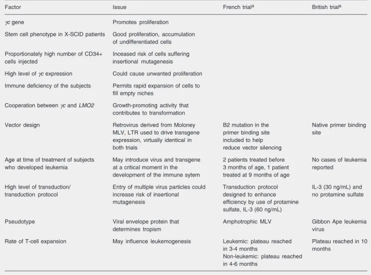
Adeno-associ-Table 2. Potential factors contributing to the development of leukemia in the French X-linked severe combined immunodeficiency (X-SCID) trial and differences, when present, compared to the British trial.
Factor Issue French triala British triala
γc gene Promotes proliferation
Stem cell phenotype in X-SCID patients Good proliferation, accumulation of undifferentiated cells
Proportionately high number of CD34+ Inceased risk of cells suffering
cells injected insertional mutagenesis
High level of γc expression Could cause unwanted proliferation
Immune deficiency of the subjects Permits rapid expansion of cells to fill empty niches
Cooperation between γc and LMO2 Growth-promoting activity that contributes to transformation
Vector design Retrovirus derived from Moloney B2 mutation in the Native primer binding
MLV, LTR used to drive transgene primer binding site site
expression, virtually identical in included to help
both trials reduce vector silencing
Age at time of treatment of subjects May introduce virus and transgene 2 patients treated before No cases of leukemia
who developed leukemia at a critical moment in the 3 months of age, 1 patient reported
development of the immune sytem treated at 9 months of age
High level of transduction/ Entry of multiple virus particles could Transduction protocol IL-3 (30 ng/mL) and
transduction protocol increase risk of insertional designed to enhance no protamine sulfate
mutagenesis efficiency by use of protamine
sulfate, IL-3 (60 ng/mL)
Pseudotype Viral envelope protein that Amphotrophic MLV Gibbon Ape leukemia
determines tropism virus
Rate of T-cell expansion May influence leukemogenesis Leukemic: plateau reached Plateau reached in 10
in 3-4 months months
Non-leukemic: plateau reached in 4-6 months
ated virus is being considered for hemato-poietic targets, though viral insertion events have been noted in hemophilia patients in a gene therapy trial (53). Lentiviruses, the fam-ily to which HIV belongs, have been devel-oped as recombinant gene transfer vectors and have also been shown, under certain conditions, to initiate oncogenesis due to insertional mutagenesis (54) or transgene activity (41). However, a recent study has suggested that lentiviral vectors may be safer than MLV when applied to hematopoietic stem cells (55). Retroviral vectors devel-oped from non-MLV sources are also being evaluated with respect to their reduced pro-pensity to integrate near promoter regions (48,56).
Assuming that no alternative to the use of the γc gene or integrating vectors is available for X-SCID gene therapy, we are left with few options other than to find a balance between the efficiency that viral vectors pro-vide and the growth-promoting properties of
γc. Improved safety for retroviral vector de-sign has been proposed and is within the reach of current technology (18,48). These strategies include inactivation of the LTR, use of an internal promoter to drive trans-gene expression, and the inclusion of a sui-cide gene to permit the selective elimination of transduced cells. However, the work of Woods et al. (41) suggests that LTR inacti-vation and the use of an internal promoter were not satisfactory.
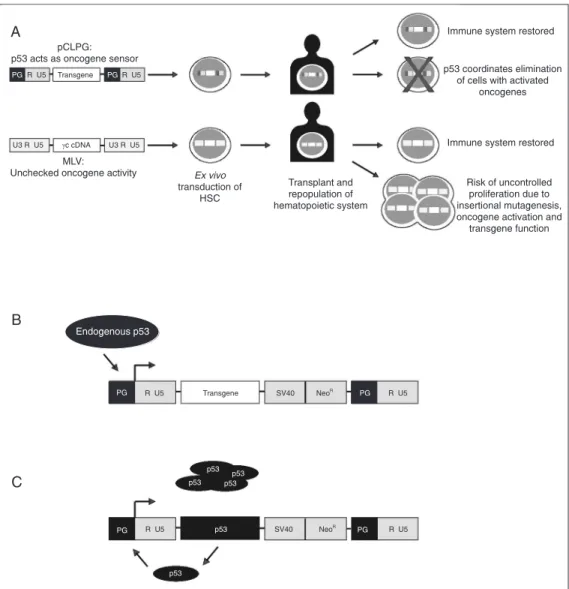
Another strategy in development includes an anti-oncogenic sensor that would elimi-nate cells destined for transformation. Our current research is related to the develop-ment of retroviral vectors with modified tran-scriptional control. Expression from one of these vectors, called pCLPG (Figure 2), is controlled by the tumor suppressor and trans-cription factor, p53 (57,58). We have initi-ated experimental work, which will take sev-eral years to complete, to determine if the employment of cellular p53 to drive viral expression might also be useful for combat-ing oncogene activation. Our expectation is that a feedback mechanism would be estab-lished between the oncogene and p53, re-sulting in the elimination of that cell by apoptosis coordinated by p53 (Figures 1 and 2). This feedback mechanism may include the activity of the transgene, such as γc or
dLNGFR, whose growth-promoting or
anti-apoptotic activity may induce the protective functions of p53. The novel transcriptional control strategy present in the pCLPG sys-tem may prove to be interesting in other gene therapy models where proliferation stimulated by the transgene is problematic.
We suspect that modifications of the LTR will have an added benefit beyond the use of the p53 tumor suppressor to drive expres-sion and sense oncogene activity. The native Moloney MLV LTR is especially active in developing thymocytes which may promote transgene and oncogene expression at a
par-Table 3. Recommendations by the American Society of Gene Therapy for hematopoietic stem cell gene transfer studies in animal models.
Follow-up of at least 12 months
Hematological analyses and autopsies performed in long-term transplant recipients
Analysis of viral integration sites related to cell type, transduction conditions, clonal expansion, duration of transgene expression, and toxicity in long-term transplant recipients
Establish database of integration sites
Test therapeutic genes in a relevant disease model
Animal studies should approximate human trial conditions
ticularly sensitive moment in T-cell matura-tion. In the pCLPG system, the Moloney MLV enhancer/promoter elements have been eliminated and thus unwanted LTR activity may be avoided during T-cell lymphopoie-sis. These ideas remain to be tested experi-mentally, a process that depends on exten-sive animal and molecular studies.
Impact on future studies
The next generation of vectors and gene therapy trials will involve higher standards. Gene therapy has been proven effective by the X-SCID trials, yet a potential risk has also been demonstrated. Unquestionably, 10
children were successfully treated by the French and British gene therapy trials and did not show any adverse effects, a point that must be underscored when considering the risk of retrovirus-mediated treatment. Clear-ly, the combination of viral integration, on-cogene activation and transgene activity were contributing factors to the leukemic events. However, a critical difference has not yet come to light between the French and British trials that could indicate how to avoid leuke-mogenesis and yet maintain the clinical ben-efit seen in both protocols. Future trials will benefit from continued study of vectors, tar-get cells, transduction strategies, and rel-evant animal models.
References
1. Gaspar HB, Thrasher AJ. Gene therapy for severe combined immu-nodeficiencies. Expert Opin Biol Ther 2005; 5: 1175-1182. 2. Srivastava A. Hematopoietic stem cell transduction by recombinant
adeno-associated virus vectors: problems and solutions. Hum Gene Ther 2005; 16: 792-798.
3. Gaspar HB, Parsley KL, Howe S, King D, Gilmour KC, Sinclair J, et al. Gene therapy of X-linked severe combined immunodeficiency by use of a pseudotyped gammaretroviral vector. Lancet 2004; 364: 2181-2187.
4. Cavazzana-Calvo M, Thrasher A, Mavilio F. The future of gene therapy. Nature 2004; 427: 779-781.
5. Schmalstieg FC, Goldman AS. Immune consequences of mutations in the human common gamma-chain gene. Mol Genet Metab 2002; 76: 163-171.
6. Malek TR, Porter BO, He YW. Multiple gamma c-dependent cyto-kines regulate T-cell development. Immunol Today 1999; 20: 71-76. 7. Rosen FS, Cooper MD, Wedgwood RJ. The primary
immunodefi-ciencies. N Engl J Med 1995; 333: 431-440.
8. Buckley RH. Primary immunodeficiency diseases due to defects in lymphocytes. N Engl J Med 2000; 343: 1313-1324.
9. Buckley RH, Schiff SE, Schiff RI, Markert L, Williams LW, Roberts JL, et al. Hematopoietic stem-cell transplantation for the treatment of severe combined immunodeficiency. N Engl J Med 1999; 340: 508-516.
10. Haddad E, Landais P, Friedrich W, Gerritsen B, Cavazzana-Calvo M, Morgan G, et al. Long-term immune reconstitution and outcome after HLA-nonidentical T-cell-depleted bone marrow transplantation for severe combined immunodeficiency: a European retrospective study of 116 patients. Blood 1998; 91: 3646-3653.
11. Hacein-Bey-Abina S, le Deist F, Carlier F, Bouneaud C, Hue C, de Villartay JP, et al. Sustained correction of X-linked severe combined immunodeficiency by ex vivo gene therapy. N Engl J Med 2002; 346: 1185-1193.
12. Cavazzana-Calvo M, Hacein-Bey S, de Saint Basile G, Gross F,
Yvon E, Nusbaum P, et al. Gene therapy of human severe combined immunodeficiency (SCID)-X1 disease. Science 2000; 288: 669-672. 13. Hacein-Bey-Abina S, Von Kalle C, Schmidt M, McCormack MP, Wulffraat N, Leboulch P, et al. LMO2-associated clonal T cell prolif-eration in two patients after gene therapy for SCID-X1. Science
2003; 302: 415-419.
14. Buckley RH. Gene therapy for SCID - a complication after remark-able progress. Lancet 2002; 360: 1185-1186.
15. Hacein-Bey-Abina S, Von Kalle C, Schmidt M, le Deist F, Wulffraat N, McIntyre E, et al. A serious adverse event after successful gene therapy for X-linked severe combined immunodeficiency. N Engl J Med 2003; 348: 255-256.
16. Nam CH, Rabbitts TH. The role of LMO2 in development and in T cell leukemia after chromosomal translocation or retroviral insertion.
Mol Ther 2006; 13: 15-25.
17. Frederickson RM. Report from the 2nd stem cell clonality and geno-toxicity retreat. Mol Ther 2005; 12: 379-383.
18. Baum C, Dullmann J, Li Z, Fehse B, Meyer J, Williams DA, et al. Side effects of retroviral gene transfer into hematopoietic stem cells.
Blood 2003; 101: 2099-2114.
19. Aberg JA, Williams PL, Liu T, Lederman HM, Hafner R, Torriani FJ, et al. A study of discontinuing maintenance therapy in human immu-nodeficiency virus-infected subjects with disseminated Mycobacte-rium avium complex: AIDS Clinical Trial Group 393 Study Team. J Infect Dis 2003; 187: 1046-1052.
20. Trono D. Virology. Picking the right spot. Science 2003; 300: 1670-1671.
21. Lund AH, Turner G, Trubetskoy A, Verhoeven E, Wientjens E, Hulsman D, et al. Genome-wide retroviral insertional tagging of genes involved in cancer in Cdkn2a-deficient mice. Nat Genet 2002; 32: 160-165.
22. Mikkers H, Berns A. Retroviral insertional mutagenesis: tagging cancer pathways. Adv Cancer Res 2003; 88: 53-99.
after transduction of murine hematopoietic stem cells is associated with methylation in vivo. Proc Natl Acad Sci U S A 1994; 91: 2567-2571.
24. Challita PM, Skelton D, el-Khoueiry A, Yu XJ, Weinberg K, Kohn DB. Multiple modifications in cis elements of the long terminal repeat of retroviral vectors lead to increased expression and decreased DNA methylation in embryonic carcinoma cells. J Virol 1995; 69: 748-755. 25. Li Z, Dullmann J, Schiedlmeier B, Schmidt M, Von Kalle C, Meyer J, et al. Murine leukemia induced by retroviral gene marking. Science
2002; 296: 497.
26. Bonini C, Ferrari G, Verzeletti S, Servida P, Zappone E, Ruggieri L, et al. HSV-TK gene transfer into donor lymphocytes for control of allogeneic graft-versus leukemia. Science 1997; 276: 1719-1724. 27. Schmidt M, Zickler P, Hoffmann G, Haas S, Wissler M, Muessig A, et
al. Polyclonal long-term repopulating stem cell clones in a primate model. Blood 2002; 100: 2737-2743.
28. Wu X, Li Y, Crise B, Burgess SM. Transcription start regions in the human genome are favored targets for MLV integration. Science
2003; 300: 1749-1751.
29. Dave UP, Jenkins NA, Copeland NG. Gene therapy insertional mutagenesis insights. Science 2004; 303: 333.
30. McCormack MP, Rabbitts TH. Activation of the T-cell oncogene LMO2 after gene therapy for X-linked severe combined immunodefi-ciency. N Engl J Med 2004; 350: 913-922.
31. Warren AJ, Colledge WH, Carlton MB, Evans MJ, Smith AJ, Rabbitts TH. The oncogenic cysteine-rich LIM domain protein rbtn2 is essen-tial for erythroid development. Cell 1994; 78: 45-57.
32. Larson RC, Osada H, Larson TA, Lavenir I, Rabbitts TH. The onco-genic LIM protein Rbtn2 causes thymic developmental aberrations that precede malignancy in transgenic mice. Oncogene 1995; 11: 853-862.
33. Larson RC, Lavenir I, Larson TA, Baer R, Warren AJ, Wadman I, et al. Protein dimerization between Lmo2 (Rbtn2) and Tal1 alters thy-mocyte development and potentiates T cell tumorigenesis in trans-genic mice. EMBO J 1996; 15: 1021-1027.
34. Foroni L, Boehm T, White L, Forster A, Sherrington P, Liao XB, et al. The rhombotin gene family encode related LIM-domain proteins whose differing expression suggests multiple roles in mouse devel-opment. J Mol Biol 1992; 226: 747-761.
35. Yamada Y, Warren AJ, Dobson C, Forster A, Pannell R, Rabbitts TH. The T cell leukemia LIM protein Lmo2 is necessary for adult mouse hematopoiesis. Proc Natl Acad Sci U S A 1998; 95: 3890-3895.
36. Berns A. Good news for gene therapy. N Engl J Med 2004; 350: 1679-1680.
37. Kustikova OS, Wahlers A, Kuhlcke K, Stahle B, Zander AR, Baum C, et al. Dose finding with retroviral vectors: correlation of retroviral vector copy numbers in single cells with gene transfer efficiency in a cell population. Blood 2003; 102: 3934-3937.
38. Modlich U, Kustikova OS, Schmidt M, Rudolph C, Meyer J, Li Z, et al. Leukemias following retroviral transfer of multidrug resistance 1 (MDR1) are driven by combinatorial insertional mutagenesis. Blood
2005; 105: 4235-4246.
39. Kustikova O, Fehse B, Modlich U, Yang M, Dullmann J, Kamino K, et al. Clonal dominance of hematopoietic stem cells triggered by
retroviral gene marking. Science 2005; 308: 1171-1174.
40. Ott MG, Schmidt M, Schwarzwaelder K, Stein S, Siler U, Koehl U, et al. Correction of X-linked chronic granulomatous disease by gene therapy, augmented by insertional activation of MDS1-EVI1, PRDM16 or SETBP1. Nat Med 2006; 12: 401-409.
41. Woods NB, Bottero V, Schmidt M, Von Kalle C, Verma IM. Gene therapy: therapeutic gene causing lymphoma. Nature 2006; 440: 1123.
42. Shou Y, Ma Z, Lu T, Sorrentino BP. Unique risk factors for inser-tional mutagenesis in a mouse model of XSCID gene therapy. Proc Natl Acad Sci U S A 2006; 103: 11730-11735.
43. Kaiser J. Gene therapy. RAC’s advice: proceed with caution. Sci-ence 2002; 298: 2113-2115.
44. Check E. Second cancer case halts gene-therapy trials. Nature
2003; 421: 305.
45. Check E. Gene therapists hopeful as trials resume with childhood disease. Nature 2004; 429: 587.
46. Friedmann T. Gene therapy’s new era: a balance of unequivocal benefit and unequivocal harm. Mol Ther 2003; 8: 5-7.
47. Thomas CE, Ehrhardt A, Kay MA. Progress and problems with the use of viral vectors for gene therapy. Nat Rev Genet 2003; 4: 346-358.
48. Nienhuis AW, Dunbar CE, Sorrentino BP. Genotoxicity of retroviral integration in hematopoietic cells. Mol Ther 2006; 13: 1031-1049. 49. Schambach A, Bohne J, Chandra S, Will E, Margison GP, Williams
DA, et al. Equal potency of gammaretroviral and lentiviral SIN vec-tors for expression of O6-methylguanine-DNA methyltransferase in hematopoietic cells. Mol Ther 2006; 13: 391-400.
50. Groth AC, Calos MP. Phage integrases: biology and applications. J Mol Biol 2004; 335: 667-678.
51. Ishikawa Y, Tanaka N, Murakami K, Uchiyama T, Kumaki S, Tsuchiya S, et al. Phage phiC31 integrase-mediated genomic inte-gration of the common cytokine receptor gamma chain in human T-cell lines. J Gene Med 2006; 8: 646-653.
52. Urnov FD, Miller JC, Lee YL, Beausejour CM, Rock JM, Augustus S, et al. Highly efficient endogenous human gene correction using designed zinc-finger nucleases. Nature 2005; 435: 646-651. 53. Check E. Harmful potential of viral vectors fuels doubts over gene
therapy. Nature 2003; 423: 573-574.
54. Themis M, Waddington SN, Schmidt M, Von Kalle C, Wang Y, Al-Allaf F, et al. Oncogenesis following delivery of a nonprimate lentiviral gene therapy vector to fetal and neonatal mice. Mol Ther
2005; 12: 763-771.
55. Montini E, Cesana D, Schmidt M, Sanvito F, Ponzoni M, Bartholo-mae C, et al. Hematopoietic stem cell gene transfer in a tumor-prone mouse model uncovers low genotoxicity of lentiviral vector integra-tion. Nat Biotechnol 2006; 24: 687-696.
56. Baum C, Schambach A, Bohne J, Galla M. Retrovirus vectors: toward the plentivirus? Mol Ther 2006; 13: 1050-1063.
57. Strauss BE, Costanzi-Strauss E. pCLPG: a p53-driven retroviral system. Virology 2004; 321: 165-172.