2018;26:e2993
DOI: 10.1590/1518-8345.2308.2993 www.eerp.usp.br/rlae
1 Emergency Medical Services Specialist, RN, Hospital 9 de Julho, São Paulo, SP, Brazil.
2 MSc, Assistant Professor, Escola Paulista de Enfermagem, Universidade Federal de São Paulo, São Paulo, SP, Brazil. 3 PhD, Full Professor, Escola Paulista de Enfermagem, Universidade Federal de São Paulo, São Paulo, SP, Brazil. 4 PhD, Assistant Professor, Escola Paulista de Enfermagem, Universidade Federal de São Paulo, São Paulo, SP, Brazil.
Results of the implementation of integrated care
after cardiorespiratory arrest in a university hospital
Evelyn Carla Borsari Mauricio
1Maria Carolina Barbosa Teixeira Lopes
2Ruth Ester Assayag Batista
3Meiry Fernanda Pinto Okuno
4Cássia Regina Vancini Campanharo
4Objectives: to identify the care measures performed after cardiorespiratory arrest (CRA) and
to relate them to the neurological status and survival at four moments: within the first 24
hours, at the discharge, six months after discharge, and one year after discharge. Method:
retrospective, analytical and quantitative study performed at the Emergency Department of a
university hospital in São Paulo. Eighty-eight medical records of CRA patients who had a return
of spontaneous circulation sustained for more than 20 minutes were included and the post-CRA
care measures performed in the first 24 hours were identified, as well as its relationship with
survival and neurological status. Results: the most frequent post-CRA care measures were use
of advanced airway access techniques and indwelling bladder catheterization. Patients who had
maintained good breathing and circulation, temperature control and who were transferred to
intensive care unit had a better survival in the first 24 hours, after six months and one year after
discharge. Good neurological status at six months and one year after discharge was associated
with non-use of vasoactive drugs and investigation of the causes of the CRA. Conclusion: the
identification of good practices in post-CRA care may help to reduce the mortality of these
individuals and to improve their quality of life.
Descriptors: Cardiac Arrest; Cardiopulmonary Resuscitation; Assistance; Critical Care; Emergency
Medical Services; Nursing.
How to cite this article
Mauricio ECB, Lopes MCBT, Batista REA, Okuno MFP, Campanharo CRV. Results of the implementation of integrated care after cardiorespiratory arrest in a university hospital. Rev. Latino-Am. Enfermagem. 2018;26:e2993. [Access ___ __ ____]; Available in: ___________________ . DOI: http://dx.doi.org/10.1590/1518-8345.2308.2993.
Introduction
In Brazil, circulatory diseases, including cardiorespiratory arrest (CRA), were the main cause of death in 2011(1). A total of 200,000 CRA events are estimated to occur every year, with approximately half occurring in hospital settings(2).
After the return of spontaneous circulation (RSC),
defined as the maintenance of myocardial contractions
capable of generating a pulse for more than 20 minutes after the completion of cardiopulmonary resuscitation (CPR), a severe clinical syndrome is started, which is responsible for about from 50 to 70% of deaths in the
first 24 to 48 hours after CRA. The post-cardiac arrest
syndrome happens due to hypoxia and reperfusion lesions during the CRA and after the RSC(3-4).
Post- care has the potential to improve early mortality rates caused by hemodynamic instability and multiple organ and system failure, and late morbidity and mortality rates resulting from neurological damage. The main goals are to improve cardiopulmonary function and systemic perfusion; transport the CRA victims from out-of-hospital settings to emergency or intensive care units; identify the precipitating cause of the CRA and prevent its recurrence; and implement measures to improve the long-term prognosis of patients and preserve their neurological function(3).
The main measures to be adopted include: early reperfusion therapy for cases of coronary thrombosis; stabilization and maintenance of hemodynamic parameters; correction of arterial gas disorders; maintenance of normal glucose values; control of water balance; administration of sedation and analgesia; prevention and treatment of seizures and temperature control(2-3).
This study is based on the low survival rates found among CRA victims and on the great risk of neurological sequel to which they are exposed when spontaneous circulation returns.
Despite the advances in emergency cardiovascular care, the need for new techniques to reverse the injury of ischemia and reperfusion is evident. In this context, it is vitally important to identify the post-CRA care measures so that strategies may be implemented with the objective of reducing mortality associated with hemodynamic instability, limiting brain damage and injury to other organs.
Thus, the objectives of this study were to identify the post-CRA care measures performed in a university hospital and to relate them to the survival and
neurological status of the patients in the first 24 hours,
at hospital discharge, six months after discharge, and one year after discharge.
Method
Study approved by the Ethics and Research Committee of the Federal University of São Paulo (CAEE: 52531315.4.0000.5505).
The study had a retrospective, analytical and quantitative approach, and it was carried out in the Emergency Room (ER) of a university hospital in the city of São Paulo (SP), Brazil.
All adult patients who had CRA in out-of-hospital settings and were taken for assistance at the ER of the above mentioned service in the year 2011, and who presented RSC sustained for more than 20 minutes were included in this study, totaling 88 patients. We excluded from this study the CRA cases assisted in other sectors of the hospital.
Data collection was performed in four different moments through the analysis of medical records. At admission, the following variables were collected: age, gender, skin color, presence of comorbidities, previous CRA events, pre-CRA neurological status, presence of consciousness, breathing and pulse at the arrival of the patient in the ER, place where the CRA occurred, if there were witnesses, presumed immediate cause, initial rhythm of CRA and interventions performed during care(5).
During the first 24 hours; the following post-CRA care measures were identified and recorded: use
of advanced airway access techniques; monitoring of respiratory rate; maintenance of respiratory rate between 10 and 12 rpm; monitoring of pulse oximetry; maintenance of oxygen saturation between 94 and 96%; maintenance of CO2 partial pressure between 40 and 45 mmHg; monitoring of capnography; maintenance of end-expiratory CO2 partial pressure between 35 and 40 mmHg; monitoring of noninvasive blood pressure (NIBP); maintenance of systolic blood
pressure (SBP) ≥ 90 mmHg; monitoring of invasive
blood pressure (IBP); maintenance of mean arterial
pressure (MAP) ≥ 65 mmHg; central venous access
puncture; monitoring of central venous pressure; maintenance of venous pressure between 8 and 12 mmHg; monitoring of venous oxygen saturation; maintenance venous oxygen saturation > 70%; administration of saline solutions; administration of vasoactive and antiarrhythmic drugs; in case
of ventricular fibrillation and pulseless ventricular
coronary intervention in cases of suspected acute coronary syndrome; realization of echocardiography;
identification and treatment of reversible causes
of CRA; monitoring of body temperature; prevention of hyperthermia; monitoring of electroencephalogram; administration of anticonvulsants; monitoring of blood glucose; maintenance of glycemia between 144 and 180mg/dl; chest X-ray; control of general exams every six hours; arterial blood gas analysis every six hours; indwelling bladder catheterization; monitoring of urine output; maintenance of urine output between 0.5 and 1 ml/kg/h; use of sedation in case of cognitive dysfunction; introduction of continuous enteral nutrition in the absence of contraindication and transfer to an Intensive Care Unit (ICU)(3).
Survival and neurological status of individuals were assessed at hospital discharge, six month later, and one year later and evaluated by the
Glasgow-Pittsburgh Cerebral Performance Categories (CPC). The
CPC is divided into five categories. Category 1 indicates
complete independence and ability to work; Category 2 indicates moderate disability, ability to work part-time and independence for the Activities of Daily Living; Category 3 indicates severe disability and total dependence on the Activities of Daily Living; category 4 indicates persistent vegetative state; and category 5 indicates brain death(6). In this study, patients diagnosed with CPC 1 and 2 were considered to be in good neurological state, and those
evaluated and classified as CPC 3, 4 and 5, were in poor
neurological state(6).
Data were analyzed in the softwares PSPP and R, version 3.3.1. Mean, standard deviation, median, minimum and maximum values were calculated for the continuous variables, and frequency and percentage for the categorical variables. The non-parametric Kruskal-Wallis test was used to correlate the survival and
neurologic status in the first 24 hours, at discharge, six
months after discharge, and one year after discharge. The non-parametric Kruskal-Wallis test was used for continuous variables. The Pearson Chi-square test was used relate the response variable with the categorical
variables. The level of significance considered in all
analyses was 5%.
Results
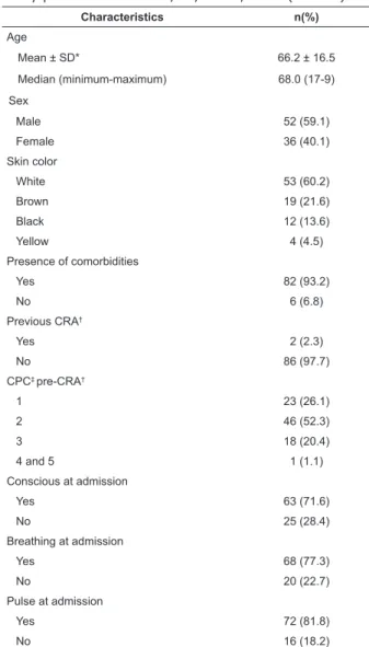
Demographic and clinical data are presented in Table 1. The mean age was 66.2 years, and there was predominance of white men, presenting at least one previous comorbidity, and independent in the activities of daily living. At admission to ER, most individuals were conscious, breathing, and had circulation.
Table 1 – Demographic and clinical characteristics of study patients. São Paulo, SP, Brazil, 2016 (N = 88)
Characteristics n(%)
Age
Mean ± SD* 66.2 ± 16.5
Median (minimum-maximum) 68.0 (17-9)
Sex
Male 52 (59.1)
Female 36 (40.1)
Skin color
White 53 (60.2)
Brown 19 (21.6)
Black 12 (13.6)
Yellow 4 (4.5)
Presence of comorbidities
Yes 82 (93.2)
No 6 (6.8)
Previous CRA†
Yes 2 (2.3)
No 86 (97.7)
CPC‡ pre-CRA†
1 23 (26.1)
2 46 (52.3)
3 18 (20.4)
4 and 5 1 (1.1)
Conscious at admission
Yes 63 (71.6)
No 25 (28.4)
Breathing at admission
Yes 68 (77.3)
No 20 (22.7)
Pulse at admission
Yes 72 (81.8)
No 16 (18.2)
*SD: standard deviation; †CRA: cardiorespiratory arrest; ‡CPC: Glasgow-Pittsburgh Cerebral Performance Categories
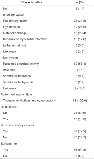
The characteristics of the CRA events and the interventions performed during CPR are presented in Table 2. Most of the events occurred in the hospital, being witnessed by the health team and with an immediate presumed cause of respiratory failure. The most prevalent rhythm was pulseless electrical activity and the most frequent interventions during the care were compressions, ventilation and medication administration.
Table 2 – Characteristics of cardiorespiratory arrest events and interventions performed during the care of the study patients. São Paulo, SP, Brazil, 2016 (N = 88)
Characteristics n (%)
Place
Intra-hospital 76 (86.3)
Extra-hospital 12 (13.6)
Witnessed
Yes 87 (98.8)
Characteristics n (%)
No 1 (1.1)
Immediate cause
Respiratory failure 28 (31.8)
Hypotension 19 (21.6)
Metabolic change 18 (20.4)
Ischemia or myocardial infarction 15 (17.0)
Lethal arrhythmia 5 (5.6)
Unknown 3 (3.3)
Initial rhythm
Pulseless electrical activity 50 (58.1)
Asystolia 9 (10.2)
Ventricular fibrillation 8 (9.1)
Ventricular tachycardia 2 (2.2)
Unknown 9 (10.2)
Performed interventions
Thoracic ventilations and compressions 88 (100.0)
Defibrillation
No 71 (80.6)
Yes 17 (19.3)
Advanced airway access
Yes 68 (77.2)
No 20 (22.7)
Epinephrine
Yes 83 (94.3)
No 5 (5.6)
The mean time between initiation of CPR and the
first shock was 7.8 minutes; between the initiation of
CPR and installation of advanced airway access was 4.1
minutes; between the initiation of CPR and the first dose
of epinephrine was 2.1 minutes; and the mean duration of CPR was 11.1 minutes.
Post-CPR care performed in the first 24 hours after
the RSC is shown in Table 3. Of the 88 medical records
analyzed, 8 did not contain sufficient information to
collect data, totaling 80 charts.
Monitoring of capnography and venous oxygen saturation and electroencephalogram were not performed in any patient.
Table 3 – Post-cardiorespiratory care after the first 24
hours in the study patients. São Paulo, SP, Brazil, 2016 (N = 80)
Care measures n (%)
Advanced airway access 77(96.2)
Indwelling bladder catheterization 60(68.1)
Systolic blood pressure ≥ 90 mmHg 52(59.0)
Investigation of the cause of cardiorespiratory arrest 51(57.9)
Vasoactive drugs 51(57.9)
Mean blood pressure ≥ 65 mmHg 50(56.8)
Care measures n (%)
Prevention of hyperthermia 43(48.8)
12-lead electrocardiogram 42(47.7)
Central venous access 40(45.4)
Crystalloid solutions 38(43.1)
Continuous sedation 38(43.1)
Chest X-ray 31 (35.2)
Transfer to intensive care unit 24(27.2)
Oxygen saturation from 94 to 98% 24(27.2)
Arterial blood gas analysis every 6 hours 17(19.3)
Urine output between 0.5 and 1 ml/kg/h 16(18.1)
Monitoring of oxygen saturation 15(17.0)
Monitoring of noninvasive blood pressure 15(17.0)
Antiarrhythmic drugs 14(15.9)
Laboratory tests every 6 hours 14(15.9)
Monitoring of respiratory rate 13(14.7)
Monitoring of temperature 12(13.6)
Capillary glycemia 144 to 180 mg/dl 10(11.3)
Electrocardiogram monitoring 9(10.2)
Hemodynamic 9(10.2)
Monitoring of urine output 7(7.9)
Enteral nutrition 7(7.9)
Monitoring of capillary glycemia 5(5.6)
Respiratory rate of 10 to 12 rpm 4(4.5)
Anticonvulsants 4(4.5)
Echocardiogram 3(3.4)
Monitoring of noninvasive blood pressure 3(3.4)
CO2* partial pressure between 40 and 45 mmHg 2(2.2)
Central venous pressure from 8 to 12 cmH2O 2(2.2)
Monitoring of central venous pressure 1(1.1)
*CO2: carbon dioxide
Of the 88 patients surveyed, 13 survived at discharge, 10 after six months, and 9 after one year. The
variables that were significantly associated with greater
patient survival are presented in Table 4.
Realization of post-CRA care was not associated
with greater survival of individuals at hospital discharge.
Table 4 – Association of post-cardiorespiratory arrest
care with survival of the studied patients in the first 24
hours, six months after discharge, and one year after discharge. São Paulo, SP, Brazil, 2016
24-hour survival (n = 35) p
Monitoring of respiratory rate 0.01
Monitoring of oxygen saturation 0.01
Oxygen saturation from 94 to 98% 0.01
Monitoring of noninvasive blood pressure 0.01
Systolic blood pressure ≥ 90 mmHg 0.01
Monitoring of invasive blood pressure 0.03
Mean blood pressure ≥ 65 mmHg 0.01
12-lead electrocardiogram 0.01
Monitoring of temperature 0.01
Prevention of hyperthermia 0.03
Table 2 - (continuation) Table 3 - (continuation)
Chest X-ray 0.01
Indwelling bladder catheterization 0.01
Urine output between 0.5 and 1 ml/kg/h 0.04
Continuous sedation 0.01
Transfer to intensive care unit 0.01
6-month survival (n = 10) p
Oxygen saturation from 94 to 98% 0.02
Vasoactive drugs 0.03
Transfer to intensive care unit 0.03
One-year survival (n = 9) p
Monitoring of respiratory rate 0.01
Monitoring of oxygen saturation 0.01
Oxygen saturation from 94 to 98% 0.01
Monitoring of noninvasive blood pressure 0.01
Vasoactive drugs 0.04
Antiarrhythmic drugs 0.02
Electrocardiogram monitoring 0.01
12-lead electrocardiogram 0.02
Hemodynamic 0.01
*Pearson’s Chi-square test (p < 0.005)
When post-CRA care was related to neurological status at discharge, six months after discharge, and one year after discharge, none of the interventions were
related to patients’ neurologic status within the first
24 hours or at hospital discharge. However, patients who did not receive vasoactive drugs and underwent investigation of the causes of the CRA presented good neurological status, CPC 1 and 2, in six months (p = 0.04) and one year (p = 0.02) after discharge hospital.
Discussion
According to the guidelines of the American Heart Association, post-CRA care aims to reduce early mortality due to hemodynamic instability and to limit later multiple organ failure and brain injury. This care includes adequate cardiopulmonary conditions and perfusion of vital organs; safe transportation to intensive care units; early recognition of the causes of the event, and treating and preventing its recurrence; controlled temperature to minimize neurological damage; diagnosis and treatment of acute myocardial ischemia; ventilatory support with mechanical ventilation to limit lung injury; reducing the risk of multiple organ failure; assessment of neurological recovery prognosis; and promotion of rehabilitation of survivors(3).
The mean age of the patients in this study was 66.2 years, as in a study carried out in Singapore by the National Emergency Ambulance System(7). There was a prevalence of conscious, white people, breathing and
with pulse at admission, and the predominant rhythm was pulseless electrical activity, a result that is different from that reported in the international literature(7). Such
findings may be associated with the fact that most events
occurred in the in-hospital setting, in more complex patients, and with other associated comorbidities(8).
In this study, maintenance of systolic blood
pressure ≥ 90 mmHg, administration of vasoactive
drugs, investigation of causes of the arrest, maintenance
of mean arterial pressure ≥ 65 mmHg, 12-lead
electrocardiogram, central venous access puncture, crystalloid administration and bladder catheterization were the most frequent care measures. These actions aim at adapting the cardiovascular conditions and organ and system perfusion, since death due to multiple organ failure is associated with a persistent low cardiac output
in the first 24 hours after CPR(2).
Advanced airway access was frequently performed in the patients in this study. In these cases, ventilation and oxygenation should be immediately optimized, thus avoiding hyperoxia, which contributes to an increase in oxidative stress and is associated with a worse neurological prognosis(2,6). A study evaluated 173 comatose patients after sudden cardiac arrest and found that those who had lower maximum partial pressure
of arterial oxygen in the first 24 hours after cardiac
arrest had higher survival rates at discharge compared to the others(9). In addition, cerebral vasoconstriction aggravated by hyperventilation potentiates ischemic brain injury(10) and reduces cardiac output at the expense of an increase in intrathoracic pressure(3).
As for prevention of brain injury, the most frequent care measure in this study was the prevention of hyperthermia and continuous sedation. Studies have shown that patients who reached temperature above 37.6ºC after the return of the spontaneous circulation had lower survival chance and worse neurological prognosis in relation to the normothermic ones(3). Evidence on prevention of post-CRA hyperthermia is still not well established, but the occurrence of fever is associated with worsening of neurological injury in patients undergoing intensive care for other conditions(11). Thus,
the fight against fever is recommended because of the
potential aggravation of ischemic brain damage(3). Other neuroprotective measures are recommended, such as the prevention of seizures and the continuous monitoring of brain activity through electroencephalogram(6).
min; 12-lead ECG and chest X-ray; indwelling bladder catheterization; continuous sedation; prevention against hyperthermia; and transfer of the patient to the ICU were related to increased survival when performed at intervals of 2 hours or less.
Six months after discharge, maintenance of oxygen saturation between 94 and 96%, non-administration of vasoactive drugs and transfer of the patient to the ICU were related to higher survival rates. In a study performed with out-of-hospital cardiorespiratory arrest patients, it was observed that increased partial oxygen pressure (PaO2), greater than 300 mmHg, during CPR were associated with higher rates of return to spontaneous circulation and better neurological outcomes when compared to normal or lower partial oxygen pressure (PaO2 of less than 60 mmHg)(12). Prevention of hypoxemia is considered more important than avoiding any potential risk of hyperoxia(3).
Regarding the administration of vasoactive drugs,
studies evaluating specific strategies to improve blood pressure comparing vasopressors and fluids are scarce.
A study performed with patients who achieved a return to spontaneous circulation after CPR found that MAP
greater than 70 mmHg in the first 6 hours after CPR was
associated with good neurological function(13). Although there was no consensus regarding the ideal values of MAP, the importance of strict monitoring to maintain effective circulation is emphasized, mainly in order to avoid hypotension in order to obtain better results after a CRA.
Transfer of post-CRA patients to the ICU may be related to greater survival rates because an ICU is a safer and better treatment environment for critical patients in view of its infrastructure with more advanced
materials and equipment, as well as qualified personnel
to provide specialized assistance(2-3).
In this study, at one year after discharge, the
variables that were significantly associated with
higher survival rates were monitoring of respiratory rate; oxygen saturation; noninvasive blood pressure; electrocardiographic tracing; maintenance of oxygen saturation between 94 and 96%; administration of antiarrhythmic drugs; performance of ECG and referral for hemodynamics in the case of acute coronary syndrome; and transfer of the patient to the ICU. After the RSC, patients have a high probability of developing multiple organ and system failure. Therefore, systemic perfusion should be adequate, metabolic homeostasis should be restored and the function of the various organs should be maintained, aiming to increase survival prospects without neurological damage over time(3).
Regarding the neurological state of the individuals,
those who did not receive vasoactive drugs had a better
six-month and one-year neurological prognosis. Brain
injury is an important cause of post-CRA morbidity
and mortality. Recognition of its pathophysiological
mechanisms and its correlation with patient
characteristics, CPR maneuvers, and post-CRA care may
improve the prognosis of these individuals(14).
Hemodynamic stabilization, MAP greater than
65 mmHg, can often only be achieved with the use of
vasoactive drugs and is critical for effective cerebral
circulation after a CRA. Good hemodynamic parameters
are related to higher survival rates at hospital discharge
and better long-term neurological outcomes(3). However,
further studies on vasoactive drugs are necessary
because, depending on the mechanism of action of such
drugs, they may lead to changes in peripheral vascular
resistance, heart rate, arrhythmias and myocardial
ischemia(15).
Differential diagnosis of the cause of CRA is
paramount for establishing definitive treatment(2),and
in this study, it was related to a higher patient survival
at six months and one year after hospital discharge.
Detecting the cause of the CRA can be difficult and
often implies frequent reassessment of the patient
through collection of information, clinical evaluation,
blood profile and imaging tests(2). More studies on
this subject are necessary to elucidate the role of new
resources to optimize the diagnosis of the causes of CRA
and their reversal, as well as measures to help in the
determination of patient prognosis(16).
The main limitation of this study was to have
been performed in a single center, which may not
represent other realities. In addition, because this was
a retrospective study, there were difficulties during
collection, such as medical records with incomplete data
and difficult to interpret.
CRA is the most severe clinical emergency and
with the worse prognosis, but it may be a transient,
reversible stage with the possibility of recovery and
returning to activities. The identification of post-CRA
care in a Brazilian referral hospital can subsidize public
policies aimed at the care of these individuals, reducing
mortality and limiting the occurrence of neurological
damage and functional disability, as well as adding key
Conclusion
The most frequent post-CRA care measures performed in the patients in this study were: use of advanced airway access techniques; indwelling bladder
catheterization; maintenance of SBP ≥ 90 mmHg and MAP ≥ 65 mmHg; investigation of the causes of the
CRA; and administration of vasoactive drugs.
Survival in the first 24 hours was greater in
patients who received the following care measures: maintenance of good breathing and circulation, temperature control, continuous sedation, chest X-ray, and transfer to intensive care unit. After 6 months,
survival was significantly greater in cases where
oxygen saturation was maintained between 94 and 96%, vasoactive drugs were not administered, and in those patients who were transferred to the ICU. After one year of hospital discharge, maintenance of good breathing and circulation, 12-lead ECG, patient referral for hemodynamic support, and transfer to ICU were the care associated with better patient survival.
Regarding neurological status, patients who did not receive vasoactive drugs and those who had the cause of CRA diagnosed survived with good neurological status at six months and one year after discharge.
References
1. Vancini-Campanharo CR, Vancini RL, de Lira CA, Lopes MC, Okuno MF, Batista RE, Atallah ÁN, Góis AF. Um ano de seguimento da condição neurológica de pacientes pós-parada cardiorrespiratória atendidos no pronto-socorro de um hospital universitário. 2015 Apr-Jun;13(2):183-8. doi: 10.1590/S1679-45082015AO3286.
2. Gonzalez MM, Timerman S, Oliveira RG, Polastri TF, Dallan LAP, Araujo S, et al. I diretriz de ressuscitação cardiopulmonar e cuidados cardiovasculares de emergência da Sociedade Brasileira de Cardiologia: resumo executivo. ArqBrasCardiol. 2013;100(2):105-13. doi: 10.5935/abc.20130022.
3. Callaway CW, Donnino MW, Fink EL, Geocadin RG, Golan E, Kern KB, et al. Part 8: post–cardiac arrest care: 2015 American Heart Association Guidelines Update for Cardiopulmonary Resuscitation and Emergency Cardiovascular Care. Circulation. 2015; 132(suppl2):465–82. doi: 10.1161/CIR.00000000000 00262
4. Nolan JP, Soar J, Cariou A, Cronberg T, Moulaert VRM, Deakin CD, et al. European Resuscitation Council and European Society of Intensive Care Medicine Guidelines for Post-resuscitation Care 2015 Section 5
of the European Resuscitation Council Guidelines for Resuscitation 2015. Resuscitation. 2015;95:202–22. doi: 10.1007/s00134-015-4051-3.
5. Avansi PA, Meneghin P. Translation and adaptation of the In-Hospital Utstein style into the Portuguese language. Rev Esc Enferm USP. 2008;42(3):504-11. doi: 10.1590/S0080-623420150000500008.
6. Rittenberger JC, Raina K, Holm MB, Kim YJ, Callaway CW. Association between Cerebral Performance Category,
Modified Rankin Scale, and Discharge Disposition after
Cardiac Arrest. Resuscitation 2011;82(8):1036-40. doi: 10.1016/j.resuscitation.2011.03.034.
7. Eng Hock Ong M, Chan YH, Anantharaman V, Lau ST, Lim SH, Seldrup J. Cardiac arrest and resuscitation epidemiology in Singapore (CARE I study). Pre hosp Emerg Care. [Internet]. 2003 [cited 2016 Nov 2];7(4): 427-33. Available from: https://www.ncbi.nlm.nih.gov/ pubmed/14582091.
8. Vancini-Campanharo CR, Vancini RL, Lira CAB, Andrade M, Góis AFT, Atallah ANA. Cohort study on the factors associated with survival post-cardiac arrest. Sao Paulo Med J. 2015;133(6):495-501. doi: 10.1590/1516 -3180.2015.00472607.
9. Janz DR, Hollenbeck RD, Pollock JS, McPherson JA, Rice TW. Hyperoxia is Associated with Increased Mortality in Patients Treated with Mild Therapeutic Hypothermia after Sudden Cardiac Arrest. Crit Care Med. 2012;40(12):3135–9. doi: 10.1097/CCM.0b013e 3182656976.
10. Phelps R, Dumas F, Maynard C, Silver J, Rea T. Cerebral performance category and long-term prognosis following out-of-hospital cardiac arrest. Crit Care Med. 2013;41(5):1252-7. doi: 10.1097/CCM.0b013e 31827ca975.
11. Bohman LE, Levine JM. Fever and therapeutic normothermia in severe brain injury: an update. Curr Opin Crit Care. 2014;20:182–8. doi: 10.1097/MCC. 0000000000000070.
12. Spindelboeck W, Schindler O, Moser A, Hausler F, Wallner S, Strasser C, Haas J, Gemes G, Prause G. Increasing arterial oxygen partial pressure during cardiopulmonary resuscitation is associated with improved rates of hospital admission. Resuscitation. 2013;84(6):770-5. doi: 10. 1016/j.resuscitation.2013.01.012.
13. Kilgannon JH, Roberts BW, Jones AE, Mittal N, Cohen E, Mitchell J, Chansky ME, Trzeciak S. Arterial blood pressure and neurologic outcome after resuscitation from cardiac arrest*. Crit Care Med. 2014 Sep;42(9):2083-91. doi: 10.1097/CCM.0000000000000406.
Received: Jun 1st 2017
Accepted: Nov 26th 2017
Copyright © 2018 Revista Latino-Americana de Enfermagem This is an Open Access article distributed under the terms of the Creative Commons (CC BY).
This license lets others distribute, remix, tweak, and build upon your work, even commercially, as long as they credit you for the original creation. This is the most accommodating of licenses offered. Recommended for maximum dissemination and use of licensed materials.
Corresponding Author:
Maria Carolina Barbosa Teixeira Lopes Universidade Federal de São Paulo Escola Paulista de Enfermagem R. Napoleão de Barros, 754 Vila Clementino
CEP: 04024-002, São Paulo, SP, Brasil E-mail: [email protected]
after cardiac arrest. Acta Neurol Colomb. [Internet]. 2013 [cited 2016 Nov 9];29(4):255-65. Available from: https://www.acnweb.org/es/acta-neurologica/volumen -29-2013/147-volumen-29-no-4/930-lesion-cerebral-posterior-a-paro-cardiorrespiratorio.html