Changes in respiratory mechanics during
respiratory physiotherapy in mechanically
ventilated patients
INTRODUCTION
Ventilatory support is provided to patients with acute respiratory failure to provide rest for the respiratory muscles and reduce the work of breathing until the acute condition is resolved. he mobilization and removal of respiratory secretions plays a key role in improving bronchial hygiene and gas exchange and in reducing the work of breathing, thus changing the mechanics of critically ill patients subjected to invasive ventilatory support.(1)
Fernanda Callefe Moreira1,2, Cassiano Teixeira3,4, Augusto Savi4, Rogério Xavier5,6
1. Centro Universitário Univates - Lajeado (RS), Brazil.
2. Respiratory Care Unit, Hospital Moinhos de Vento - Porto Alegre (RS), Brazil.
3. Department of Internal Medicine, Universidade Federal de Ciências da Saúde de Porto Alegre - Porto Alegre (RS), Brazil.
4. Intensive Care Center, Hospital Moinhos de Vento - Porto Alegre (RS), Brazil.
5. Universidade Federal do Rio Grande do Sul - Porto Alegre (RS), Brazil.
6. Experimental Laboratory of Airways and the Lung, Hospital de Clínicas de Porto Alegre - Porto Alegre (RS), Brazil.
Objective: To evaluate the
changes in ventilatory mechanics and hemodynamics that occur in patients dependent on mechanical ventilation who are subjected to a standard respiratory therapy protocol.
Methods: his experimental and
prospective study was performed in two intensive care units, in which patients dependent on mechanical ventilation for more than 48 hours were consecutively enrolled and subjected to an established respiratory physiotherapy protocol. Ventilatory variables (dynamic lung compliance, respiratory system resistance, tidal volume, peak inspiratory pressure, respiratory rate, and oxygen saturation) and hemodynamic variables (heart rate) were measured one hour before (T-1), immediately after (T0) and one hour after (T+1) applying the respiratory physiotherapy protocol.
Results: During the period of data collection, 104 patients were included in the study. Regarding the ventilatory variables, an increase in dynamic lung compliance (T-1 = 52.3 ± 16.1mL/cmH2O versus T0 = 65.1 ± 19.1mL/cmH2O;
Conflicts of interest: None.
Submitted on December 15, 2014 Accepted on May 20, 2015
Corresponding author:
Fernanda Callefe Moreira
Rua Ramiro Barcelos, 910 - Bairro Floresta Zip code: 90035-001- Porto Alegre (RS), Brazil E-mail: [email protected]
Responsible editor: Carmen Valente Barbas
Alterações da mecânica ventilatória durante a isioterapia
respiratória em pacientes ventilados mecanicamente
ABSTRACT
Keywords: Respiratory therapy;
Respiration, artificial; Respiratory mechanics
p < 0.001), tidal volume (T-1 = 550 ± 134mL versus T0 = 698 ± 155mL; p < 0.001), and peripheral oxygen saturation (T-1 = 96.5 ± 2.29% versus T0 = 98.2 ± 1.62%; p < 0.001) were observed, in addition to a reduction of respiratory system resistance (T-1 = 14.2 ± 4.63cmH2O/L/s versus T0 = 11.0 ± 3.43cmH2O/L/s; p < 0.001), after applying the respiratory physiotherapy protocol. All changes were present in the assessment performed one hour (T+1) after the application of the respiratory physiotherapy protocol. Regarding the hemodynamic variables, an immediate increase in the heart rate after application of the protocol was observed, but that increase was not maintained (T-1 = 88.9 ± 18.7 bpm versus T0 = 93.7 ± 19.2bpm versus T+1 = 88.5 ± 17.1bpm; p < 0.001).
Conclusion: Respiratory therapy
leads to immediate changes in the lung mechanics and hemodynamics of mechanical ventilation-dependent patients, and ventilatory changes are likely to remain for at least one hour.
In most intensive care units (ICUs), respiratory physiotherapy is an integral part of the management of critically ill patients who require invasive ventilatory support. Respiratory physiotherapy techniques include postural drainage (PD), mobilization, vibration, percussion, manual hyperinlation (MH), and aspiration
of the airways.(2) he routine combination of these
techniques primarily aims to prevent complications such as tracheal prosthesis obstruction, ventilator-associated pneumonia (VAP), and patient-ventilator asynchrony.(3-14)
Numerous respiratory therapy protocols have been described for the care of mechanical ventilation (MV)-dependent patients, but with conlicting results.(10-12,15,16) he aim of this study was to evaluate the changes in ventilatory mechanics and hemodynamics that occur in MV-dependent patients undergoing a standard respiratory physiotherapy protocol.
METHODS
his was an experimental and prospective study conducted at two mixed ICUs: the Intensive Care Center
of the Hospital Moinhos de Vento in Porto Alegre and
the ICU of the Hospital de Pronto-Socorro de Canoas in
Canoas, both in the state of Rio Grande do Sul. he study was approved by the Research Ethics Committees of both hospitals (Project no. 2004/18 and 05-407, respectively), and patients and close family members participated in the study after signing an informed consent form.
All patients dependent on MV for ≥ 48 hours and who were prescribed respiratory physiotherapy during the period from February to September 2014 were consecutively included in the study after signing the informed consent form. he exclusion criteria consisted of patients with hemodynamic instability (change in vasopressor dose in the last two hours, mean blood pressure ≥ 120mmHg or ≤ 60mmHg, heart rate (HR) ≥ 130bpm or ≤ 50bpm, and the presence of serious ventricular arrhythmias); patients with ventilatory instability, i.e., the need for a fraction of inspired oxygen (FiO2) ≥ 0.8 or the need for positive end-expiratory pressure (PEEP)
≥ 15cmH2O; dying patients (as deined by the medical
staf); patients with radiological evidence of fracture of two or more ribs; and patients with severe bronchospasms.
Respiratory physiotherapy protocol
he following protocol was applied to all patients. First, the use of inhaled or intravenous bronchodilators, as well as changes in the MV parameters 120 minutes before and after respiratory physiotherapy care, were
not allowed. hen, chest compression-vibrations were performed for ive minutes in right lateral decubitus and ive minutes in the left lateral decubitus. his technique consists of manually compressing the chest during expiration and releasing the compression at the end of expiration, facilitating active inspiration to mobilize pulmonary secretions and improve alveolar ventilation. Next, manual hyperinlation (MH) was performed by the instillation of 10ml of saline solution (0.9% saline) in the endotracheal tube and the use of a manual resuscitation bag for one minute, followed by aspiration of bronchial secretions for a maximum of 15 seconds. Finally, the patient was placed in a 30º-decubitus position.
Data collected
All patients were mechanically ventilated using Savina®
, Evita-2®
, or Evita-4®
ventilators (Drager, Lübek, Germany) with the capacity to analyze the pressure, low, and volume curves. he ventilatory variables collected consisted of the ventilation mode, dynamic lung compliance (Cdyn), respiratory system resistance (Rsr) in patients receiving constant-low, volume-controlled ventilation, tidal volume (VT), peak inspiratory pressure (PIP), and PEEP. he hemodynamic and oxygenation variables continuously recorded by the multiparameter
monitors (Siemens SC 7000®
and SC 9000®
(Siemens, Sweden)) consisted of the heart rate, respiratory rate
(RR), and peripheral oxygen saturation (SpO2). All
data were collected at three timepoints relative to the application of the respiratory physiotherapy protocol: (a) pre-protocol - the variables were analyzed one hour before
applying the respiratory physiotherapy protocol (T-1);
immediate post-protocol - immediately after applying the respiratory physiotherapy protocol (T0); and (c) late post-protocol - one hour after applying the respiratory
therapy protocol (T+1). Pulmonary radiological data
from the ICU admission day (radiologist report), the causes of acute respiratory failure, preexisting conditions, and MV parameters, as well as demographic data, were also acquired.
Statistical analysis
performed on the same patient, the ANOVA test for repeated measures was used with Bonferroni multiple comparisons to identify potential diferences between the assessment timepoints. Statistical Package for the Social Sciences (SPSS) 12.0 was used for data analysis, and the signiicance level was set at 5%.
RESULTS
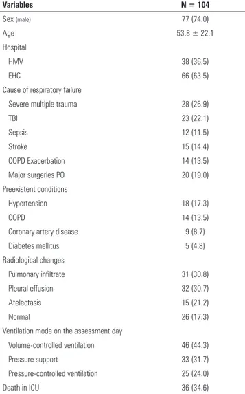
During data collection, 104 patients were included in the study. Each patient was included only once in the analysis. he mean age of patients was 53 ± 22 years, and the main causes of respiratory failure were multiple trauma (26.9%) and traumatic brain injury (22.1%), as shown in table 1.
Table 2 shows an increase in the Cdyn, VT, RR, and SpO2, and a decrease in the Rsr immediately after applying the respiratory physiotherapy protocol. he changes observed remained during the assessment performed one hour after the end of the respiratory therapy protocol. An immediate increase in the heart rate, which was not sustained, is shown in table 2.
Table 1 - Sample characterization
Variables N = 104
Sex (male) 77 (74.0)
Age 53.8 ± 22.1
Hospital
HMV 38 (36.5)
EHC 66 (63.5)
Cause of respiratory failure
Severe multiple trauma 28 (26.9)
TBI 23 (22.1)
Sepsis 12 (11.5)
Stroke 15 (14.4)
COPD Exacerbation 14 (13.5)
Major surgeries PO 20 (19.0)
Preexistent conditions
Hypertension 18 (17.3)
COPD 14 (13.5)
Coronary artery disease 9 (8.7)
Diabetes mellitus 5 (4.8)
Radiological changes
Pulmonary infiltrate 31 (30.8)
Pleural effusion 32 (30.7)
Atelectasis 15 (21.2)
Normal 26 (17.3)
Ventilation mode on the assessment day
Volume-controlled ventilation 46 (44.3)
Pressure support 33 (31.7)
Pressure-controlled ventilation 25 (24.0)
Death in ICU 36 (34.6)
HMV - Hospital Moinhos de Vento; HPSC - Hospital de Pronto-Socorro de Canoas; TBI - traumatic brain injury; COPD - chronic obstructive pulmonary disease; PO - postoperative; ICU - intensive care unit. The results are expressed as N (%) and means ± SD.
Table 2 - Behavior of ventilatory and hemodynamic variables at the three assessment timepoints, i.e., one hour before (T-1) and immediately (T0) and one hour after (T+1) application of the respiratory therapy protocol
Variables T-1 T0 T+1
Cdyn(ml/cmH2O) 52.3 ± 16.1
a 65.1 ± 19.1b 64.7 ± 20.2b
VT(ml)
# 550 ± 134a 698 ± 155c 672 ± 146b
PIP (cmH2O)
## 22.2 ± 5.54b 21.6 ± 5.71ab 21.5 ± 5.24a
RSR(cmH2O/l/s)
## 14.2 ± 4.63b 11.0 ± 3.43a 11.2 ± 3.68a
RR (bpm) 20.8 ± 5.40b 21.9 ± 5.89c 19.4 ± 4.97a SpO2(%) 96.5 ± 2.29a 98.2 ± 1.62c 97.8 ± 1.79b
Heart rate (bpm) 88.9 ± 18.7a 93.7 ± 19.2b 88.5 ± 17.1a
Cdyn - dynamic lung compliance; VT - tidal volume; PIP - peak inspiratory pressure; Rsr - respiratory system resistance; RR - respiratory rate; SpO2 - peripheral oxygen saturation. The results are expressed as means ± standard deviations. * ANOVA for repeated measures. a,b,c The same letter indicates no difference according to the Bonferroni test (p < 0.001). # Values for the 58 patients who received pressure-controlled ventilation; ## Values for the 46 patients who received constant-flow, volume-controlled ventilation.
DISCUSSION
he indings of this study suggest that, in MV-dependent patients, signiicant hemodynamic and ventilatory changes occur immediately after respiratory therapy; however, only the ventilatory changes persisted for at least one hour.
Respiratory therapy is part of the multidisciplinary care of critically ill patients dependent on MV because pulmonary complications resulting from the depression of the cough relex, reduction in mucociliary clearance, and increase in bronchial mucus production can lead to the retention of bronchial secretions, atelectasis formation,
and the development of nosocomial pneumonia.(2-5,17)
Particularly in intubated, sedated, and MV-dependent patients, respiratory therapy techniques facilitate the mobilization and elimination of bronchial secretions.(1-3,18)
and mobilize and remove secretions from the airway with the help of gravity.(7) Manual chest compression provides an expansion of collapsed lung areas, thus improving the V/Q ratio, in addition to acting as a facilitating stimulus
of thoracic mobility, which can be decreased.(12) he
chest compression-vibrations applied to the expiratory phase of the respiratory cycle allow better lung emptying, thus facilitating bronchial hygiene.(2,6) MH favors the displacement of accumulated secretions in the airways
and reduces pulmonary shunting.(6,8-19) his technique,
performed on patients with spontaneous ventilation, aims to prevent alveolar collapse, expand collapsed alveoli, improve oxygenation and lung compliance, and minimize the risk of hypoxemia, as well as stimulate coughing in MV-dependent patients.(9,11,12) Our study protocol utilized the application of two physiotherapy techniques (manual chest compression and MH).
Although most authors compare those techniques alone, protocols combining diferent techniques are part of the physiotherapy practice in the ICU and have a primary
goal of the removal of respiratory secretions. Fink(14)
studied the alternate application of the techniques TA,
PD, and chest percussion and observed improved SpO2
and airway resistance (Raw) in all sessions performed in the four days of the study. Mackenzie et al.(16) applied manual chest techniques in 19 patients and found increased Cdyn up to two hours after inishing the application of the
maneuver. In our study, the increase in SpO2 and Cdyn
and decrease in Raw started immediately after applying the protocol and were sustained for the next hour. However, the changes in the pulmonary mechanics did not exceed 30 minutes.(8,10)
Regarding the changes in the oxygenation indices, Barker and Adams(5) showed no diferences in oxygenation (PaO2) or alveolar ventilation (PaCO2) in 17 patients with acute respiratory distress syndrome when comparing three treatment protocols: TA, TA + PD, and TA + PD + MH.
In our patients, SpO2 measurements showed improved
oxygenation immediately after applying the respiratory physiotherapy protocol, which lasted at least one hour. Other authors(20,21) have found similar results of improved oxygenation, however, with shorter duration.
Few studies(20) have evaluated the changes in PIP
during and after physiotherapy treatment. Our data showed a persistent reduction of PIP after completion of the protocol.
In a study of diicult-to-wean patients, Hodgson et al.(9) and Maa et al.,(19), in 18 and 23 patients, respectively,
showed improvement of static lung compliance (Cstat)
in the group that underwent MH. Choi and Jones(15)
conirmed that a physiotherapy protocol (MH + TA) could increase Cstat and decrease Rsr for at least 30 minutes. In our study, which included the evaluation of patients who were receiving pressure support ventilation (PSV), the chest compliance changes were demonstrated by measuring the Cdyn.
Regarding techniques for tracheal secretion removal, a protocol consisting of MH + PD + TA increased the amount (weight) of mucus removed.(9,23) Mackenzie et al.(16) showed that after completion of a bronchial hygiene protocol, a reduction of 20% in the intrapulmonary
shunt, an increase of Cdyn by 14%, and an improvement
of gas exchange within two hours after physiotherapy were observed. Hodgson et al.(9) showed an increased removal of respiratory secretions and an increase of 30% of Cdyn after the use of MH, compared to TA alone, in 18 mechanically ventilated patients. Unoki et al.(12) found no diferences when comparing those two techniques in 31 mechanically ventilated patients. Stiller et al.(22) found greater eicacy in the resolution of atelectasis and improvement in oxygenation when using a protocol that combined PD, chest compression-vibrations, MH, and TA compared with a protocol consisting of MH and TA.
Regarding the changes in hemodynamics, Hodgson et al.(9) and Paratz et al.(11) showed a 10% decrease in the mean blood pressure when MH techniques were applied, but no changes in the heart rate were observed, which contradicts our indings.
Ntoumenopulos et al.(23) evaluated the clinical
assessment of each protocol technique; and (c) the heterogeneity of the sample studied (e.g., evaluation of patients under diferent ventilation conditions), which did not allow evaluation according to the ventilation mode used or the cause of respiratory failure (hypoxemic versus hypercapnic patients), among others.
CONCLUSION
he physiotherapy protocol applied was efective in improving the respiratory mechanics of patients dependent on mechanical ventilation. It is worth noting that these efects were measured after 60 minutes and remained present in most patients.
Objetivo: Avaliar as alterações da mecânica ventilatória e da hemodinâmica que ocorrem em pacientes dependentes de ventilação mecânica submetidos a um protocolo padrão de isioterapia respiratória.
Métodos: Estudo experimental e prospectivo realizado em duas unidades de tratamento intensivo, nas quais pacientes dependentes de ventilação mecânica por mais de 48 horas foram alocados, de forma consecutiva, e submetidos a um protocolo estabelecido de manobras de isioterapia respiratória. Variáveis ventilatórias (complacência pulmonar dinâmica, resistência do sistema respiratório, volume corrente, pressão de pico inspiratório, frequência respiratória e saturação periférica de oxigênio) e hemodinâmicas (frequência cardíaca) foram mensuradas 1 hora antes (T-1), imediatamente (T0) e após 1 hora (T+1) da realização do protocolo de manobras de isioterapia respiratória.
Resultados: Durante o período de coleta dos dados, 104
pacientes foram incluídos no estudo. Quanto às variáveis ven-tilatórias, houve aumento da complacência pulmonar dinâmica
(T-1 = 52,3 ± 16,1mL/cmH2O versus T0 = 65,1 ± 19,1mL/cmH2O; p < 0,001), do volume corrente (T-1 = 550 ± 134mL versus T0 = 698 ± 155mL; p < 0,001) e da saturação periférica de oxigê-nio (T-1 = 96,5 ± 2,29% versus T0 = 98,2 ± 1,62%; p < 0,001), além de redução da resistência do sistema respiratório (T-1 = 14,2 ± 4,63cmH2O/L/s versus T0 = 11,0 ± 3,43cmH2O/L/s; p < 0,001) logo após a realização das manobras de isioterapia respiratória. Todas as alterações se mantiveram na avaliação re-alizada 1 hora (T+1) após as manobras de isioterapia respirató-ria. Já com relação às variáveis hemodinâmicas, houve elevação imediata, porém não sustentada da frequência cardíaca (T-1 = 88,9 ± 18,7bpm versus T0 = 93,7 ± 19,2bpm versus T+1 = 88,5 ± 17,1bpm; p < 0,001).
Conclusão: Manobras de isioterapia respiratória geram
mudanças imediatas na mecânica pulmonar e na hemodinâmica dos pacientes dependentes da ventilação mecânica, e as alterações ventilatórias provavelmente permanecem por pelo menos 1 hora.
RESUMO
Descritores: Terapia respiratória; Respiração artiicial; Me-cânica respiratória
REFERENCES
1. Stiller K. Physiotherapy in intensive care: towards and evidence-based practice. Chest. 2000;118(6):1801-13. Review.
2. Imle PC. Percussão e vibração. In: Mackenzie CF, Ciesla N, Imle PC, Klemic N. Fisioterapia respiratória em unidade de terapia intensiva. São Paulo: Panamericana; 1998. p. 89-98.
3. David CM, Machado M, Vianna A, Marinho JM. Complicações da ventilação mecânica. J Pneumol. 2000;26(Supl 2):45-54.
4. Guglielminotti J, Desmonts JM, Dureuil B. Effects of tracheal suctioning on respiratory resistances in mechanically ventilated patients. Chest. 1998;113(5):1335-8.
5. Barker M, Adams S. An evaluation of a single chest physiotherapy treatment on mechanically ventilated patients with acute lung injury. Physiother Res Int. 2002;7(3):157-69.
6. Denehy L. The use of manual hyperinflation in airway clearance. Eur Respir J. 1999;14(4):958-65. Review.
7. Unoki T, Mizutani T, Toyooka H. Effects of expiratory rib cage compression and/or prone position on oxygenation and ventilation in mechanically ventilated rabbits with induced atelectasis. Respir Care. 2003;48(8):754-62.
8. Berney S, Denehy L. A comparison of the effects of manual and ventilator hyperinventilator hyperinflation on static lung compliance and sputum production in intubated and ventilated intensive care patients. Physiother Res Int. 2002;7(2):100-8.
9. Hodgson C, Denehy L, Ntoumenopoulos G, Santamaria J, Carroll S. An investigation of the early effects of manual lung hyperinflation in critically ill patients. Anaesth Intensive Care. 2000;28(3):255-61.
10. Berney S, Denehy L, Pretto J. Head-down tilt and manual hyperinflation enhance sputum clearance in patients who are intubated and ventilated. Austr J Physiother. 2004;50(1):9-14.
11. Paratz J, Lipman J, McAuliffe M. Effect of manual hyperinflation on hemodynamics, gas exchange, and respiratory mechanics in ventilated patients. J Intensive Care Med. 2002;17(6):317-24.
12. Unoki T, Kawasaki Y, Mizutani T, Fujino Y, Yanagisawa Y, Ishimatsu S, et al. Effects of expiratory rib-cage compressionon oxigenatuion, ventilation, and airway-secretion removal in patients receiving mechanical ventilation. Respir Care. 2005;50(11):1430-7.
14. Fink JB. Positive pressure techniques for airway clearance. Respir Care. 2002;47(7):786-96.
15. Choi JS, Jones AY. Effects of manual hyperinfl ation and suctioning in respiratory mechanics in mechanically ventilated patients with ventilator-associated pneumonia. Aust J Physiother. 2005;51(1):25-30.
16. Mackenzie CF, Shin B. Cardiorespiratory function before and after chest physiotherapy in mechanically ventilated patients with post-traumatic respiratory failure. Crit Care Med. 1985;13(6):483-6.
17. Clini E, Ambrosino N. Early physiotherapy in the respiratory intensive care unit. Respir Med. 2005;99(9):1096-104.
18. Rosa FK, Roese CA, Savi A, Dias AS, Monteiro MB. Comportamento da mecânica pulmonar após a aplicação de protocolo de fisioterapia respiratória e aspiração traqueal em pacientes com ventilação mecânica invasiva. Rev Bras Ter Intensiva. 2007;19(2):170-5.
19. Maa SH, Hung TJ, Hsu KH, Hsieh YI, Wang KY, Wang CH, et al. Manual hyperinflation improves alveolar recruitment in difficult-to-wean patients. Chest. 2005;128(4):2714-21.
20. Clarke RC, Kelly BE, Convery PN, Fee JP. Ventilatory characteristics in mechanically ventilated patients during manual hyperventilation for chest physiotherapy. Anaesthesia. 1999;54(10):936-40.
21. Ciesla ND. Chest physical therapy for patients in the intensive care unit. Phys Ther. 1996;76(6):609-25.
22. Stiller K, Geake T, Taylor J, Grant R, Hall B. Acute lobar atelectasis. A com-parison of two chest physioteraphy regimens. Chest. 1990;98(6):1336-40. 23. Ntoumenopoulos G, Presneill JJ, McElholum M, Cade JF. Chest