Rev Odontol UNESP. 2017 Sept-Oct; 46(5): 299-306 © 2017 - ISSN 1807-2577
ORIGINAL ARTICLE
Doi: http://dx.doi.org/10.1590/1807-2577.02917
Assessment of patient’s anxiety and expectation
associated with hemodynamic changes during surgical
procedure under local anesthesia
Avaliação da ansiedade e da expectativa do paciente associadas às alterações hemodinâmicas durante
procedimento cirúrgico sob anestesia local
Marcos Vinícius Mendes DANTAS
a*, Bianca NESSO
a, Daniel Sagawa MITUUTI
a,
Marisa Aparecida Cabrini GABRIELLI
aaFaculdade de Odontologia de Araraquara, UNESP – Universidade Estadual Paulista, Araraquara, SP, Brasil
Resumo
Introdução: A ansiedade e a expectativa do paciente odontológico podem alterar de forma significativa seus
sinais vitais. Da mesma forma, o uso de anestésico local associado a um vasoconstrictor também alteram os sinais vitais desses pacientes, promovendo alterações hemodinâmicas que podem resultar em situações de emergência.
Objetivo: Avaliar a influência da ansiedade de pacientes submetidos à exodontia de terceiros molares e do uso
de diferentes substâncias anestésicas com adrenalina sobre seus sinais vitais (saturação de oxigênio, pulsação e pressão arterial sistólica e diastólica) em diferentes momentos operatórios. Material e método: Quarenta pacientes responderam os questionários da Escala de Ansiedade Dentária (Escala de Corah) e de medo (Escala de Kleinknecht) e foram submetidos à exodontia dos terceiros molares em dois tempos cirúrgicos para utilização de articaína ou mepivacaína, ambos associados com adrenalina. Os resultados obtidos foram analisados por ANOVA seguido do teste post hoc de Tukey, t de Student e coeficientes de correlação de Pearson (α=0,05). Resultado: Não houve alteração significativa na saturação nem na frequência cardíaca. A pressão arterial apresentou variações significantes nos tempos aferidos para os dois anestésicos, entretanto a mepivacaína resultou em maior tempo de pós-operatório para o restabelecimento da pressão arterial. Os pacientes com muita ou moderada ansiedade e alto índice de medo foram os que tiveram correlações positivas com os maiores números pressóricos aferidos. Conclusão: A ansiedade e medo influenciam positivamente no aumento da pressão arterial. A mepivacaína promoveu uma maior resistência ao retorno da normalidade dos sinais vitais, especialmente dos níveis pressóricos.
Descritores: Ansiedade; medo; sinais vitais; pressão sanguínea; anestésico local.
Abstract
Introduction: The dental patient’s anxiety and expectation may significantly alter their vital signs. The use of local
anesthetics associated with a vasoconstrictor may also alter the vital signs of these patients, promoting hemodynamic changes that may result in emergency situations. Objective: To evaluate the influence of anxiety of patients submitted to third molar extraction and the use of different anesthetic substances with adrenaline on their vital signs (oxygen saturation, heart rate, and systolic and diastolic blood pressure) in different moments. Material and method: Forty patients answered the questionnaire of the Dental Anxiety Scale (Corah’s Scale) and fear (KleinKnecht’s Scale) and were submitted to third molar extraction in two surgical times for the use of articaine or mepivacaine, both associated with adrenaline. The results were analyzed by ANOVA followed by Tukey post hoc test, Student’s t test, and Pearson’s correlation coefficients (α=0.05). Result: There was no significant differences in saturation or heart rate. The blood pressure showed significant variations during time for both anesthetics, however mepivacaine resulted in a longer postoperative time to restore blood pressure. Patients with high or moderate anxiety and high fear index were those who had positive correlations with the highest blood pressure values. Conclusion: Anxiety and fear positively influence the increase in blood pressure. Mepivacaine promoted a greater resistance to the return of normal vital signs, especially blood pressure levels.
INTRODUCTION
he basic monitoring of vital signs is commonly used in the practice of ambulatory care. Its use allows to obtain important information about the patient’s current state, such as blood pressure, heart rate, oxygen saturation and respiratory rate. hese signs can be obtained in a non-invasive way through easily manipulated devices, sphygmomanometer and pulse oximeter. his practice has been increasingly used because of the growing number of high-risk patients in oral surgeries1, mainly because many of them come to the clinic to undergo dental treatments without knowing if they have any type of systemic problem. he identiication and control of these physiological indicators are some of the factors responsible for the promotion of successful treatments, avoiding emergencies that expose the patient to risky situations2. herefore, monitoring has three major advantages: the ability to detect, evaluate and prevent emergencies in clinical practice.
In dental surgical procedures, usually medium- or long-term local anesthetics are used to promote anesthesia during the transoperative period. Local anesthetics cause depression of the excitation of the nerve endings or inhibition of the conduction process in the peripheral nerves in a certain circumscribed area of the body3. In addition, anesthetic salts cause peripheral vasodilatation4, causing an increase in the blood volume in the site and consequently there is more bleeding during the surgical procedure. herefore, the duration and efectiveness of the anesthetic become reduced. Among these anesthetic solutions, mepivacaine hydrochloride and articaine hydrochloride are the most commonly indicated for surgical procedures due to their longer duration.
Mepivacaine, an amide group local anesthetic, has been used in dentistry since 1960. It is biotransformed in the liver and has a discrete vasodilator property, so its duration is 2 to 3 hours when associated with a vasoconstrictor. Its half-life is 1.9 hour with onset of action of 2 minutes. Articaine has been used as a local anesthetic in dentistry in Brazil since 1999. It also belongs to the amide group, but, unlike other anesthetics, it has a thiophene ring as a chemical chain radical, being the only local anesthetic of the amide group containing an ester group. herefore, its biotransformation begins in blood plasma and tissues, and later part of it is metabolized in the liver, presenting a half-life of 27 minutes and low toxicity5. Due to the presence of the thiophene ring, which gives it greater solubility, articaine exhibits high penetration and difusion in the tissues, including bone tissue. Studies show that this anesthetic has a latency time of around 3 minutes and, when associated with adrenaline, duration of approximately 71 minutes4,6.
he association of vasoconstrictors with local anesthetics increases the anesthetic duration and efectiveness, and decreases its toxicity, since it becomes possible to use a lower volume of these solutions, in addition to the advantage of reducing local bleeding in the surgical procedure. However, the use of vasoconstrictors in dental practice is still an issue of high controversy because many dentists state that the use of substances such as adrenaline and noradrenaline cause signiicant hemodynamic changes, especially to blood pressure. In fact, the use of adrenaline in local anesthetics
bears the risk of systemic absorption when used in large amounts, resulting in undesirable cardiovascular efects that can alter patients’ hemodynamics5. On the other hand, several studies4,7-9 show that the amount of vasoconstrictors present in the local anesthetic is insuicient to cause any signiicant alteration. Mepivacaine and articaine, when associated with adrenaline 1:100,000 or 1:200,000 and administered in therapeutic doses, oten do not promote major changes to blood pressure, heart rate, respiratory rate and oxygen saturation4,7-9.
he substances used in anesthesia are not the only ones capable of promoting hemodynamic changes10. Factors inherent to the patient can also trigger fear and decrease the pain threshold10. Since the patient is frequently in a state of anxiety and fear during the procedure, these emotional issues can imply changes to vital signs and in turn cause transoperative and postoperative complications. In 2001, Malamed3 described that the stress to the patient caused by minor oral surgeries causes endogenous catecholamines (adrenaline and noradrenaline) to be released from their original site in an amount 40 times greater than the adrenaline present in the local anesthetic tube. In 2012, Costa et al.2 showed in their study that the patient’s discomfort during the surgical procedure relects on behavioral and physiological changes, which are extremely important, as they promote signiicant changes to the patient’s vital signs. In 2002, Chaia et al.11 analyzed that the expectation and fear can promote changes to blood pressure and cause tachycardia. In addition to these variations, altered emotional state can lead to a reduction in pain tolerance, raising the level of anxiety, thus establishing a vicious circle where local anesthetic agents could not act eiciently because they do not interfere directly with anxiety and stress12. hus, ensuring the patient’s comfort and safety so he/she is in a less apprehensive state is more efective to decrease the possible hemodynamic variations throughout the surgical procedure than avoiding the use of vasoconstrictors, since stress is also generated by the pain of an inadequate anesthesia.
Considering that the great majority of patients who arrive at the dental oice have some type of fear or anxiety about dental treatments, Corah et al.13 in 1978 developed a questionnaire containing only four questions to rate the patients’ level of anxiety, thus allowing the dentist to prepare himself/herself to deal with certain types of patients. Similarly, in 1978, Kleinknecht, Bernstein14 developed a questionnaire simulating various situations in the daily routine of a dental appointment, from the time of scheduling to the anesthesia, in order to rate the level of fear felt by the patient in certain situations, allowing the dentist to know in advance when his/her patient will require special attention.
MATERIAL AND METHOD
Sample Selection
his prospective study was conducted with patients who underwent surgical procedures, under local anesthesia, for extraction of included third molars. hese patients were treated in “Oral and Maxillofacial Surgery and Traumatology” clinic of the Araraquara School of Dentistry - UNESP, and were operated by a single experienced surgeon.
For this study, 40 healthy ASA I and II patients (American Society of Anesthesiologists, physical status 1), of both genders, aged 18-45 years, who had the four third molars indicated for extraction, and mandatorily followed these exclusion criteria, were selected: smoker; presence of infection sites involving third molars; presence of uncontrolled systemic diseases or psychiatric problems that can counter indicate the surgical procedure or interfere with the methodology; history of radiation in the head and neck region or use of chemotherapy; patients on chronic use of corticosteroids; female patients in menopause; pregnant patients; patients with cardiac alterations using (or not) medications; patients allergic to the study anesthetics; patients with systemic alterations or using medications that change bone repair; and immunosuppressed patients.
All patients included in this study signed the informed consent form. In addition, we submitted the research work to the approval by the Ethics Committee on Human Research (CAAE: 30245314.0.0000.5416).
Preoperative Evaluation
We used a standardized health form of the “Oral and Maxillofacial Surgery and Traumatology” clinic of the Araraquara School of Dentistry to perform the anamnesis and clinical exam of the patients. Imaging evaluation was performed with panoramic radiographs to surgical planning and the type of impaction of the third molar was not considered for patient inclusion (or not) in this study.
Experimental Groups
he forty patients were divided into two groups, according to the type of local anesthetic used:
Group M (n=20): Patients submitted to surgery using local anesthetic containing mepivacaine hydrochloride 2% (36mg) with epinephrine 1:100,000 (18μg)/1.8ml tube (DFL-Rio de Janeiro-RJ).
Group A (n=20): Patients submitted to surgery using local anesthetic containing articaine hydrochloride 4% (72mg) with epinephrine 1:100,000 (18μg)/1.8ml tube (DFL-Rio de Janeiro-RJ).
Pre-surgical Procedures
Prior to the surgical procedure, in a quiet room separated from the surgical center, and ater the patient was seated in the room for about 5 minutes, the vital signs were checked and noted on a standardized form. Aterwards, the patient completed a Corah’s Scale13 and Kleinknecht’s Scale form14.
In addition, at that time, each patient received 1g of amoxicillin, 500mg of sodium dipyrone and 20mg of omeprazole. Patients allergic to penicillin received 600mg of clindamycin, and those allergic to dipyrone received 750mg of paracetamol.
Vital Signs
We noted the values of vital signs of blood pressure (BP) expressed in millimeters of mercury (mmHg), heart rate (HR) expressed in beats per minute (bpm), respiratory rate (RR) expressed in breaths per minute (bpm) and oxygen saturation (SaO2) expressed in percentage (%), using the digital monitor FOXIMETER (MD 300C21): at 5 diferent time points:
T0: Before the beginning of the procedure and outside the surgical environment
T1: Five minutes ater the end of local anesthetic application T2: Halfway through the surgical procedure
T3: Immediately ater the end of surgery and T4: Fiteen minutes ater the end of surgery
All values obtained were recorded in tables and the data inserted in spreadsheets for analysis. A single trained and calibrated examiner recorded the vital signs.
Anxiety and Fear Scales
Prior to the surgical procedure, we instructed each patient to complete two anxiety and fear assessment forms, described below:
1 - Corah’s Dental Anxiety Scale (DAS)13 - Corah’s scale of anxiety for dental treatment [the peak was 15, and over 12 was considered intense anxiety]. he scale is composed of three questions. Each question has ive alternatives: a (value 1), b (value 2), c (value 3), d (value 4), and e (value 5).
2 - Kleinknecht’s Dental Fear Scale (DFS)14 - Kleinknecht’s Scale for the evaluation of fear of dental treatment [composed of six diferent situations with 20 alternatives].
Pain Scale
We used the visual analog scale, ranging from 0 (no pain) to 10 (intense pain) to obtain the pain intensity in the transoperative period (completed by the patient at the end of the surgical) and postoperative period.
Surgical Procedures
In the irst surgical time point, we removed the lower and upper third molars from one of the sides of the jaws, randomly chosen (e.g. teeth 18 and 48 or 28 and 38). Likewise, we randomly selected the anesthetic for the procedure of the irst surgical time point. he surgery side and anesthetic type were randomly selected to ensure an unbiased representation for each experimental group, not compromising the results (for example, the side where the anesthetic was more easily applied could be favored and the side where the teeth were greatly impacted could be disfavored).
other two (upper and lower) third molars, using a diferent anesthetic from the one used in the irst time point, in order to guarantee that both anesthetic solutions were used in the same patient.
We noted the duration of surgical procedures as well as the number of anesthetic tubes used for each patient from groups M and A.
In each surgical time point, we prescribed in the postoperative period: amoxicillin or clindamycin; sodium dipyrone or paracetamol; nimesulide and omeprazole (orally) and chlorhexidine for use in mouthwashes.
Statistical Analyses
For the data correlated with the changes to physiological signs as for the type of anesthetic used and pain sensitivity, we applied ANOVA followed by Tukey post hoc test (α=0.05). To evaluate the results obtained for anxiety, we applied the Student’s statistical t-test and Pearson’s correlation coeicients (α=0.05).
RESULT
In this study, the mean age of the patients was 22.4 years, with minimum age of 18 years and maximum age of 30 years. he population consisted of 26 women (65%) and 14 men (35%).
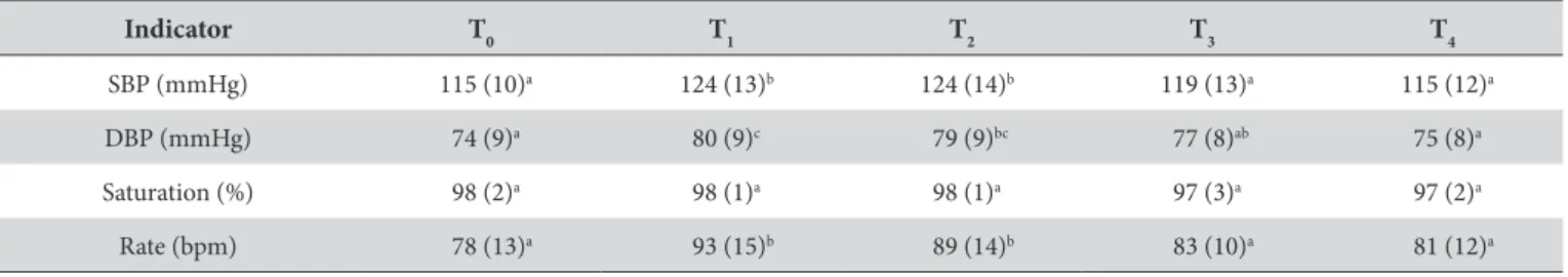
Tables 1 and 2 show the means and standard deviations for the physiological indicators: systolic and diastolic blood pressure, oxygen saturation and heart rate, recorded at ive diferent time points from preoperative to postoperative period for patients submitted to third molar extraction and anesthetized, respectively, with articaine 4% associated with adrenaline (1:100,000) and mepivacaine 2% associated with adrenaline (1:100,000).
From the tables we can observe that there were changes to the vital signs during the surgical procedure for both anesthetics.
Oxygen saturation was the only physiological indicator to remain stable during all recorded time points, with no signiicant variations for the results. We observed that the changes to heart rate and blood pressure were similar, and at the time point T1 both of them increased signiicantly and remained at this level until halfway through the surgical procedure. At the time point T4 the values returned to the initial value. he return of the indicators to the initial level was more efective when using articaine, occurring at the end of the surgical procedure, while for mepivacaine it took iteen minutes ater surgery.
he analyses of variance found no diferences between means of saturation during monitoring (p=0.388 for articaine and p=0.143 for mepivacaine). In all other cases there was evidence of a signiicant variation of the means during monitoring (p<0.05). he Tukey test was applied to explain these variations of means of physiological indicators and the results are summarized in Tables 1 and 2.
Table 3 shows the means and standard deviations of pain intensity monitored at certain postoperative time points for both anesthetic solutions used in this study. he analyses of variance revealed diferences between means in the monitoring period of pain (p<0.05). he results of the Tukey test are summarized in Table 3. We observed that the means of pain intensity experienced by the patients were higher in the irst 12 postoperative hours and decreased progressively up to 36 postoperative hours, remaining at this level in the next evaluation, ater 48 hours. However, throughout the monitoring period, the intensity of pain ater surgical intervention with articaine always remained below the levels following intervention with mepivacaine.
Regarding the emotional factor, Figure 1 shows the percentages in each category of level of fear experienced by the 40 patients in certain situations, according to the questions proposed by Kleinknecht, Bernstein14. We observed that 20% of the patients
Table 1. Means (standard deviations) of physiological indicators monitored in subjects under anesthesia with articaine, according to the 5 monitoring periods
Indicator T0 T1 T2 T3 T4
SBP (mmHg) 115 (10)a 124 (13)b 124 (14)b 119 (13)a 115 (12)a
DBP (mmHg) 74 (9)a 80 (9)c 79 (9)bc 77 (8)ab 75 (8)a
Saturation (%) 98 (2)a 98 (1)a 98 (1)a 97 (3)a 97 (2)a
Rate (bpm) 78 (13)a 93 (15)b 89 (14)b 83 (10)a 81 (12)a
Means with equal letters in the same line are not signiicantly diferent by the Tukey test (p>0.05).
Table 2. Means (standard deviations) of physiological indicators monitored in subjects under anesthesia with mepivacaine, according to the 5 monitoring periods
Indicator T0 T1 T2 T3 T4
SBP (mmHg) 114 (13)a 126 (14)c 126 (15)bc 121 (11)b 114 (11)a
DBP (mmHg) 72 (10)a 78 (8)c 79 (9)c 77 (8)bc 74 (7)ab
Saturation (%) 97 (2)a 98 (2)a 98 (1)a 97 (2)a 97 (2)a
Rate (bpm) 77 (16)a 97 (18)d 90 (14)cd 85 (13)bc 82 (13)ab
Table 3. Intensity of pain at the speciied time points following the surgical procedure
Anesthetic Post-operative period (h)
12 24 36 48
Articaine 4(3)c 3 (2)b 2 (2)a 1 (2)a
Mepivacaine 5 (3)c 4 (3)bc 3 (3)ab 2 (2)a
Means with equal letters in the same line are not signiicantly diferent by the Tukey test (p>0.05).
reported they always felt tense during the consultation with the dentist. No patient reported to experience nausea (100%) during consultations. In addition, the patients reported to have more fear in the following time points: when he/she sees and feels the anesthesia needle and when he/she hears and feels the motor.
In the evaluation of the anxiety index through the Corah’s Scale13, we obtained the means of 10.1 (standard deviation 3.5) for females and 9.5 (standard deviation 9.5) for males. he patients could be rated into four levels of anxiety according to the score obtained by the scale: <9 for low anxious patients; 9-12 for moderately anxious patients; 13-14 for very anxious patients; and >15 for severely anxious patients. Both men and women participating in the study were classifed as moderately anxious. No signiicant diference was observed between genders for the means of anxiety (Student’s t-test - p=0.800). We also observed no signiicant correlation between the anxiety rates and any of the variables studied, for neither articaine nor mepivacaine. Pearson’s correlation coeicients ranged from -0.219 to 0.355, all of which were not signiicantly diferent from zero (p>0.05).
DISCUSSION
Surgery for the extraction of third molars is one of the most common occurrences among all surgical procedures and, mainly, under local anesthesia. his study evaluated the possible changes to vital signs that occurred during this surgical procedure using two local anesthetics with vasoconstrictor. Furthermore, the degree of anxiety and fear and their possible relation with the changes to vital signs, as part of changes to hemodynamics, were also evaluated.
he population of this study was composed of young people who are the age group who most seek this type of surgical procedure. his conirms the research potential, which was performed with patients (exclusively ASA I) under 40 years of age who did not present any type of health impairment or use medications routinely. hese data, as described above, support the data reported in other studies in the literature, including a larger proportion of female patients, relecting the gender that most seeks treatment2,15,16.
Adrenaline is the most used vasoconstrictor in dentistry, being an important additive in local anesthetic solutions, mainly for use in surgical procedures. he major advantages of using adrenaline are slowing the absorption rate and decreasing the systemic blood level of the anesthetic drug in addition to prolonging the duration of anesthesia. On the other hand, vasoconstrictors are not devoid of complications. Like the anesthetic substance, the vasoconstrictor is absorbed into the bloodstream and can reach levels that inluence the hemodynamics of the heart and blood vessels. he increase in adrenaline plasma levels has a dose-dependent linear relationship and persists for hours3.
he results showed that, generally, the highest increase in blood pressure, especially SBP, occurred within ive minutes of iniltration of the anesthetic substance (T1), when using both articaine and mepivacaine associated with adrenaline. Cardiac hemodynamics seems to be afected early in the beginning of the vasoconstrictor absorption. A previous study by Brkovic et al.17, using lidocaine associated with adrenaline, observed that ten minutes (on average)
following the application of the anesthesia there was an increase in the heart rate and pressure values and these are maintained on a prolonged basis. In fact, in our sample, this increase, mainly in blood pressure, was observed and maintained until halfway through the surgical procedure (T2). In all treated patients, the surgeon initiated the surgical procedure by removing the upper third molar and ended by removing the lower third molar, on the treated side at each surgical time point. In all patients of this sample, for the removal of the upper third molar, we used, on average, one and a half tubes of mepivacaine 2% with epinephrine 1:100,000 or articaine 4% with epinephrine 1:100,000. herefore, we injected the same amount of vasoconstrictor as the diferent anesthetic solutions, that is, on average, 0.030mg of adrenaline. he same increased measure of blood pressure was maintained until the T2 period, corresponding to half the time of the surgical procedure for the extraction of the upper and lower third molars on one side of the jaws. his means that at T2 the anesthesia for the removal of the lower tooth was already performed and, on average, another two and a half tubes of anesthetic solution were used, adding more 0.050mg of adrenaline. Despite the addition of a greater amount of adrenaline, it did not cause an even greater increase in systolic or diastolic pressure, as we can see from the results described in Tables 1 and 2. In addition, we must take into account that the endogenous catecholamines corresponding to the stress of the surgical procedure are released.
Absorption of adrenaline into the bloodstream results in some cardiac hemodynamic change related to both heart rate and blood pressure. Despite these changes, in healthy patients, it is not enough to exhibit a clinically signiicant efect18. Although increased heart rate and blood pressure were observed in all patients in the sample, none of them showed symptoms that clinically revealed the changes in these parameters.
When analyzing the values found in Tables 1 and 2 it is possible to observe that both anesthetics caused an increase in blood pressure that was almost reestablished at the end of the procedure. Of the 40 patients evaluated, with the use of articaine, 10 patients had systolic blood pressure between 130 and 145 and diastolic blood pressure of 90 to 111.5. When using mepivacaine, 11 patients had blood pressure change (values mentioned above). However, by using mepivacaine, the blood pressure levels remained longer with a high blood pressure pattern and required a longer rest period to match the initial value. Possibly, this fact is due to the vasodilation power of the anesthetic substance. In this case, the vasodilation capacity of mepivacaine is considered lower than that of articaine, allowing the associated vasoconstrictor to be partially inhibited according to the vasodilatory power of the anesthetic substance. In addition, it is possible that articaine had less prolonged efect on cardiac changes.
more than 12 hours is that the patient experienced less initial pain and, therefore, presented less postoperative stress and, consequently, it inluenced the pain threshold, providing articaine with higher efectiveness. he transoperative diiculties must also be considered as the result of greater intensity and duration of pain. In this sense, we can consider the anatomical diiculties, such as the need for bone removal or odontosection, which make dental extraction and postoperative period more complex16.
For patients who, at the end of the surgery, remained with high systolic and diastolic pressure, we observed that coincidentally they are the same subjects who (when using mepivacaine) took a longer time from the end of surgery until blood pressure was normalized. For these patients the Corah’s13 and Kleinknecht’s14 scales show that they can be classified as anxious or highly anxious and apprehensive people, with a high degree of fear of the proposed treatment. Of the 11 patients previously mentioned who presented a more significant pressure change, 7 were classified as anxious and only 1 as extremely anxious. These data are the same as those found in the studies by Costa et al.2 and Maggirias, Locker19. On the Kleinknecht’s scale14, 15 patients with high fear, 1 patient with very high fear and 1 patient with phobia (extreme fear) were found, making up a total of 30% of moderately to very anxious patients and 50% classified as fearing the procedure. In addition, the presence of anxiety and fear may
have contributed to the increase in blood pressure. This becomes clear in cases in which the initial pressure values were already changed.
he idea of surgical procedure undoubtedly causes more anxiety than any other type of dental care15. Eli et al.20, who evaluated the relationship of anxiety and pain during and ater the installation of dental implants, observed that women had higher relation of high anxiety levels with pain than men. In our study, the same correlation cannot be made because the number of female patients in the sample was higher (65% females/35% males). Even so, we observed a higher correlation between postoperative exacerbated pain and anxiety in female patients than in male patients.
CONCLUSION
According to the information above, we can conclude that:
1) he presence of anxiety and fear positively inluenced the increase in blood pressure;
2) he need for diagnosis and anxiety control was evident in patients who will undergo surgical procedures in order to avoid or prevent risk situations;
3) Mepivacaine promoted a greater resistance to the return to normal vital signs, especially blood pressure levels.
REFERENCES
1. Arrigoni J, Lambrecht JT, Filippi A. Cardiovascular monitoring and its consequences in oral surgery. Schweiz Monatsschr Zahnmed. 2005;115(3):208-13. PMid:15832655.
2. Costa RR, Silva PVR, Iwaki L Fo, Takeshita WM, Farah GJ. Avaliação da influência da expectativa e da ansiedade do paciente odontológico submetido a procedimento cirúrgico a partir de seus sinais vitais. Rev Odontol UNESP. 2012 Jan-Fev;41(1):43-7.
3. Malamed SF. Manual de anestesia local. Rio de Janeiro: Guanabara Koogan; 2001.
4. Palma FR, Lins LHS, Branco FP, Wygladala LG. Verificação da variação da pressão arterial pelo uso de anestésicos locais com vasoconstritor. Rev Odonto Ciência. 2005 Jan-Mar;20(47):35-9.
5. Schertzer ER Jr. Articaine vs. lidocaine. J Am Dent Assoc. 2000 Sep;131(9):1248-50.
6. Oertel R, Rahn R, Kirch W. Clinical pharmacohinetics of articaine. Clin Pharmacokinet. 1997 Dec;33(6):417-25. PMid:9435991. http:// dx.doi.org/10.2165/00003088-199733060-00002.
7. Tolas AG, Pflug AE, Halter JB. Arterial plasma epinephrine concentrations and hemodynamic responses after dental injection of local anesthetic with epinephrine. J Am Dent Assoc. 1982 Jan;104(1):41-3. PMid:6948029. http://dx.doi.org/10.14219/jada.archive.1982.0114. 8. Zottis D, Bernardes R, Wannmacher L. Efeito de vasoconstritor usado em anestesia local sobre a PA sistêmica e FC durante o atendimento
odontológico. Rev ABO Nac. 1999;7:289-93.
9. Dantas MVM, Gabrielli MAC, Hochuli-Vieira E. Efeito da mepivacaínia 2% com adrenalina 1:100.000 sobre a pressão arterial. Rev Odontol UNESP. 2008;37(3):223-7.
10. Alemany-Martínez A, Valmaseda-Castellón E, Berini-Aytés L, Gay-Escoda C. Hemodynamic changes during the surgical removal of lower third molars. J Oral Maxillofac Surg. 2008 Mar;66(3):453-61. PMid:18280377. http://dx.doi.org/10.1016/j.joms.2007.06.634.
11. Chaia A, Mandarino SCA, Gandelmann JH, Cavalcante MA, Alencastro VC. Análise da média aritmética da pressão arterial, frequência cardíaca e saturação de oxigênio durante as cirurgias de terceiros molares inclusos sob anestesia local e sedação prévia. Rev Bras Implant. 2002;8(4):29-31.
12. Meyer FU. Haemodynamic changes under emotional stress following a minor surgical procedure under local anaesthesia. Int J Oral Maxillofac Surg. 1987 Dec;16(6):688-94. PMid:3125267. http://dx.doi.org/10.1016/S0901-5027(87)80054-1.
13. Corah NL, Gale EN, Illig SJ. Assessment of a dental anxiety scale. J Am Dent Assoc. 1978 Nov;97(5):816-9.
15. Sirin Y, Humphris G, Sencan S, Firat D. What is the most fearful intervention in ambulatory oral surgery? Analysis of an outpatient clinic. Int J Oral Maxillofac Surg. 2012 Oct;41(10):1284-90. PMid:22832662. http://dx.doi.org/10.1016/j.ijom.2012.06.013.
16. Aznar-Arasa L, Figueiredo R, Valmaseda-Castellón E, Gay-Escoda C. Patient anxiety and surgical difficulty in impacted lower third molar extractions: a prospective cohort study. Int J Oral Maxillofac Surg. 2014 Sep;43(9):1131-6. PMid:24837553. http://dx.doi.org/10.1016/j. ijom.2014.04.005.
17. Brkovic B, Gardasevic M, Roganovic J, Jovic N, Todorovic L, Stojic D. Lidocaine+clonidine for maxillary infiltration anaesthesia: parameters of anaesthesia and vascular effects. Int J Oral Maxillofac Surg. 2008 Feb;37(2):149-55. PMid:17822879. http://dx.doi.org/10.1016/j. ijom.2007.07.019.
18. Salonen M, Forssell H, Scheinin M. Local dental anaesthesia with lidocaine and adrenaline. Effects on plasma cathecolamines, heart rate and blood pressure. Int J Oral Maxillofac Surg. 1988 Dec;17(6):392-4. PMid:3145958. http://dx.doi.org/10.1016/S0901-5027(88)80071-7. 19. Maggirias J, Locker D. Five-year incidence of dental anxiety in an adult population. Community Dent Health. 2002 Sep;19(3):173-9.
PMid:12269464.
20. Eli I, Schwartz-Arad D, Baht R, Ben-Tuvim H. Effect of anxiety on the experience of pain in implant insertion. Clin Oral Implants Res. 2003 Feb;14(1):115-8. PMid:12562373. http://dx.doi.org/10.1034/j.1600-0501.2003.140115.x.
CONFLICTS OF INTERESTS
he authors declare no conlicts of interest.
*CORRESPONDING AUTHOR
Marcos Vinícius Mendes Dantas, Departamento de Diagnóstico e Cirurgia, Faculdade de Odontologia de Araraquara, UNESP – Universidade Estadual Paulista, Rua Humaitá, 1680, Centro, 14801-903 Araraquara - SP, Brasil, e-mail: [email protected]