Universidade de Lisboa
Faculdade de Farmácia
Epigenetic Regulation of Signalling Pathways in Pancreatic Cancer
Inês Correia da Silva Pires Faleiro
Dissertação orientada pelo Professor Doutor Pedro Castelo-Branco e pela
Professora Doutora Elsa Rodrigues.
Mestrado em Ciências Biofarmacêuticas
Universidade de Lisboa
Faculdade de Farmácia
Epigenetic Regulation of Signalling Pathways in Pancreatic Cancer
Inês Correia da Silva Pires Faleiro
Dissertação orientada pelo Professor Doutor Pedro Castelo-Branco e pela
Professora Doutora Elsa Rodrigues.
Mestrado em Ciências Biofarmacêuticas
I
De acordo com o disposto no ponto 1 do artigo nº 45 do Regulamento de Estudos Pós Graduados da Universidade de Lisboa, deliberação nº 4624/2012, publicada em Diário da República – II Série nº 65 – 30 de Março de 2012, o Autor desta dissertação declara que participou na concepção e execução do trabalho experimental, interpretação dos resultados obtidos e redação dos manuscritos.
II
The studies presented in this thesis were performed at the Center for Biomedical Research (CBMR), Algarve University, under the scientific supervision of Professor Pedro Castelo-Branco, and at the Faculty of Pharmacy, University of Lisbon, under the scientific supervision of Professor Elsa Rodrigues.
III Agradecimentos
Em primeiro lugar agradeço ao meu orientador, o professor Pedro Castelo-Branco por acreditar em mim, por tudo o que me ensinou e por todo o apoio que me deu que me permitiu chegar até aqui. Muito obrigada.
Agradeço à minha orientadora, à professora Elsa Rodrigues por me ter acompanhado ao longo de todo o percurso do mestrado, pela motivação transmitida e pela sua disponibilidade e ajuda em tudo o que precisei.
À Joana e à Célia, pela sua amizade e por desde o inicio me terem apoiado e integrado tão bem na equipa. Obrigada por estarem sempre presentes.
Quero também agradecer à Gabriela por ter trabalhado ao meu lado ao longo deste ano e me ter ajudado sempre que precisei e a todos os membros do laboratório, não poderia pedir melhor ambiente para evoluir cada vez mais.
Um agradecimento especial à Vânia, cuja ajuda na escrita e planeamento do projeto foi essencial para que isto fosse possível. Obrigada.
A todos os meus amigos pelo apoio ao longo deste ano, em especial à Rita, que esteve sempre presente, que me ajudou em tudo o que podia e tantas vezes teve de me ouvir falar do projeto.
A toda a minha família e em particular à minha tia Paula, por me motivar e encorajar a seguir os seus passos na investigação cientifica, por tentar sempre guiar-me na direção certa e por estar presente em tudo o que eu faça.
O agradecimento mais especial de todos ao Cláudio, aos meus irmãos e aos meus pais pelo encorajamento e por todo o apoio para que pudesse dedicar-me por completo a este projeto. Sem vocês não teria conseguido, obrigada não chega.
IV Abstract
Pancreatic cancer is one of the most lethal malignancies worldwide. Deregulation of epigenetic marks is known to alter the expression of crucial genes for cancer development. Replicative immortality, achieved mainly through telomerase reactivation, and aberrant activation of the PI3K/Akt, Wnt, Notch and Hedgehog pathways triggers transduction cascades that potentiate cell proliferation.
The aim of this study is to evaluate epigenetic alterations of genes related with these biological processes as potential pancreatic cancer biomarkers.
Gene selection of the PI3K/Akt, Wnt, Notch and Hedgehog pathways was based on differential expression between normal and malignant tissue using the pancreatic expression database and the miRDB database was used to uncover miRNAs targeting the selected genes. Methylation and miRNA analysis was performed using The Cancer Genome Atlas (TCGA) data on the cohort of pancreatic cancer and the impact of DNA methylation and miRNA expression on patient’s outcome was also analysed.
Most of the selected genes presented higher expression in tumour tissue. Our results reveal that the majority of the CpGs sites analysed were hypermethylated in tumour tissue. Methylation levels of the selected genes allowed the distinction between normal and malignant tissue even in initial stages of the disease revealing its potential as a diagnostic tool for pancreatic cancer.
The methylation levels of the TERT, ITGA4, SFN, ITGA2, PIK3R1 and SFRP2 genes could act as independent prognostic indicators of patients’ survival with higher sensitivity and specificity than the currently implemented biomarker. Additionally, differential methylation of the TERT, SFN and PIK3R1 genes were also associated with recurrence of the patients.
Moreover, analysis of the expression of miRNAs involved in the regulation of the TGFBR1, PTEN, EIF4EBP1, AKT3, JAG1 and CSNK1A1 have demonstrated the ability to discriminate between groups of patients with different outcomes when comparing the patients with highest and lowest expression of each miRNAs.
Despite the promising results in this area no epigenetic biomarkers have reached the clinic yet. Our results reveal that the methylation levels of genes involved in pancreatic carcinogenesis could be used to predict the outcome of pancreatic cancer patients with high sensitivity and specificity.
These results provide new evidences of the potential of epigenetic alterations as pancreatic cancer biomarkers for disease screening and management.
Keywords
V Resumo
O cancro do pâncreas é um dos mais letais em todo o mundo e a desregulação do padrão epigenético das células influencia a expressão de genes cruciais para o desenvolvimento e progressão da doença. A capacidade ilimitada de autorrenovação celular resultante principalmente da reativação da enzima telomerase e a ativação das vias de sinalização PI3K/Akt, Wnt, Notch e Hedgehog desencadeia cascatas de transdução de sinal que potenciam a proliferação celular contribuindo para o processo carcinogénico.
O principal objetivo deste estudo é avaliar alterações epigenéticas de genes relacionados com estes processos biológicos como potenciais biomarcadores para o cancro do pâncreas.
A selecção dos genes envolvidos nas vias de sinalização PI3K/Akt, Wnt, Notch e Hedgehog foi baseada na expressão diferencial dos genes entre tecido normal e maligno usando a base de dados Pancreatic Expression Database e para a selecção de miRNAs envolvidos na regulação dos genes de interesse recorremos à base de dados miRDB. Os níveis de metilação e de expressão dos miRNAs foram analisados usando os dados do The Cancer Genome Atlas (TCGA) relativos à coorte de pacientes com cancro do pâncreas e o seu potencial para discriminar entre pacientes com diferentes desfechos clínicos e para identificar os pacientes que se encontram em maior risco de progressão da doença assim como uma menor sobrevivência dos pacientesfoi avaliado.
A maioria dos genes selecionados apresentou maior expressão no tecido tumoral. Os nossos resultados revelam que a maioria das regiões genómicas analisadas estão hipermetiladas no tecido tumoral e que os níveis de metilação dos genes selecionados permitem a distinção entre tecido normal e maligno mesmo nos estadios iniciais da doença, revelando seu potencial como biomarcador de diagnóstico para o cancro de pâncreas.
A metilação dos genes TERT, ITGA4, SFN, ITGA2, PIK3R1 e SFRP2 permitiram discriminar entre grupos de pacientes com diferentes desfechos clínicos considerando o seu tempo de sobrevida com maior sensibilidade e especificidade que o biomarcador atualmente implementado na clinica para este tipo de cancro. Adicionalmente, a metilação diferencial dos genes TERT, SFN and PIK3R1 revelou estar também associada com a recorrência dos pacientes.
Além disso, a análise de expressão de miRNAs envolvidos na regulação dos genes TGFBR1, PTEN, EIF4EBP1, AKT3, JAG1 and CSNK1A1 permitiu diferenciar grupos de pacientes com diferentes tempos de sobrevivência comparando os pacientes com maior e menor expressão de cada miRNA.
Os resultados obtidos demonstram o potencial da análise de metilação do DNA e dos níveis de miRNAs como indicadores de prognóstico com elevada sensibilidade e especificidade. Este estudo revela novas evidências sobre o potencial das alterações epigenéticas como biomarcadores de diagnóstico e prognóstico para o cancro do pâncreas de modo a contribuir para uma melhoria da qualidade e esperança de vida de pacientes com esta doença.
Palavras-chave
VI
Index
Abstract ... IV Resumo ... V List of Tables ... VIII List of Figures ... IX Abbreviations ... XI
1. CHAPTER 1 - GENERAL INTRODUCTION ... 2
1.1 Pancreatic Cancer ... 2
1.2 Disease subtypes ... 3
1.3 Therapeutic Strategies ... 4
1.4 Molecular Pathogenesis of Pancreatic Cancer ... 5
1.4.1 Genetics of pancreatic ductal adenocarcinoma ... 5
1.4.2 Genetics of pancreatic neuroendocrine tumours ... 6
1.5 Epigenetics ... 7
1.5.1 Histone Modifications ... 8
1.5.2 DNA methylation ... 9
1.5.3 MicroRNAs (miRNAs) ... 10
1.6 Epigenetic Alterations in Pancreatic Cancer ... 11
1.6.1 Epigenetic therapy ... 11
1.6.2. Epigenetic alterations as potential biomarkers ... 13
1.7 Objectives ... 14
2. CHAPTER 2 - METHODS ...16
2.1 Bioinformatic Resources ... 16
2.1.1 Pancreatic Expression Database ... 16
2.1.2 The Cancer Genome Atlas ... 16
2.1.3 miRDB ... 16
2.2 Gene Selection ... 17
2.3 Epigenetic Alterations Analysis ... 17
2.3.1 Data collection ... 17
2.3.2 Methylation analysis ... 19
2.3.3 miRNA expression analysis ... 19
2.3.4 Clinical data analysis ... 20
2.3.5 Statistical analysis ... 20
2.4 Study Pipeline ... 21
CHAPTER 3 - RESULTS AND DISCUSSION ...22
3.1 The TERT hypermethylated Oncologic Region (THOR) predicts recurrence and survival in pancreatic cancer ... 22
3.1.1 Introduction ... 23
VII
3.1.3 THOR methylation distinguishes normal from tumour tissue in pancreatic cancer ... 25
3.1.4 THOR methylation predicts outcome in patients with pancreatic cancer ... 26
3.1.5 Discussion ... 27
3.2 Epigenetic alterations of PI3K/Akt, Wnt, Hedgehog and Notch signalling pathways are associated with survival of pancreatic cancer patients ... 28
3.2.1. Introduction ... 29
3.2.2 Results ... 31
3.2.3 Discussion ... 56
CHAPTER 4 - CONCLUSIONS AND FUTURE PERSPECTIVES ...58
REFERENCES ...61
VIII List of Tables
Table 1 – Characteristics of the patients from the TCGA pancreatic cancer cohort. ...18 Table 2 - Differentially expressed genes in pancreatic cancer. Genes related with the PI3K/Akt, Notch, Hedgehog and Wnt pathways that present differential expression between malignant and healthy pancreatic tissue in the data available on the Pancreatic Expression Landscape integrated in the PED database ...32 Table 3 – Differential methylated genes in the TCGA pancreatic cancer cohort. ...35 Table 4 – Correlation between DNA methylation and gene expression levels in the TCGA pancreatic cancer cohort. ...36 Table 5 – Correlation between miRNA and gene expression levels in the TCGA pancreatic cancer cohort. ...38
IX List of Figures
Figure 1 – Hallmarks of Cancer ... 3
Figure 2 – Representation of the signalling pathways involved in pancreatic cancer ... 6
Figure 3 – Implication of epigenetics in pancreas development, function and disease ... 8
Figure 4 – Schematic representation of the TERT promoter ... 24
Figure 5 – Hypermethylation of THOR in pancreatic cancer ... 24
Figure 6 – THOR discriminates between normal tissue and malignant pancreatic tissue ... 25
Figure 7 – THOR can predict survival and recurrence in pancreatic cancer ... 26
Figure 8 – CpGs differentially methylated in pancreatic cancer ... 37
Figure 9 – ITGA4 methylation can predict survival in pancreatic cancer ... 40
Figure 10 – SFN methylation can predict survival in pancreatic cancer ... 42
Figure 11 – SFN methylation can predict recurrence in pancreatic cancer ... 43
Figure 12 – ITGA2 methylation can predict survival in pancreatic cancer ... 44
Figure 13 – PIK3R1 methylation can predict survival and recurrence in pancreatic cancer ... 45
Figure 14 – SFRP2 methylation can predict survival in pancreatic cancer ... 46
Figure 15 – Methylation of SFN and SFRP2 genes is associated with patients’ history of chronic pancreatitis ... 47
Figure 16 – Methylation of the ITGA4, ITGA2 and PIK3R1 genes discriminates between normal and malignant tissue of different pathological stages ... 48
Figure 17 – Methylation of the SFN gene discriminates between normal and malignant tissue of different pathological stages ... 49
Figure 18 – Methylation of the SFRP2 gene discriminates between normal and malignant tissue of different pathological stages ... 50
Figure 19 – Methylation of the ITGA4, ITGA2 and PIK3R1 genes differs between distinct histological subtypes of pancreatic cancer ... 51
Figure 20 – Methylation of the selected genes differs between distinct histological subtypes of pancreatic cancer ... 52
X
Figure 21 – Methylation of the selected genes differs between distinct histological subtypes of
pancreatic cancer ... 53
Figure 22 – The expression levels of miRNAs targeting the TGFBR1, PTEN, EIF4EBP1 and AKT3
genes can predict survival in pancreatic cancer patients ... 55
Figure 23 – The expression levels of miRNA targeting the JAG1 and CSNK1A1 genes can predict
XI Abbreviations
5hmC – 5-hydroxymethylcytosine 5mC – 5- methylcytosine
ADM – Acinar to ductal metaplasia
ALT – Alternative lengthening of telomeres mechanism AUC – Area under curve
CA19-9 – Cancer antigen 19-9 DNMT – DNA methyltransferase
EMT – Epithelial to mesenchymal transition H3K27ac – Acetylation of histone 3 at lysine 27 H3K27me – Methylation of histone 3 at lysine 27 H3K36me – Methylation of histone 3 at lysine 36 H3K4me – Methylation of histone 3 at lysine 4 H3K4me2 – Di-methylation of histone 3 at lysine 4 H3K4me3 – Tri-methylation of histone 3 at lysine 4 H3K9ac – Acetylation of histone 3 at lysine 9 H3K9me – Methylation of histone 3 at lysine 9 H3K9me3 – Tri-methylation of histone 3 at lysine 9 HAT – Histone acetyltransferase
HDAC – Histone deacetylase HDM – Histone demethylase HTM – Histone methyltransferase
IPMN – Intraductal papillary-mucinous neoplasm OS – Overall survival
XII PanIN – Pancreatic intraepithelial neoplasia PanNETs – Pancreatic neuroendocrine tumours PCA – Pancreatic cancer
PDAC – Pancreatic ductal adenocarcinoma PED – Pancreatic expression database PI3K – Class I phosphoinositidine 3- kinases RFS – Recurrence-free survival
ROC – Receiver operator curve TCGA – The Cancer Genome Atlas
TERT – Telomerase reverse transcriptase gene TET – Ten-eleven translocation enzymes
1 CHAPTER 1 - GENERAL INTRODUCTION
2 1. CHAPTER 1 - GENERAL INTRODUCTION 1.1 Pancreatic Cancer
Cancer is defined as a group of diseases characterized by an abnormal and uncontrolled division of cells that can ultimately proliferate and invade other tissues [1,2]. Hananan and Weinberg have proposed six biological processes as the hallmarks of cancer: sustained proliferative signalling, replicative immortality, bypass of growth suppressors regulation, resistance to cell death, induction of angiogenesis and ability to metastasize and invade other tissues (Fig. 1A) [2]. Recently, two more processes were proposed to the list as critical events to tumour development: cell reprogramming of energy metabolism and the ability to evade the control mediated by our immune system (Fig. 1B) [2].
It is a complex disease marked by aberrant expression of tumor promoting genes, named oncogenes, and tumor suppressor genes [2]. Altered transcriptional patterns confer cells specific capabilities that allow them to escape the regulatory mechanisms responsible for maintaining cell homeostasis.
The main cause of death by cancer is recurrence after treatment. The tumour mass is composed by a heterogeneous group of cells with distinct sensibility to therapeutic agents making the treatment of this disease more difficult [3]. Therapeutic resistance is related to the existence of cancer stem cells and several mechanisms of resistance have already been described affecting multiple cellular pathways [2,3].
An additional layer of complexity must be considered as an increasingly number of studies have highlighted the importance of the group of cells present in the tumor microenvironment including cancer-associated fibroblasts, endothelial cells, pericytes and infiltrating cells of the immune system [2]. This special subgroup of cells is involved in cellular processes that enable tumor growth and development.
All these factors result in a highly complicated disease to treat being the second most common cause of death [4].
Pancreatic cancer (PCA) is one of the most mortal malignancies worldwide with a 5-year survival rate of 7% [4]. Known risk factors associated with pancreatic cancer include family history, age, cigarette smoking, and history of pancreatitis or diabetes mellitus [5,6].
The early symptoms of pancreatic cancer can be vague and unrecognized including weight loss, abdominal and mid back pain, jaundice, indigestion and loss of appetite. Frequently, these symptoms are not enough to make a clear diagnose of pancreatic cancer as they can also be associated with other diseases as gallstones, gastritis, irritable bowel syndrome, gastroenteritis, indigestion or liver disease leading to misdiagnosis [6]. A late diagnosis and a poor response rate to treatment make the discovery and validation of novel biomarkers for screening and management of PCA an absolute necessity [5].
3
Figure 1. Hallmarks of Cancer. A. Illustration of the six first established hallmarks of cancer. B.
Recently proposed emerging hallmarks and characteristics that contribute to the malignant transformation of cells. From Hananan et al, 2011.
1.2 Disease subtypes
PCA can be divided into two main groups, depending from which pancreatic cell type it arises. Almost all PCAs are classified as exocrine tumours since the affected cells are exocrine cells (responsible for enzyme production) and can arise from acinar cells, connective tissue or from the ductal epithelium where pancreatic ductal adenocarcinomas (PDAC) represent about 80% of all pancreatic cancers [5].
About 60-70% of all PCAs originate from cells localized in the head of the pancreas while 20-25% arise in the body and the tail of the pancreas [7]. The remaining minority of the
A
4
cases diffusely affect different pancreatic cells [7]. Tumours in the head of the pancreas normally present obstruction of the common bile duct and/or the pancreatic duct and develop specific symptoms that allow a more rapid diagnose compared with other pancreatic cancer [7].
Only about 5% of all cases develop from pancreatic endocrine cells, the islet cells, which are responsible for hormone production [8]. These tumours tend to be less aggressive than the exocrine ones [8].
Pancreatic neuroendocrine tumours (PanNETs) can be classified as functional or non-functional tumours. Non-non-functional PanNETs represent the majority of neuroendocrine tumours of the pancreas and are associated with a poorer prognosis as the disease is generally diagnosed at an advanced stage [8]. Functional PanNETs present hormone excess syndrome which leads to specific clinical symptoms due to increased hormone production facilitating the diagnosis of the disease [8,9].
The islets of Langerhans are formed by the endocrine cells of the pancreas: β-cells, α-cells, γ-cells and δ-cells. These cells are responsible for the production of several hormones such as insulin, glucagon, pancreatic polypeptide and somastotatin, respectively. Accordingly, functional tumours of the pancreas are defined by the hormone secreted being named insulinomas, glucagonomas, VIPomas and somastinomas, [8,10].
1.3 Therapeutic Strategies
Currently the therapeutic options available are surgical resection, radiotherapy and chemotherapy. However, only 15-20% of the patients are eligible for surgery since by the time of diagnosis the majority of patients present the disease at advanced stage [5].
While surgery remains the principal therapeutic strategy for pancreatic cancer, medical treatment of patients with advanced disease differs according to the distinct disease subtypes [7,11].
For resectable PDAC the standard of care is surgery followed by adjuvant chemotherapy with fluorouracil and leucovorin or gemcitabine. In borderline cases, resectable tumours are treated with neoadjuvant therapy and/or radiotherapy followed by surgery and posterior administration of adjuvant therapy or chemotherapy depending on the surgery success [6,7].
In locally advanced and metastatic PDAC surgical removal of the tumour is no longer a viable option. For these patients, the administrated regimens consist in FOLFIRINOX (5-fluorouracil, leucovorin, irinotecan and oxaliplatin) and gemcitabine plus nab-paclitaxel or gemcitabine alone for patients with reduced tolerance to the treatment and best supportive care [6,7].
For PanNETs patients with advanced disease, the pharmacological agents available include somastotin analogs, chemotherapy and targeted therapy using everolimus (a mammalian target of rapamycin (mTOR) inhibitor) and sutinib (a multikinase inhibitor) [11].
Despite efforts of the scientific community to develop new therapeutic approaches for pancreatic cancer no effective therapies have reach the clinic and the mortality rates for this type of cancer have remain almost unaltered with tendency to increase [4,6].
Most patients present progressive disease and recurrence after treatment with a response rate to chemotherapy inferior to 20% making it necessary the discovery and validation of novel biomarkers for diagnosis, prognosis and prediction of therapeutic response [5].
5
Currently, the only FDA approved biomarker for PCA is the Cancer-Antigen 19-9 (CA19-9, a modified Lewis(a) blood group antigen) with a sensitivity of 60-70% and a specificity of 70-85% [12]. However, this biomarker is not useful as a diagnostic tool since other diseases can also cause an increase in the plasma levels of this protein. Additionally, in a subset of pancreatic cancer patients not harbouring a functional Lewis enzyme required for CA19-9 production the levels of this protein are very low or undetectable leading to false negatives [13]. Despite not being useful as a diagnostic biomarker, CA19-9 presents significant value as a prognostic tool and can be used to monitor patients’ response to treatment, helping the clinicians to adopt the best therapeutic approach [12]. Until now, no biomarker revealed to significantly outperform CA19-9 in the clinic.
Our study will be focused mainly on pancreatic ductal adenocarcinomas however, other histological subtypes will also be included and therefore the molecular pathogenesis of both PDAC and PanNETs will be addressed.
1.4 Molecular Pathogenesis of Pancreatic Cancer
The development and progression of pancreatic cancer is driven by successive accumulation of somatic mutations and epigenetic alterations [6,14,15].
1.4.1 Genetics of pancreatic ductal adenocarcinoma
The most important genetic events in PDAC, the most common type of PCA, are the activation of the oncogene KRAS and the inactivation of the tumour suppressor genes cyclin dependent kinase inhibitor 2A (CDKN2A), tumour protein p53 (TP53) and SMAD4 [16].
Several lesions such as intraductal papillary-mucinous neoplasm (IPMN), pancreatic intraepithelial neoplasia (PanIN) and mucinous cystic neoplasm can progress and originate PDACs [16]. The precursor lesions mostly associated with tumour development are PanINs which consist of epithelial neoplasms that occur in pancreatic ducts [16].
One of the cellular processes implicated in PCA is the process of acinar to ductal metaplasia (ADM). ADM is a cellular mechanism required for pancreatic tissue regeneration after inflammation or injuries [17]. These ADM lesions can progress to PanIn lesion and eventually progress to pancreatic ductal adenocarcinoma in response to oncogenic signalling [18].
Alterations in the most important PDAC driver genes are also observed in precursor lesions such as PanIN with activation of the oncogene KRAS as an early event of lesion development, which then leads to inactivation of CDKN2A, characteristic of PanIN2 stage. With the progression of the lesion and the establishment of a PanIN3 lesion, TP53 and SMAD4 are inactivated, which are the most commonly observed genetic alterations in PDAC. This is consistent with the progression of these lesions to a malignant state [16,19].
Cancer heterogeneity is one of the main reasons of therapeutic failure comprising inter and intra-individual changes between malignant cells. Gene mutations differ from individual to individual and the initial alteration responsible for the induction of a malignant state was not identified yet. However, global genomic analysis revealed that the deregulation of some signalling pathways including the phosphatidylinositol 3-kinase (PI3K)/Akt, Wnt, Notch and Hedgehog pathways is deeply involved in PDAC development [20,21]. A detailed description of these signalling pathways can be found in Section 3.2.1 (a simplified representation of those signalling pathways is illustrated in Fig. 2).
6
Figure 2 – Representation of the signalling pathways involved in pancreatic cancer. A. PI3K/Akt pathway: In the PI3K/Akt pathway, extracellular ligands bind to cell surface receptors leading to class I
phosphoinositidine 3- kinases (PI3Ks) activation. When activated, PI3K generates PIP3 that activates phosphoinositide-dependent protein kinase 1 (PDK1) and leads to subsequent Akt activation. Upon activation, Akt phosphorylates several proteins that control multiple cellular processes that contribute to cancer development including cell survival and proliferation. B. Hedgehog pathway: Binding of Hedgehog ligands to Patched receptor (PTCH1) disrupts the repression of SMO proteins that are then capable of activating downstream proteins such as GLI proteins that act as transcriptional activators inducing the expression of several genes that can contribute to cancer progression and development.
C. Notch pathway: Binding of Delta and Jagged Notch ligands induces receptors cleavage by
γ-secretase. Release and translocation to the nucleus of the cytoplasmic domain allows its association with transcription factors promoting expression of genes associated with cell growth and proliferation.
D. Wnt pathway: In the canonical Wnt pathway, β-catenin is phosphorylated and targeted for
degradation by the proteasome (off state). Binding of the Wnt, a secreted protein, to its receptor, Frizzled, leads to stabilization of β-catenin by downstream proteins of the pathway (on state). This leads to β-catenin translocation to the nucleus where it forms a complex with transcription factors (TCF-Lef) to induce gene expression. Activation of the pathway is represented by an arrow (→) and repression of the pathway is represented by a blocked-line (Ͱ).
1.4.2 Genetics of pancreatic neuroendocrine tumours
The genetic alterations behind PanNETS development and progression affect distinct genes in comparison to PDAC [22]. The most affected genes in PanNETs are the menin 1
(MEN1) gene, Death Domain Associated Protein (DAXX) gene, ATRX chromatin remodeler
7
(ATRX) gene and Mtor gene [22,23]. Indeed, a study of whole-genome sequencing using
PanNETs samples revealed four pathways commonly altered in this type of pancreatic: DNA damage repair, chromatin remodelling, telomere maintenance and mTOR signalling [23].
Resistance to therapeutic agents is one of the major factors that contribute to the discouraging survival rates of the patients. One of the causes of treatment failure is poor diffusion of drugs which is influenced by the formation of a desmoplastic reaction creating a unique and protective microenvironment surrounding cancer cells [18]. This microenvironment is composed of extracellular matrix proteins, fibroblasts and immune cells as well as its secreted factors affecting cells behaviour and its ability to metastasize and resist to the effects of chemotherapeutic agents [18].
1.5 Epigenetics
Epigenetic modifications are somatically heritable and reversible changes that contribute to gene expression regulation without altering the DNA sequence. Among these are histone post-translational modifications, DNA methylation and non-coding RNAs, especially miRNAs [24]. All cells in the human body have essentially the same genome, however cells from different tissues have distinct structures and functions as the result of differential gene expression. The process of cell differentiation during the embryonic development is due to epigenetic reprogramming of the cells allowing each cell to express only certain genes conferring them specific functions [25,26]. Also, the epigenome can be influenced by various factors including diet, exercise or medication mediating the interplay between the environment and gene expression [26].
Cancer is one of the leading causes of death worldwide and cancer development and progression comprises wide alterations in gene expression that result not only from genetic but also from epigenetic modifications.
Epigenetic regulation is implicated in the normal development and function of the pancreas and deregulation of these mechanisms can result in the development of pancreatic diseases including pancreatic cancer (Figure 3) [27].
8
Figure 3. Implication of epigenetics in pancreas development, function and disease. DNA
methylation, histone modifications and the action of non-coding RNAs are crucial for pancreas normal development and are deeply involved in the regulation of pancreas functions. Deregulation of these mechanisms leads to an impairment in pancreas functions resulting in the development of several pancreatic diseases. From Quilichini et al, 2015. DNMT: DNA methyltransferase; HAT: histone acetyltransferase; HDAC: histone deacetyltransferase; HMT: histone methyltransferase; HDM: histone demethyltransferase.
1.5.1 Histone Modifications
There are several histone post-translational modifications that can affect the compaction state of chromatin, which influences the folding, position and organization of DNA, thereby affecting gene expression. Some of these alterations include histone methylation, acetylation, phosphorylation, ubiquitylation and sumoylation [28,29]. Among these, the best described histone modifications with important roles in epigenetic deregulation in cancer are histone acetylation and methylation [24].
Histone acetylation can promote disruption of the electrostatic interactions between histones and DNA by reducing the positive charge of histone tails, which leads to a more uncondensed chromatin state. Addition of the acetyl group is carried out by histone acetyltransferases (HATs), and its removal is catalysed by histone deacetylases (HDACs) [30]. In addition to influencing DNA-histone interactions, histone acetylation can promote recruitment of the chromatin remodeling complex SWI/SNF (SWItch/Sucrose Non-Fermentable) which activity is associated with transcription activation [31,32].
Histone methylation can occur on lysine or arginine residues and, depending on the target, it can lead to activation or repression of gene expression [33]. Unlike acetylation and phosphorylation, however, histone methylation does not alter the charge of the histone protein. Furthermore, there is an added level of complexity when considering this modification; lysines
9
may be mono-, di- or tri-methylated, whereas arginines may be mono-, symmetrically or asymmetrically di-methylated [33].
The regulation of histone methylation levels is achieved by the activity of histone demethylases (HDMs) and histone methyltransferases (HTMs) [33].
As previously mentioned these post-translational modifications in histones can be associated with active or repressive chromatin. Modifications such as histone 3 methylation at lysine 4 (H3K4me), histone 3 di-methylation at lysine 4 (H3K4me2), histone 3 tri-methylation at lysine 4 (H3K4me3), histone 3 acetylation at lysine 9 (H3K9ac), histone 3 methylation at lysine 9 (H3K9me) or histone 3 acetylation at lysine 27 (H3K27ac) are associated with active chromatin whereas histone 3 tri-methylation at lysine 36 (H3K36me3), histone 3 tri-methylation at lysine 9 (H3K9me3) or histone 3 methylation at lysine 27 (H3K27me) are associated with repressive chromatin [34].
1.5.2 DNA methylation
DNA methylation results from the addition of a methyl group on the 5-carbon of a cytosine residue in a CpG site by a DNA methyltransferase (DNMT) [35]. The enzyme forms a complex with the DNA molecule to allow the transfer of the methyl group to the cytosine residue of CpG dinucleotides [35]. DNA methylation is involved in processes such as genomic imprinting, inactivation of the X chromosome and silencing of repetitive DNA sequences [25]. There are 3 active DNMTs in eukaryotes: DNMT1 is responsible for the maintenance of methylation patterns after DNA replication whereas de novo methylation is carried out by DNMT3A and DNMT3B [30].
The mechanisms of de novo methylation and how DNMTs target specific DNA sequences remain unknown. However, it is known that DNMTs can bind to DNA through a conserved PWWP domain, named after a conserved Pro-Trp-Trp-Pro motif, and interact between them or with methyl-binding domain proteins (MBDs) and transcription factors (TFs) to reinforce DNA methylation and influence gene transcription [36–39].
The idea that DNA methylation is a more dynamic process than previously believed is starting to arise with the possibility of directed DNA methylation and the recent discovery of enzymes capable to erase DNA methylation [40,41]. While the mechanism of DNA methylation and the enzymes involved have been well characterized, the mechanism of DNA demethylation remains controversial [42]. DNA demethylation is the removal or modification of the methyl group from 5-methylcytosine (5mC) [42]. The discovery of the ability of the ten-eleven translocation (TET) family enzymes to oxidize 5mC to 5-hydroxymethylcytosine (5hmC) using molecular oxygen as substrate has revolutionized this area [42]. In fact, overexpression of these enzymes resulted in a decreased of genomic 5mC levels [42].
If these modifications could directly influence DNA-protein interactions or if the biological effect is only due through disruption of 5mC patterns has not yet been scrutinized but it is believed that 5hmC is a key intermediate in active demethylation pathways [42].
Several pathways have been proposed following 5mC oxidation: the oxidized base can be passively removed during replication or actively through enzymatic reactions by enzymes involved in DNA repair mechanisms [42].
Considering the CG content of the human genome the abundance of CpG sites across the genome is relative lower than the expected [43]. However, the human genome contains sequences in most gene promoters with high content of CpG sites known as CpG islands [44].
In these regions, DNA methylation can contribute to changes in chromatin conformation influencing gene expression by affecting DNA exposure to transcription factor binding [45].
10
Generally, gene promoter DNA methylation is recognized as an event leading to transcriptional repression. However, there are several exceptions to this classical view of epigenetics related to DNA methylation, with increased gene expression associated with promoter DNA hypermethylation [46–48]. Gene activation through DNA methylation can be due to activation of an alternative TSS or to inhibition of repressive protein binding [49].
Increased gene expression associated with promoter CpG hypermethylation is also associated with an increase in H3K4me3, a histone mark characteristic of gene activation, reinforcing the interplay between these epigenetic mechanisms in transcriptional regulation [49]. Indeed, the interplay between proteins involved in both histone modifications and DNA methylation consolidate the effect of these epigenetic mechanisms. Histone acetylation and H3K4me are marks associated with active gene transcription and are typically associated with unmethylated DNA [50]. Histones marks associated with transcriptional repression as H3K9m and H3k27me are often associated with DNA methylation [50].
Any alteration that affects the activity of epigenetic-modifying enzymes can lead to an imbalance in gene expression regulation and provide the basis, or contribute, to the initiation of carcinogenesis.
1.5.3 MicroRNAs (miRNAs)
miRNAs are small endogenous ncRNAs with 21-25 nucleotides that participate in the regulation of gene expression by targeting specific mRNAs for translation repression or degradation.
miRNA genes can be localized in specific locus or in introns of other genes and are mainly transcribed by RNA polymerase II originating a primary transcript, the pri-miRNA [51], which is then processed by the RNase DROSHA. This enzyme and its cofactor Di George syndrome critical region 8 (DGR8) constitute the microprocessor responsible for the cleavage of the pri-miRNA to generate a pre-miRNA that contains a stem loop structure [51]. This structure is recognized and processed by DICER, after pre-miRNA exportation to the cytoplasm by exportin-5 [51]. DICER cleavage originates a RNA duplex, which will then associate with the RNA-induced silencing complex (RISC). Only the guide strand that consists of the mature miRNA remains incorporated into the complex while the other RNA strand is degraded [51]. The mature miRNA will then guide the RISC complex to the 3’ untranslated region (3’UTR) of a specific mRNA that depending on their complementarity level the mRNA is degraded or its translation blocked [51].
Previous studies have shown that miRNA expression patterns differ in normal and tumour tissue and that miRNAs can act either as tumour suppressors or oncogenes depending on their target [51]. Downregulation of a miRNA that targets an oncogene or upregulation of a miRNA that targets a tumour suppressor gene can promote carcinogenesis [51].
11 1.6 Epigenetic Alterations in Pancreatic Cancer
Disruption of the normal epigenetic patterns of the cells is associated with the development of PCA [52]. Also, deregulation of epigenetic-modifying enzymes disturbs normal epigenetic patterns and is associated with cancer development and progression.
For instance, in pancreatic cancer, the histone deacetylases HDAC2 and HDAC7 and the histone methyltransferase EZH2 are upregulated [15]. EZH2 upregulation is associated to cell proliferation, invasion and migration and is an indicator of a worst prognosis [15].
Specific histone modifications have also been associated with pancreatic cancer [53]. Analysis of two cohorts of patients revealed that low levels of the histone marks H3K4me2, H3K9me2, or H3K18ac are independent factors associated with poor prognosis and lower survival probability [53].
The DNA methyltransferases DNMT1, DNMT3A, DNMT3B are also upregulated in pancreatic cancer [54]. In fact, increased expression of DNMT1 was associated with disease progression suggesting the role of epigenetic regulators in PCA development [54]. Interestingly, several groups have performed genome-wide DNA methylation analysis of pancreatic cancer [55–57]. Those studies revealed aberrant patterns of DNA methylation of genes involved in important signalling pathways implicated in pancreatic carcinogenesis including transforming growth factor beta (TGF-β), Wnt, integrin signalling, cell adhesion, stellate cell activation, axon guidance and genes involved in stem cell pluripotency [55–57]. Recently, Mishra and coworkers have analysed the data of The Cancer Genome Atlas (TCGA) and found three distinct subtypes considering the genome-wide methylation patterns [58].
Epigenetic changes such as hypermethylation of CpG islands in several genes have been found in pancreatic cancer and in cancer precursor lesions such as PanINs. The methylation levels increase during the progression of these lesions. So, a higher level of methylation is probably associated with the progression of these lesions to cancer [59]. It has been shown that methylation levels can be detected through the analysis of pancreatic juice samples therefore, it has the potential to become a biomarker for the early detection of premalignant lesions [59].
Finally, miRNA levels are also altered in pancreatic cancer, affecting the expression of genes involved in cell cycle control, apoptosis, migration, and invasion and have the potential to be used as biomarkers for diagnosis and prognosis [15].
Several authors have described combinatory panels of miRNAs that can potentially be used for pancreatic cancer diagnosis [15]. Importantly, those panels were evaluated using serum samples from the patients proving the feasibility of miRNA profiling as a non-invasive method for PCA diagnosis. Results showed that several miRNAs have altered expression and were associated with disease progression and with worse prognosis. Of these miRNAs, miR-155 was also upregulated in pancreatic juice samples of patients with IPMN, a PCA precursor lesion [15].
1.6.1 Epigenetic therapy
Deregulation of epigenetic marks leads to changes in gene expression that, in cancer cells, can result in activation of oncogenes or inactivation of tumour suppressor genes, both of
12
which can contribute to cancer. Unlike genetic mutations, however, epigenetic changes are reversible. Therefore, the development of drugs capable of restoring the normal epigenetic patterns of cells has great therapeutic potential.
Two strategies for epigenetic therapy are currently in use: small molecules that inhibit epigenetic-modifying enzymes and manipulation of miRNA expression.
Amongst the small molecule inhibitors are HDAC inhibitors and DNMT inhibitors. HDAC inhibitors (HDACi) are classified into 4 groups according to their chemical structures: hydroxamates (SB393, Vorinostat, Panobinostat), cyclic peptides (Romidepsin), benzamides (Entinostat and Mocetinostat) and aliphatic fatty acids (Valproic Acid) [60]. The majority of HDACi inhibit zinc-dependent HDACs by interacting with the zinc ion. In cancer cells, the inhibition of histone deacetylation restores expression of tumour suppressor genes that were previously silenced by epigenetic mechanisms [60,61].
DNMT inhibitors are divided into nucleoside analogues and non-nucleoside analogs [30]. Nucleoside analogues, such as Azacitidine, Decitabine and FdCyd, are cytosine analogues modified at the C5 position. Inside the cell these compounds are metabolized and incorporated into DNA [30]. DNA methyltransferases can bind to these modified nucleotides but their modification at C5 prevents their methylation. It also prevents the dissociation of the enzyme thereby reducing DNMT activity at other sites [30]. Non-nucleoside analogues, such as Hydralazine, Procainamide and MG98, inhibit methylation by binding to the catalytic region of the enzyme [30].
Another focus of epigenetic therapy is the manipulation of miRNA expression and activity. Several strategies have been employed to silence miRNAs that are overexpressed in cancer. These include anti-miRNA oligonucleotides (AMOs), peptide nucleic acids (PNAS), miRNA-masking antisense oligonucleotides (miR-mask) and miRNA sponges [62]. Restoration of miRNA expression that has been downregulated in cancer is achieved by administration of synthetic miRNAs or by induced expression of miRNA coding genes using viral constructs, such as adenovirus-associated vectors [62].
To investigate the efficacy of this new therapeutic approach in cancer, our group has performed a comprehensive review where we analysed the results of clinical trials testing epigenetic therapies in urologic cancers [63] (Annex 1).
The best results were achieved when using epigenetic drugs as part of multidrug therapy regimens. Despite the obvious importance of epigenetics in the development of cancer, few epigenetic therapies have thus far reached advanced clinical testing. As our study demonstrates, pre-clinical data has not translated into the hoped-for clinical responses. This is likely secondary to the nonspecific actions of epigenetics drugs and the consequent toxicities associated with their administration. Many of the epigenetic therapies being tested have global epigenetic effects on both cancerous and non-cancerous tissues. Moreover, some of them have additional non-epigenetic effects that limit their efficacy.
A better knowledge of the specific mechanism of action of these agents is essential to overcoming their clinical limitations and improving therapeutic success.
As for other cancers, epigenetic therapy can be a new therapeutic option for pancreatic cancer patients as inhibition of epigenetic enzymes showed promising results in preclinical trials using PCA models [64,65]. Specifically, inhibition of HDAC1 using PDAC cell lines resulted in an increase in apoptosis and inhibition of HMTs presented synergistic effects when combined with gemcitabine [64].
Some clinical trials are currently evaluating the potential of epigenetic therapy in PCA but so far, most of them failed to reproduce the results observed in pre-clinical studies [64,65].
13
However, studies using research models of the disease continue to present promising results and hopefully, epigenetic therapies will be used to treat patients with pancreatic cancer in the near future.
1.6.2. Epigenetic alterations as potential biomarkers 1.4.2 Epigenetic alterations as potential biomarkers
Development of efficient biomarkers for disease screening and management represents a major challenge in PCA. A biomarker is defined as any substance, structure or process that can be measured in the body and evaluated as an indicator of presence/absence of a pathological state, outcome of the disease or pharmacologic response to therapy [66].
The development of a biomarker with clinical applicability is the result of several phases of scientific research [67]. First, a set of samples is analysed to identify changes between patients’ samples and controls that can potentially be used as a disease biomarker (discovery cohort) [67]. The alterations that revealed to be the most relevant in predicting the presence or absence of disease or that can distinguish patients according to different clinical parameters are then validated in a larger group of samples (validation cohort) [67]. The candidate alterations validated in this cohort are subsequently evaluated in independent cohorts of patients to confirm the sensitivity and specificity of the test in different populations. The optimization of the test in a clinical context and its applicability regarding socio-economic terms is then evaluated [67].
Recent efforts and investments in this area resulted in the development of new and innovative technologies with the potential to be used in clinical laboratories for epigenetic profiling [68,69].
Epigenetic biomarkers hold some advantages over genetic and protein based biomarkers. Contrarily to genetic studies that require the analysis of all gene length for mutations profiling, examination of DNA methylation is generally focused on specific CpG sites covering a smaller region [68]. Additionally, DNA methylation profiling might contribute to increased sensitivity as generally this epigenetic alteration in observed in a higher percentage of tumours [68]. The establishment of assays for the analysis of histone alterations in the clinic is more challenging since those epigenetic modifications present lower stability compared with DNA methylation [68].
Profiling of miRNAs expression levels is another promising alternative to incorporate epigenetic analysis in clinical tests. miRNAs present high stability in tissues and its possible its detection using low amounts of biological samples [70].
Understanding the epigenetic alterations associated with cancer progression can lead to the development of novel biomarkers for an early diagnosis, improving the chances of survival for many patients.
14 1.7 Objectives
The main goal of this work is to discovery novel PCA biomarkers, focusing on epigenetic alterations, namely DNA methylation and miRNAs.
Our study focused essentially on two hallmarks of cancer: the competence of cells to replicate indefinitely and the ability to activate signalling pathways that promote cell proliferation [2].
Firstly, we have analysed the methylation pattern of a specific region of the telomerase (TERT) gene promoter and its correlation with gene expression and patients’ outcome. This gene has been shown to be involved in replicative immortality and was previously established by our group as differentially methylated in several types of cancer with the potential to be a pan-cancer biomarker. Additionally, TERT is involved in multiple other mechanisms beyond telomere maintenance that allow cells to acquire a malignant phenotype [2,71]. Specifically, TERT is a cofactor implicated in Wnt signalling pathway activation [2,71].
This reveals the importance of TERT in pancreatic carcinogenesis and with this initial approach, we intended to validate our methodology and to improve the study pipeline for subsequent analysis.
Secondly, we explored alterations in DNA methylation in genes related with the pancreatic cancer signalling pathways: PI3K/Akt, Notch, Wnt and Hedgehog. Moreover, we aimed at uncovering miRNAs that target genes involved in the former signalling pathways and understand if those miRNAs can themselves be potential pancreatic cancer biomarkers.
To achieve that goal, we propose an in silico approach to evaluate epigenetic changes characteristic of pancreatic cancer cells that could be used as biomarkers and/or therapeutic targets. The in silico analysis was based on a discovery cohort with patient data publicly available through the TCGA data for pancreatic cancer, and on the analysis of DNA methylation, gene and miRNA expression of pancreatic cancer patients using that database.
Finally, to assess the potential of the most relevant genes as novel alternative biomarkers for pancreatic cancer we have combine gene expression/methylation changes with clinical outcomes including disease progression and tumour recurrence. The ultimate goal of our work is to develop to a personalized medicine approach in which treatment is projected to the specific needs of each patient to obtain better outcomes.
15 CHAPTER 2 - METHODS
16
2. CHAPTER 2 - METHODS
In order to evaluate epigenetic alterations involved in PCA we performed multi-dimensional analysis of data from publicly available datasets. The bioinformatic tools and resources used in this project are described below.
2.1 Bioinformatic Resources
2.1.1 Pancreatic Expression Database
The Pancreatic Expression Database (PED) is a public repository of genomic, transcriptomic and epigenomic data obtain from biopsies and body fluids collected from patients and healthy individuals freely accessible at www.pancreasexpression.org [72].
PED contains data not only derived from pancreatic cancer patients and pancreatic cancer cell lines but also from patients with benign disease and cancer precursor lesions such as IPMNs, PanINs and mucinous cystic neoplasms [72].
The Pancreatic Expression Landscape integrated in the PED database is the result of a comprehensive meta-analysis of pancreatic gene expression data extracted from numerous published studies [72]. PED allows the analysis of differential gene expression considering different samples comparison (e.g. Pancreatic Adenocarcinoma vs Healthy donor), Log Fold-change, p-value or cell pathway to identify gene deregulation considering specific biological functions [72,73].
2.1.2 The Cancer Genome Atlas
The Cancer Genome Atlas (TCGA) project results from a collaboration between the National Cancer Institute (NCI) from the National Institute of Health (NIH) and the National Human Genome Research Institute (NHGRI). [74]. This extensive and collaborative effort have generated a wide variety of genomic and epigenomic data in large cohorts of 33 distinct human cancers, including pancreatic cancer [74].
The TCGA is a publicly available database (https://cancergenome.nih.gov) with the purpose to stimulate the scientific investigation by the cancer research community in order to improve the methods of diagnosis and the therapeutic strategies of cancer [74].
2.1.3 miRDB
To date, around thousands of human miRNAs have been discovered and several databases with miRNA annotations have been created allowing researchers to predict miRNA regulation of multiple genes [75,76]. However, some of the miRNAs identified from high-throughput studies may not have any functional relevant role. Most of the publicly available databases do not distinguish between functional and non-functional miRNAs which may result in false positive associations and eventually hide real biological findings [75,76].
The miRDB is an open-access database for microRNA data analysis accessible at
http://mirdb.org that allows miRNA target prediction and contains miRNA functional
annotations [75,76]. The users can search by individual or multiple target genes or by specific miRNAs [75,76]. Additionally, miRDB also allows the users to query by miRNAs involved in specific biological pathways [75,76]. The results page displayed after the query includes information about the miRNA and the targeted gene with all the targets having a prediction
17
score ranging between 50 and 100, highlight of the target sites and links to other miRNAs related databases including the TarBase [75,76].
In miRDB, the criteria for the selection of functional miRNA is based on four parameters: literature search, sequence conservation, expression profile and functional annotations by miRbase. For each miRNA is attributed a score according to the number of associated Pubmed records, level of conservation and number of orthologous, number of normalized read counts from RNA-seq experiments and previously classification by miRbase [75,76]. The final combination of the scores designated for each parameter yields the functional score for a specific miRNA. miRNAs with higher scores present more evidence of having a relevant biological and functional role.
2.2 Gene Selection
In this study, we included only the genes related with the PI3K/Akt, Wnt, Notch and Hedgehog pathways that present differential expression levels between malignant and healthy pancreatic tissue. To perform gene selection we relied on the data available on the Pancreatic Expression Landscape integrated in the PED database [72].
We performed a query using the online web resource available at
https://pancreasexpression.org/includes/PancreaticCancerLandscape.html. A fold-change
equal or bigger than 1.5 and a p-value lower than 0.05 were established as including criteria in our study. The expression data available in this database results from the Affymetrix HG U133 Plus 2.0 array platform were one gene can be represented by multiple probes and gene-pathway association is based on Ingenuity Pathway Analysis (www.ingenuity.com) version 8.5 [72].
2.3 Epigenetic Alterations Analysis
After gene selection we assessed epigenetic changes in the genes of interested considering its methylation status and the expression of regulatory miRNAs using the data from the TCGA cohort of pancreatic cancer.
2.3.1 Data collection
Patient data was retrieved from the TCGA data portal via the UCSC Cancer Genome Browser (http://xena.ucsc.edu/). Data was specifically extracted from the pancreatic cancer cohort with data for 196 samples. Incomplete information was available for some patients and thus the sample sizes differ between analyses. For that reason, the sample size is explicitly stated for each analysis in the respective figure and figure legend. Moreover, we only included data from patients with no history of neoadjuvant therapy (n=193), as this parameter could independently influence methylation levels.
Normal tissue in the TCGA pancreatic cancer collection is derived from uninvolved tissue surrounding the pancreas including adipose, omentum, subcutaneous tissue or small intestine. We will henceforth refer to those samples collectively as “normal tissue”. Data processing was conducted according to the TCGA data access policies.
To investigate if epigenetic alterations could be potential biomarkers in pancreatic cancer we have analysed the methylation status of genes related with signalling pathways as well as alterations in associated miRNAs and compared it with several clinical parameters.
18
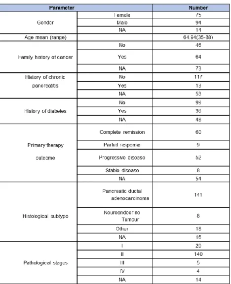
In Table 1 are represented the characteristics of the patients from the TCGA pancreatic cancer cohort considered for this study: approximately 55% of the patients are male against 45% female, patients present a mean age of 64.94 years ranging from 35 to 88 years. Many of the patients revealed a positive family history of cancer while few had previously history of chronic pancreatitis and diabetes, known risk factors for PCA development (Table 1).
Considering the patients’ outcome after primary therapy administration, 60 patients presented complete remission, 9 revealed a partial response to the treatment while 8 patients had stable disease and 53 patients presented progressive.
Regarding the histological classification of the tumour, 142 were classified as pancreatic ductal adenocarcinomas, the most common type of pancreatic cancer with 8 tumours being classified as neuroendocrine tumours (Table 1). The remaining samples presented discrepancies in the classification or were classified as distinct types of pancreatic cancer. However, the number of samples for each type did not reach a significant value to include the different subtypes separately.
Most of the patients in this cohort that were considered in our analysis are representative of early pathological stages of the disease with the majority (n=140) belonging to the stage II of disease. This will allow us to search for epigenetic biomarkers for diagnosis and prognosis of initial stages of the disease.
19 2.3.2 Methylation analysis
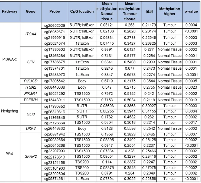
In order to evaluate the methylation status of the selected genes, level 3 methylation data derived from the Illumina Infinium HumanMethylation450K array was analysed for the pancreatic cancer cohort (normal tissue (n=8) and primary tumour (n=170)). The methylation score ranges from unmethylated (0) to completely methylated DNA (1). Differences between the two groups were evaluated by statistical analysis. We considered as differentially methylated the CpGs sites with a methylation delta beta absolute value (|Δβ|) equal or bigger than 0.2 and a p-value lower than 0.05.
The effect of DNA methylation in gene expression regulation is influenced by CpG location thus, characterization of the CpGs differentially methylated according to gene region was also performed. CpGs were annotated according to the manifest file for the Infinium
HumanMethylation450 version 1.2 CSV format (23/05/2013) available at
https://support.illumina.com/downloads.html.
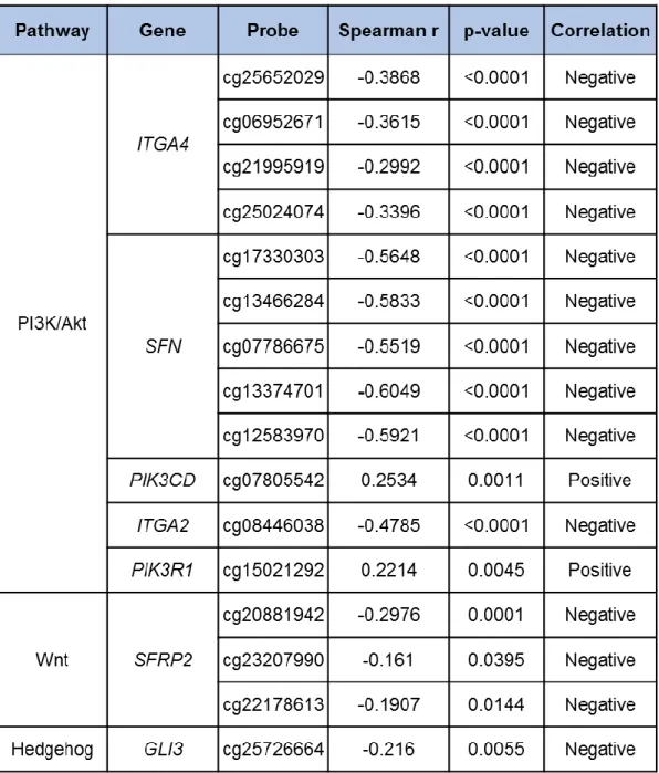
To investigate the relationship between DNA methylation and gene expression we have assessed the correlation between these two parameters. Gene expression data (level 3 data, RNA-seq Version 2 Illumina; gene-level transcription estimates, as in log2(x+1) transformed RSEM normalized count) and clinical data from the pancreatic cancer cohort was retrieved from the TCGA data portal and mapped to corresponding THOR methylation status using the unique TCGA identifier barcodes. Due to the lack of expression data from control samples in the TCGA cohort, the DNA methylation/gene expression correlation analysis was performed considering only data from pancreatic tumour tissue samples.
2.3.3 miRNA expression analysis
miRNA data for the TCGA pancreatic cancer cohort (level 3 data,
IlluminaHiSeq_miRNASeq, log2(RPM+1)) was analysed to investigate differences of miRNA expression levels between patients and controls.
Since the regulation of gene expression mediated by miRNA is an epigenetic mechanism not directly dependent on DNA methylation we have considered for this analysis all the genes that presented differential expression levels (n=35), independently of the methylation differences between the two groups. The following inclusion criteria were established: the miRNA targets a gene related with the pathways previously selected, presents a target score superior to 90 (the higher the score the higher the statistical confidence in the mRNA-miRNA complementarity result), and has functional annotation according to the miRDB database [75,76]. Due to the lack of information regarding miRNA expression levels in normal samples, we were only able to investigate the correlation between miRNA and gene expression with the data derived from malignant tissue.
Additionally, the absence of data from normal pancreatic tissue did not allow us to perform the receiver operator (ROC) curve to establish the cut-off value for the analysis of the impact of miRNA expression levels on patient’s outcome.
Alternatively, we assessed the survival time of the patients comparing the groups of patients with the highest and lowest levels of miRNA expression. Unfortunately, without the ROC curve results we were not able to assess the sensitivity and specificity values of the test.
20 2.3.4 Clinical data analysis
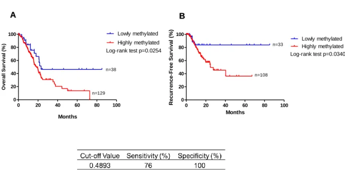
The clinical and pathological parameters evaluated were the following: history of chronic pancreatitis and diabetes, primary therapy outcome, histological classification, pathological stage and familiar history of cancer. Histological classification and pathological stage were used to investigate the impact of DNA methylation of the selected genes on disease prognosis by crossing the data from HumanMethylation450K array regarding DNA methylation status with the clinical information for each patient. Patient overall survival and recurrence were also analysed to determine the clinical significance of the observed epigenetic alterations and their potential as a biomarker and therapeutic target. Methylation cut-offs for each probe were established by performing ROC curve analysis considering an area under the ROC curve (AUC) with a minimum value of 0.8 to distinguish between healthy and malignant tissue. Only the cut-offs values that presented sensitivity and specificity values comparable or higher to the values of the CA19.9, the current biomarker for pancreatic cancer management, were selected for analysis. The patients with methylation values below and above the cut-off value are defined as lowly methylated and highly methylated, respectively.
2.3.5 Statistical analysis
The statistical analysis was performed using the unpaired t-test for data from a normal distribution. Otherwise the two-tailed Mann-Whitney test was applied, with a confidence interval of 95% for two groups comparisons. Correlation analysis was performed using the Spearman correlation coefficient. To analyse the differences between more than two groups we used one-way ANOVA, followed by Kruskal-Wallis test and Dunn's Multiple Comparison Test. Overall survival (OS) and recurrence-free survival (RFS) was determined by Kaplan-Meier Survival curves and comparisons were done with the log-rank test. All statistical analysis was performed using GraphPad Prism5.0.
21 2.4 Study Pipeline
Correlation between methylation/miRNAs levels and clinical features: history of chronic pancreatitis and diabetes, primary therapy outcome, histological classification, pathological stage, familiar history of cancer and patient overall survival and recurrence
Genes related with the PI3K/Akt, Wnt, Notch and Hedgehog pathways that presented differential expression between malignant and
healthy tissue
Fold-change ≥ 1.5 p-value < 0.05 Pancreatic Expression Database
35 genes Gene Selection Clinical Correlation mirDB database miRNA Analysis
miRNAs targeting the 35 genes of interest Target score>90 Have functional annotation 9 miRNAs Differential methylated CpG sites
|∆β|≥0.2 p-value < 0.05 The Cancer Genome Atlas
10 genes 28 probes 8 genes 17 probes Methylation Analysis p-value < 0.05 Correlation with gene expression Correlation with gene expression
22 CHAPTER 3 - RESULTS AND DISCUSSION
3.1 The TERT hypermethylated Oncologic Region (THOR) predicts recurrence and survival in pancreatic cancer
Faleiro, I, Apolónio, JD, Price, AJ, Andrade de Mello, R, Roberto, VP, Tabori, U, Castelo-Branco, P (2017). Future Oncology. In press.
23 3.1.1 Introduction
The defining feature of cancer cells replicative immortality is attained by telomere maintenance [77,78]. Ordinarily, telomere attrition occurs with each round of cell division due to the end replication problem. In non-malignant tissues, this phenomenon of telomere shortening imposes a ceiling on proliferative capacity in any given cell lineage. With the exception of the early developmental period, and select stem cell populations, telomeres are not reconstructed after shortening. In order to restore telomere length, 85-90% of cancers reactivate the telomerase reverse transcriptase enzyme while only about 10% are dependent on the alternative lengthening of telomeres (ALT) mechanism [79]. Telomerase reactivation in cancer is intimately related with expression of the telomerase reverse transcriptase (TERT) gene, which also serves as a prognostic factor [80,81].
Both genetic and epigenetic events have been found to deregulate TERT expression in cancer [79,82]. In this regard, point mutations and DNA methylation have gained special attention. The mechanisms underlying mutational TERT activation are unambiguous. Indeed, mutations (C228T and C250T) in the TERT core promoter [83] are known to generate binding motifs for E-twenty-six (ETS) transcription factors [83,84], and thereby upregulate TERT expression.
DNA methylation of the TERT promoter, on the other hand, has generated conflicting mechanistic hypotheses. This controversy is likely due to inconsistencies in the precise definition of the regions that constitute the epigenetically vulnerable portions of the TERT promoter [85]. The general consensus at the moment is that methylation of the TERT core promoter as a whole decreases TERT expression while methylation of a specific region upstream of the core promoter increases TERT expression [46,86–90]. The notion of epigenetic upregulation of an oncogene is counterintuitive given the traditional silencing effect exerted by hypermethylation. This region, termed TERT Hypermethylated Oncologic Region (THOR), was shown to be associated with TERT expression and disease progression in childhood brain tumours and biochemical recurrence in prostate cancer [46,91]. Studies from international groups have corroborated this correlation in brain tumours, hepatocellular, gastric and medullary thyroid carcinomas, to name a few [86–90]. To our knowledge, promoter mutations were not found in pancreatic adenocarcinoma until now [92].
To investigate if THOR could be a potential biomarker in pancreatic cancer we have analysed THOR methylation status and compared it with several clinical parameters.
3.1.2 THOR is hypermethylated in pancreatic cancer
To investigate if THOR is methylated in pancreatic cancer the CpG site targeted by the probe cg11625005 (chr5:1,295,737; position -575 in relation to the transcription start site (TSS)), localized within the THOR region (-591/-159), was analysed (Fig. 4). Pancreatic cancer revealed differential methylation at cg11625005 (a surrogate for the THOR region) with increased methylation levels in primary tumour tissue compared to normal tissue with mean values of 0.5579 and 0.3588, respectively (p<0.0001, Fig. 5A). THOR hypermethylation in pancreatic cancer is in concordance with previous results observed for other types of cancer such as prostate cancer [91].