Epigenetic Alterations in Fanconi Anaemia:
Role in Pathophysiology and Therapeutic
Potential
Hélio Belo
1,2, Gabriela Silva
1,2, Bruno A. Cardoso
1,2, Beatriz Porto
3, Jordi Minguillon
4,
José Barbot
5, Jorge Coutinho
5, Jose A. Casado
6, Manuela Benedito
7, Hema Saturnino
7,
Emília Costa
5, Juan A. Bueren
5, Jordi Surralles
4, Antonio Almeida
1,2*
1Unidade de Investigação em Patobiologia Molecular, Instituto Português de Oncologia de Lisboa
Francisco Gentil, E.P.E., Lisboa, Portugal,2CEDOC, Faculdade de Ciências Médicas, Universidade Nova
de Lisboa, Lisboa, Portugal,3Laboratório de Citogenética do Instituto de Ciências Biomédicas de Abel
Salazar, Porto, Portugal,4Center for Biomedical Network Research on Rare Diseases (CIBERER) and Department of Genetics and Microbiology, Universitat Autonoma de Barcelona, Barcelona, Spain,5Unidade de Hematologia Pediátrica do Centro Hospitalar do Porto, Porto, Portugal,6Hematopoiesis and Gene Therapy Division, CIEMAT, Madrid, Spain,7Serviço de hematologia do Centro Hospitalar e Universitário de Coimbra, Coimbra, Portugal
Abstract
Fanconi anaemia (FA) is an inherited disorder characterized by chromosomal instability.
The phenotype is variable, which raises the possibility that it may be affected by other
fac-tors, such as epigenetic modifications. These play an important role in oncogenesis and
may be pharmacologically manipulated. Our aim was to explore whether the epigenetic
pro-files in FA differ from non-FA individuals and whether these could be manipulated to alter
the disease phenotype. We compared expression of epigenetic genes and DNA
methyla-tion profile of tumour suppressor genes between FA and normal samples. FA samples
exhibited decreased expression levels of genes involved in epigenetic regulation and
hypo-methylation in the promoter regions of tumour suppressor genes. Treatment of FA cells with
histone deacetylase inhibitor Vorinostat increased the expression of
DNM3T
β
and reduced
the levels of
CIITA
and
HDAC9
,
PAK1
,
USP16
, all involved in different aspects of epigenetic
and immune regulation. Given the ability of Vorinostat to modulate epigenetic genes in FA
patients, we investigated its functional effects on the FA phenotype. This was assessed by
incubating FA cells with Vorinostat and quantifying chromosomal breaks induced by DNA
cross-linking agents. Treatment of FA cells with Vorinostat resulted in a significant reduction
of aberrant cells (81% on average). Our results suggest that epigenetic mechanisms may
play a role in oncogenesis in FA. Epigenetic agents may be helpful in improving the
pheno-type of FA patients, potentially reducing tumour incidence in this population.
a11111
OPEN ACCESS
Citation:Belo H, Silva G, Cardoso BA, Porto B, Minguillon J, Barbot J, et al. (2015) Epigenetic Alterations in Fanconi Anaemia: Role in Pathophysiology and Therapeutic Potential. PLoS ONE 10(10): e0139740. doi:10.1371/journal. pone.0139740
Editor:Hiromu Suzuki, Sapporo Medical University, JAPAN
Received:May 6, 2015
Accepted:September 15, 2015
Published:October 14, 2015
Copyright:© 2015 Belo et al. This is an open access article distributed under the terms of theCreative Commons Attribution License, which permits unrestricted use, distribution, and reproduction in any medium, provided the original author and source are credited.
Data Availability Statement:All relevant data are within the paper and its Supporting Information files.
Funding:This project was funded by a project grant 2011-2012 from Associação Portuguesa Contra a Leucemia and from a project grant from Liga Portuguesa contra o Cancro/Fundação Terry Fox 2013-2014. The funders had no role in study design, data collection and analysis, decision to publish, or preparation of the manuscript.
Introduction
Fanconi anaemia (FA) is an inherited disorder characterized by developmental abnormalities,
bone marrow failure, leukemic progression and solid tumours, especially head and neck. At the
cellular level, FA is characterized by impaired DNA repair and increased chromosomal
fragil-ity, a feature used in its diagnosis [
1
]. Mutations in 17 genes have been described, all with
simi-lar phenotypes, suggesting that all FA proteins function in common DNA repair pathway [
2
].
However, the severity of the disease varies even amongst patients from the same family and
with the same mutation [
3
,
4
].
It is therefore plausible that, alongside genetic mutations in FA genes, other factors may
contribute to disease severity and increase the risk of neoplastic transformation.
Epigenetic modifications are important mechanisms by which cells regulate gene
expres-sion. DNA methylation and posttranslational modifications of histones affect chromatin
struc-ture, modulating gene expression and changes in cellular physiology and behavior [
5
]. There is
ample evidence implicating epigenetic changes in the pathophysiology of MDS, AML and solid
tumours [
6
–
11
]. In these malignancies, abnormal DNA methylation and histone deacetylation
have been shown to silence tumour suppressor genes, and change normal expression of
onco-genes, tumour suppressor genes and genes associated with several key cellular functions like
DNA damage repair, cell cycle regulation, adhesion, motility, apoptosis and also signaling
pathways [
6
,
8
,
12
]. For example, in ovarian and cervical cancers, hypermethylation of FANCF
leads to its inactivation and to the disruption of the FA-BRCA pathway [
13
].
These epigenetic changes may be pharmacologically manipulated with DNA
hypomethylat-ing agents and histone deacetylase inhibitors (HDACi) [
12
,
14
–
16
].
Vorinostat is an HDACi approved for the treatment of cutaneous T-cell-lymphoma.
Vori-nostat promotes protein acetylation, leading to the activation of genes involved in the control
of cell cycle progression, differentiation and apoptosis. It also affects the expression of
epige-netic regulator genes, contributing to their normal expression. In clinical trials it has shown
promising clinical activity against hematological and solid tumours [
17
].
The aim of this study was to investigate whether epigenetic mechanisms could play a role in
the pathophysiology of oncogenesis in FA and explore the potential of HDACi to improve the
phenotype in FA.
Materials and Methods
Blood samples
Anonymized blood samples were obtained from twelve confirmed Fanconi anemia (FA)
patients following written informed consent. Blood from healthy blood donors was used for
normal controls. The study was approved by the Ethical Committee of Instituto Português de
Oncologia de Lisboa, Francisco Gentil, EPE and all samples treated according to the
Declara-tion of Helsinki.
In vitro
cell cultures
Peripheral blood mononuclear cells (PBMC) from blood samples were separated with Ficcol
(Sigma) and cultured in RPMI
—
1640 medium supplemented with 10% fetal bovine serum
(GIBCO), 2mM L-glutamine and 100
μ
g/ml penicillin/streptomycin (all from Gibco).
Treat-ments were performed with 1
μ
M Vorinostat (Selleck Chemicals) or vehicle for the indicated
Gene Expression analysis by real time PCR(qPCR)
Total RNA was isolated from cells using the RNeasy Mini Kit (Qiagen), treated with DNase
(Qiagen) and reverse-transcribed into cDNA using RT
2First Strand Kit (Qiagen) according to
the manufacturer
’
s protocol. qPCR was performed on Roche LightCycler 480 with 84 gene
spe-cific primers for Human Epigenetic Chromatin Modification Enzymes (PAHS-085G,
SABios-ciences, Qiagen). Data was analyzed according to manufacturer
’
s instructions.
In silico
analysis
Bioinformatic analysis of gene expression in FA was performed using the expression array data
published in Vanderwerf
I
[
18
].
Analysis of DNA Methylation
Genomic DNA was extracted from primary cells (1x10
7/ml) using Citogene kit (Citomed),
treated with RNase (Citomed) and digested using EpiTect Methyl DNA Restriction Kit
(Qia-gen) according to the manufacturer
’
s protocol. qPCR was performed on Roche LightCycler
480 with 94 gene specific primers for Human Tumour Suppressor Genes (EAHS-3550ZG,
Qia-gen). Samples from 2 FA patients were compared with 2 Healthy donors. Data analysis was
performed according to manufacturer
’
s instructions.
Chromosomal instability assay
Whole blood (0,5ml) was cultured in RPMI
–
1640 (supplemented as above and cultures were
stimulated with 5
μ
g/ml of phytohemaglutinin (GIBCO) during 24h. Thereafter, the cultures
were treated with 1
μ
M Vorinostat or vehicle for an additional 24h at which point 0.05
μ
g/ml of
1,2:3,4-diepoxybutane (DEB, Sigma) or vehicle was added to the cultures for 48h. After 96h of
culture, cells were treated with 2
μ
g/ml of colcemid (GIBCO) for 1h, spread on slides, subjected
to hypotonic lysis with 75mM KCl [
19
,
20
] and fixed in solution of 3:1 volumes of methanol:
acetic acid [
21
].
Slides were stained with 0,5M Leishamn (Sigma) in phosphate buffer, pH 6.8. Fifty
meta-phases per sample were analysed for chromosome aberrations including chromosome and
chromatid breaks, acentric fragments and chromosome and chromatid-type exchange. Gaps
were excluded and rearrangements were scored as two breaks for the calculation of percentage
of cells with aberrations.
Cellular viability assays.
Viability was assessed by flow cytometry with Annexin-V- FITC
(Biolegend) and Propidium Iodide (PI) (Sigma-Aldrich).
Statistical analysis.
Populations were compared using unpaired 2-tailed Student
’
s t test or
One-way ANOVA, when appropriate (a p
<
0.05 was considered significant) using the
Graph-Pad Prism version 5.00 for Windows (GraphGraph-Pad Software).
Results
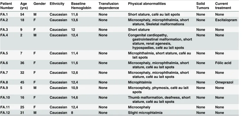
The clinical characteristics of the patients whose samples were used in these experiments are
detailed in
Table 1
.
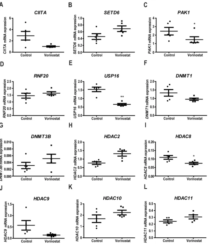
Fanconi anemia patients exhibit different expression of epigenetic genes
compared to healthy donors
in PBMC from 12 FA and compared these to PBMC from 14 healthy donors. We found that 13
genes were differentially expressed in FA as compared to normal cells (
Fig 1
). These included
genes encoding DNA methyltransferases (DNMT1, DNM3T
β
) and genes encoding histone
modifying enzymes: acetylases (CIITA), phosphorylases (PAK1), ubiquitinases (RNF20),
dea-cetylases (HDAC2, HDCA8, HDAC9, HDAC10, HDAC11) and also methyltransferases
(SETD6). This differential expression between normal and FA individuals was confirmed
bioinformatically from data from Vanderwerf
et
.
al
[
18
] for
PAK1
,
USP16
,
DNMT1
,
DNMT3B
,
HDAC2
,
HDAC9
,
CIITA
,
HDAC10
and
HDAC11
(
S1 Fig
).
FA cells exhibit DNA hypomethylation of tumour suppressor genes
In order to ascertain whether the findings from the gene expression assays translated into a
dif-ference in epigenetic patterns in FA compared to normal subjects, we studied the pattern of
DNA methylation in FA PBMC. For this we used the Human Tumour Suppressor gene PCR
Array to assess promoter DNA methylation of 94 tumour suppressor genes in 2 PBMC of FA
patients and 2 healthy donors. This revealed a global aberrant hypomethylation of tumour
sup-pressor genes in FA cells as compared to healthy donors. Six of 94 genes were differentially
methylated in FA relative to healthy donors. These included genes whose function are related
to apoptosis (
CADM1
,
SFRP1
), cell cycle (
ING1
), motility (
CDH13
), oxidative stress (
LOX
) and
angiogenesis/transcription (
CDX2
) (
Fig 2
).
Vorinostat modifies the expression of epigenetic genes
Having observed differences in epigenetic regulator gene expression and in epigenetic patterns
between FA and normal subjects, we tested whether these could be normalized using epigenetic
agents. We chose Vorinostat to test this effect as it is a wide HDACi which able to modulate
epigenetic and gene expression patterns[
8
] and with proven clinical efficacy. We tested its
Table 1. Summary of clinical data in FA patients.
Patient Number
Age (yrs)
Gender Ethnicity Baseline
Hemoglobin
Transfusion dependence
Physical abnormalities Solid
Tumors
Current treatment
FA.1 54 M Caucasian 11,6 None Short stature, café au lait spots None None
FA.2 18 F Caucasian 13,6 None Microcephaly, microphthalmia, short
stature, Skeletal malformations
None Escitalopram
FA.3 9 F Caucasian 12 None Short stature None None
FA.4 2 M Caucasian 12,4 None Congenital cardiopathy,
gastrointestinal malformation, short stature, renal agenesis,
hypospadias, café au lait spots
None None
FA.5 7 F Caucasian 11,4 None Microphthalmia, short stature, café au
lait spots
None None
FA.6 36 F Caucasian 11,6 None Microcephaly, microphthalmia, short
stature, café au lait spots
None Fólic acid
FA.7 32 F Caucasian 12,6 None Microcephaly, microphthalmia, short
stature, café au lait spots
None None
FA.8 45 F Caucasian 12,4 None Microphtalmia None Omeprazol
FA.9 5 M Caucasian 10,9 None Microcephaly, phymosis, café au lait
spots
None None
FA.10 16 F Caucasian 14,6 None Thumb malformation, deafness, short
stature, café au lait spots
None None
FA.11 25 F Caucasian 12,4 None Microcephaly None None
FA.12 31 M Caucasian 8 None Slight microphtalmia None None
effect on the expression of epigenetic chromatin modification genes which expression was
altered in PBMC from FA patients (
Fig 1
). Following treatment of FA PBMC with Vorinostat
for 8h and 16h (
Fig 3
) there was an increase in the expression levels of the
DNMT3
β
gene.
Interestingly, Vorinostat treatment reduced the expression of
CIITA
and
HDAC9
, involved in
Fig 1. FA patients have decreased expression of epigenetic chromatin modification enzymes.RNA was isolated from PBMC from 12 FA patients and 14 healthy controls and the expression of epigenetic regulator genes was quantified using the Human Epigenetic Chromatin Enzymes PCR array
(SABiosciences). Each panel (A-L) represents the expression of the indicated genes in FA patients and control samples as described in the materials and methods section.(*0.05>p;**0.01>p;***0.001>p). In
the immune response,
PAK1
, regulator of the
MAPK
signaling pathway, and
USP16
, involved
in regulating the activity of histone H2A. The expression levels of
HDAC10
,
HDAC11
,
HDAC2
,
SETD6
,
RNF20
and
DNMT1
genes were not significantly altered by Vorinostat.
Vorinostat reduces chromosomal breaks in FA cells
Given the capacity of Vorinostat to modulate the expression of epigenetic regulator genes in
FA samples, we investigated its effect on the
in vitro
phenotype of FA cells. This functional
effect of Vorinostat in FA was tested by assessing its effect on chromosome breaks induced by
DEB. The percentage of aberrant cells induced by DEB was assessed on metaphases obtained
from peripheral blood lymphocytes of patients with FA. Vorinostat reduced the percentage of
aberrant cells (81% ± 21%, p = 0,06) in 6 patients with Fanconi anemia (
Table 2
,
Fig 4A, 4B,
4C, 4D and 4E
). There was no reduction in the number of spontaneous chromosomal breaks in
FA cells following treatment with Vorinostat (
Table 2
).
In this experimental system Vorinostat did not significantly reduce the viability of cells (
S2
Fig
).
Discussion
Fanconi anaemia is an inherited disease caused by defective DNA repair. Whereas the skeletal,
genitourinary and morphological abnormalities are rarely life-threatening, the development of
bone marrow failure, leukemia and solid cancers are frequently lethal. The aggressiveness of
these complications are compounded by the low tolerance FA patients have to chemotherapy
and radiotherapy [
6
]. Therefore, a treatment option which could improve the DNA repair
Fig 2. DNA methylation pattern on tumour suppressor genes in peripheral blood mononuclear cells of FA patients.PBMC from FA patients and healthy donor were isolated as in A, DNA was isolated and methylation profile was identified in 2 FA samples and 2 healthy donors samples using EpiTect Methyl II Complete PCR array (SABiosciences). Samples CTR1 and CTR2 represent control and FA1 and FA2 represent FA patients. Each row represents a tumour-supressor genes and each column represent a single DNA sample. The methylation degree are represented by the level of intensity of the square, red representing greater than 10% promoter hypermethylation, and green representing less than 10% promoter methylation (unmethylated) alleles for the tumour-suppressor gene.
defect and reduce the incidence of mutations and secondary malignancies would be highly
desirable.
Epigenetic modifications consist in the addition or removal of small molecules onto DNA
or DNA-associated proteins, i.e. histones, and enable eukaryotic cells to alter their gene
expres-sion without altering the DNA sequence [
8
,
22
]. The molecules most commonly implicated in
Fig 3. Effect of vorinostat on epigenetic patterns on chromatin epigenetic modifications enzymes differentially expressed in FA mononuclear cells.PBMC from FA patients in culture were treated with 1μM vorinostat as indicated or vehicle (control) for 8h and gene expression quantified by qPCR.
epigenetic regulation are methyl and acetyl groups and DNA methylation status is closely
related and plays a role in regulating to histone acetylation [
7
,
10
,
22
]. In addition to the
regula-tion of gene expression, these modificaregula-tions, in particular DNA methylaregula-tion, play an important
role in maintaining DNA and chromatin stability [
17
].
Table 2. Number of breaks per cell in cultured lymphocytes from FA patients.
Mean number of breaks per cell
Patients Spontaneous breaks (n) Vor 1μM % reduction DEB 0.05μM (n) DEB 0.05μM + Vor 1μM (n) % reduction
FA1 0.28 (50) 0.32 (50) 0.00 2.04 (50) 0.24 (50) 88.24
FA2 0.08 (50) 0.14 (50) 0.00 0.88 (50) 0.14 (50) 84.09
FA3 0.18 (50) 0.18 (50) 0.00 1.38 (50) 0.7 (50) 49.28
FA4 0.8 (50) 0.32 (50) 60.00 7.96 (50) 0.24 (50) 96.98
FA5 0.44 (50) 0.34 (50) 22.72 1.58 (50) 0.88 (50) 44.3
FA6 0.12 (50) 0.25 (50) 0.00 0.74 (50) 0.16 (50) 79.38
Mean±SD 0.23±0.269 0.28±0.083 1.48±2.750 0.24±0.3151
The effect of the Vorinostat was calculated by the percentage of reduction of the number of breaks per cell in the Vorinostat treatments relatively to Spontaneous breaks and DEB.
doi:10.1371/journal.pone.0139740.t002
Fig 4. Effect of vorinostat on DEB-induced chromosome fragility of FA lymphocytes.(A) Lymphocytes metaphase without treatment. (B) Lymphocyte metaphase treated with DEB. (C) Lymphocyte metaphase treated with DEB after Vorinostat treatment. The red arrows indicate aberrant chromosomes characteristic of FA cells. (D) Number of breaks per cell after treatment with Vorinostat. (E) Number of breaks per cell after treatment with Vorinostat on DEB-induced breaks. The p values are indicated.
Aberrant epigenetic patterns have been implicated in the pathophysiology of a variety of
haematological and solid tumours [
23
]. These have been successfully manipulated
pharmaco-logically with promising therapeutic results [
8
,
24
,
25
].
Our aim was to investigate whether the epigenetic machinery in FA differs from that of
unaffected individuals and whether its manipulation could somehow affect the FA phenotype.
In the present study, we show that FA cells present decreased levels of several genes involved
in epigenetic regulation as compared to cells from healthy subjects (
Fig 1
) Gene expression
studies revealed that DNMT1 and DNMT3
β
expression, involved in DNA methylation, were
significantly reduced in FA patients both in PBMCs and BM. DNMT1 is responsible for
copy-ing DNA methylation patterns established durcopy-ing embryonic development and their
subse-quent maintenance. DNMT3
β
is a tumour-suppressor gene with a critical role in DNA
methylation playing a major role in the establishment and maintenance of genomic
methyla-tion patterns. Reduced activity of DNMT1 and DNMT3
β
may lead to DNA hypomethylation,
inducing genomic instability and disruption of proto-oncogenes and is directly associated with
tumour formation[
26
–
28
]. Abnormal expression of DNMT1 and DNMT3
β
has been reported
in several tumours, including lung, liver, breast, ovarian, colorectal, meningiomas and
lympho-mas [
29
,
30
].Reduced expression of CIITA was observed in PBMCs but not in BM. This gene
regulates the expression of MHCII gene by recruiting the transcriptional machinery of basic
proteins, acetyltransferases, histone deacetylases (HDAC), and other proteins involved in
chro-matin remodeling. CIITA expression is decreased in several types of hematological and solid
tumours and is associated with decreased tumour immune recognition [
31
].
The expression of SETD6, a gene that may also impair anti-tumour immune response by
dysregulation of the NF-kB pathway [
32
], was also reduced in FA PMBCs but remain roughly
the same in BM.
RNF20, which is essential for the regulation of normal levels of p53 [
33
], is also
underex-pressed in FA. Its reduced expression causes chromosome instability and has been described in
several types of tumours [
34
–
48
]. The decreased expression of RNF20 enhances the
transcrip-tional effects of EGF, leading to an increase in transformation, migration and metastasis of
can-cer cells and tumourigenesis [
5
,
23
,
49
].
We also found reduced expression of histone deacetylases HDAC 2, 8, 9, 10 and 11 in FA
PMBCs and confirmed HDAC2 and HDAC9 reduced expression in FA BM samples. HDACs
regulate chromatin remodeling and regulate many genes involved both in the initiation and
progression of a variety of cancers.
Despite some discrepancy between our data, obtained from blood samples and the
bioinfor-matics analysis of gene expression data, obtained from bone marrow samples, both concur that
there is altered expression of epigenetic regulating genes in FA. A larger number of samples
would be required to reach firm conclusions.
The reduced expression of these genes in FA, suggests that these patients have altered
epige-netic regulation, which may be involved in the neoplastic complications of this disease. This
hypothesis is corroborated by our finding showing increased DNA hypomethylation at tumor
suppressor gene loci in FA as compared to normal cells (
Fig 2
). Liu
et al
have described
aber-rant expression of tumor suppressor and tumor-related genes in FA, corroborating our data
[
50
]. It is possible that this hypomethylation may increase genomic instability and have an
additive effect on the DNA repair defect of FA, increasing the oncogenic potential in FA
tissues.
In fact, treatment of FA cells with Vorinostat induced expression of the DNMT3
β
, involved
in the maintenance of physiological DNA methylation (
Fig 3
). The suppression of expression
of CIITA and HDAC9 are consistent with a reduction in inflammatory response, which may
play a role in oncogenesis in the context of DNA repair defects.
Of particular relevance was our finding that Vorinostat treatment reduced chromosomal
breaks in FA cells (
Fig 4
). This may have been due to DNA stabilization through induction of
DNMT3
β
or mediated by another mechanism yet to be investigated. Whichever mechanism at
play, our findings are very suggestive that Vorinostat exerts a protective effect on the
chromo-somal breaks induced by cross-linking agents. This effect may have an important clinical
coun-terpart as it may translate into greater tolerability to chemotherapeutic agents in FA patients.
These results are the first report of a potential improvement in FA phenotype with an
epige-netic agent. They warrant further pre-clinical testing in animal models with a view to initiating
clinical trials in FA if the results remain promising.
Supporting Information
S1 Fig. FA patients have altered expression of epigenetic chromatin modification enzymes.
Each panel (
A-J
) represents the expression of selected genes of interest in FA and control
sam-ples as described in materials and methods section. With the exception of DNMT3B, there is
significant difference in the expression of these genes in FA compared to normal samples (
0.05
>
p;
0.01
>
p;
0.001
>
p).
(TIF)
S2 Fig. Vorinostat has no effect on the viability of DEB-treated cells.
Viability PBMC of FA
patients as determined by Annexin V/PI staining following treatment with Vorinostat and
DEB in cell culture medium as used to test for chromosomal fragility.
(TIF)
Acknowledgments
The authors would like to thank volunteers that provided blood samples.
Author Contributions
Conceived and designed the experiments: HB GS BC AA JS. Performed the experiments: HB
GS BC JM. Analyzed the data: HB GS BC AA JM. Contributed reagents/materials/analysis
tools: BP JB JC JAC MB HS EC JB JS. Wrote the paper: HB GS BC JS AA.
References
1. Auerbach AD. Fanconi anemia and its diagnosis. Mutat Res. 2009; 668(1–2):4–10. Epub 2009/07/23. doi:10.1016/j.mrfmmm.2009.01.013S0027-5107(09)00053-0 [pii]. PMID:19622403; PubMed Central PMCID: PMC2742943.
2. Sawyer SL, Tian L, Kahkonen M, Schwartzentruber J, Kircher M, Majewski J, et al. Biallelic mutations in BRCA1 cause a new Fanconi anemia subtype. Cancer Discov. 2015; 5(2):135–42. Epub 2014/12/ 05. doi:10.1158/2159-8290.CD-14-11562159-8290.CD-14-1156 [pii]. PMID:25472942; PubMed Cen-tral PMCID: PMC4320660.
3. Soulier J. Fanconi anemia. Hematology Am Soc Hematol Educ Program. 2011; 2011:492–7. Epub 2011/12/14. doi:10.1182/asheducation-2011.1.4922011/1/492 [pii]. PMID:22160080.
4. Geiselhart A, Lier A, Walter D, Milsom MD. Disrupted Signaling through the Fanconi Anemia Pathway Leads to Dysfunctional Hematopoietic Stem Cell Biology: Underlying Mechanisms and Potential Thera-peutic Strategies. Anemia. 2012; 2012:265790. Epub 2012/06/08. doi:10.1155/2012/265790PMID:
5. Feinberg AP, Tycko B. The history of cancer epigenetics. Nat Rev Cancer. 2004; 4(2):143–53. Epub 2004/01/21. doi:10.1038/nrc1279nrc1279 [pii]. PMID:14732866.
6. Ganesan A, Nolan L, Crabb SJ, Packham G. Epigenetic therapy: histone acetylation, DNA methylation and anti-cancer drug discovery. Curr Cancer Drug Targets. 2009; 9(8):963–81. Epub 2009/12/23. PMID:20025605.
7. Issa JP. Epigenetic changes in the myelodysplastic syndrome. Hematol Oncol Clin North Am. 2010; 24 (2):317–30. Epub 2010/04/03. doi:10.1016/j.hoc.2010.02.007S0889-8588(10)00028-6 [pii]. PMID:
20359628; PubMed Central PMCID: PMC2848959.
8. Jones PA, Baylin SB. The epigenomics of cancer. Cell. 2007; 128(4):683–92. Epub 2007/02/27. S0092-8674(07)00127-4 [pii] doi:10.1016/j.cell.2007.01.029PMID:17320506; PubMed Central PMCID: PMC3894624.
9. Plass C, Oakes C, Blum W, Marcucci G. Epigenetics in acute myeloid leukemia. Semin Oncol. 2008; 35(4):378–87. Epub 2008/08/12. doi:10.1053/j.seminoncol.2008.04.008S0093-7754(08)00118-8 [pii]. PMID:18692688; PubMed Central PMCID: PMC3463865.
10. Seligson DB, Horvath S, Shi T, Yu H, Tze S, Grunstein M, et al. Global histone modification patterns predict risk of prostate cancer recurrence. Nature. 2005; 435(7046):1262–6. Epub 2005/07/01. nature03672 [pii] doi:10.1038/nature03672PMID:15988529.
11. Stintzing S, Kemmerling R, Kiesslich T, Alinger B, Ocker M, Neureiter D. Myelodysplastic syndrome and histone deacetylase inhibitors: "to be or not to be acetylated"? J Biomed Biotechnol. 2011; 2011:214143. Epub 2011/06/02. doi:10.1155/2011/214143PMID:21629744; PubMed Central PMCID: PMC3100562.
12. Jain N, Rossi A, Garcia-Manero G. Epigenetic therapy of leukemia: An update. Int J Biochem Cell Biol. 2009; 41(1):72–80. Epub 2008/10/25. doi:10.1016/j.biocel.2008.10.006S1357-2725(08)00416-0 [pii]. PMID:18948224; PubMed Central PMCID: PMC3833715.
13. Taniguchi T, Tischkowitz M, Ameziane N, Hodgson SV, Mathew CG, Joenje H, et al. Disruption of the Fanconi anemia-BRCA pathway in cisplatin-sensitive ovarian tumors. Nat Med. 2003; 9(5):568–74. Epub 2003/04/15. doi:10.1038/nm852nm852 [pii]. PMID:12692539.
14. Griffiths EA, Gore SD. DNA methyltransferase and histone deacetylase inhibitors in the treatment of myelodysplastic syndromes. Semin Hematol. 2008; 45(1):23–30. Epub 2008/01/09. doi:10.1053/j.
seminhematol.2007.11.007S0037-1963(07)00164-3 [pii]. PMID:18179966; PubMed Central PMCID: PMC2234265.
15. Narayan G, Arias-Pulido H, Nandula SV, Basso K, Sugirtharaj DD, Vargas H, et al. Promoter hyper-methylation of FANCF: disruption of Fanconi Anemia-BRCA pathway in cervical cancer. Cancer Res. 2004; 64(9):2994–7. Epub 2004/05/06. PMID:15126331.
16. Wang Z, Li M, Lu S, Zhang Y, Wang H. Promoter hypermethylation of FANCF plays an important role in the occurrence of ovarian cancer through disrupting Fanconi anemia-BRCA pathway. Cancer Biol Ther. 2006; 5(3):256–60. Epub 2006/01/19. 2380 [pii]. PMID:16418574.
17. Siegel D, Hussein M, Belani C, Robert F, Galanis E, Richon VM, et al. Vorinostat in solid and hemato-logic malignancies. J Hematol Oncol. 2009; 2:31. Epub 2009/07/29. doi:10.1186/1756-8722-2-31
1756-8722-2-31 [pii]. PMID:19635146; PubMed Central PMCID: PMC2731787.
18. Vanderwerf SM, Svahn J, Olson S, Rathbun RK, Harrington C, Yates J, et al. TLR8-dependent TNF-(alpha) overexpression in Fanconi anemia group C cells. Blood. 2009; 114(26):5290–8. Epub 2009/10/ 24. doi:10.1182/blood-2009-05-222414blood-2009-05-222414 [pii]. PMID:19850743; PubMed Cen-tral PMCID: PMC2796134.
19. Silva G, Cardoso BA, Belo H, Almeida AM. Vorinostat induces apoptosis and differentiation in myeloid malignancies: genetic and molecular mechanisms. PLoS One. 2013; 8(1):e53766. Epub 2013/01/16. doi:10.1371/journal.pone.0053766PONE-D-12-19513 [pii]. PMID:23320102; PubMed Central PMCID: PMC3540071.
20. Richon VM, Garcia-Vargas J, Hardwick JS. Development of vorinostat: current applications and future perspectives for cancer therapy. Cancer Lett. 2009; 280(2):201–10. Epub 2009/02/03. doi:10.1016/j.
canlet.2009.01.002S0304-3835(09)00002-0 [pii]. PMID:19181442.
21. Kennedy RD, D'Andrea AD. DNA repair pathways in clinical practice: lessons from pediatric cancer susceptibility syndromes. J Clin Oncol. 2006; 24(23):3799–808. Epub 2006/08/10. 24/23/3799 [pii] doi:
10.1200/JCO.2005.05.4171PMID:16896009.
22. Andreoli F, Barbosa AJ, Parenti MD, Del Rio A. Modulation of epigenetic targets for anticancer therapy: clinicopathological relevance, structural data and drug discovery perspectives. Curr Pharm Des. 2013; 19(4):578–613. Epub 2012/09/29. CPD-EPUB-20120926-1 [pii]. PMID:23016851; PubMed Central PMCID: PMC3529403.
2006; 66(17):8462–9468. Epub 2006/09/05. 66/17/8462 [pii] doi:10.1158/0008-5472.CAN-06-0293 PMID:16951157.
24. Nosho K, Shima K, Irahara N, Kure S, Baba Y, Kirkner GJ, et al. DNMT3B expression might contribute to CpG island methylator phenotype in colorectal cancer. Clin Cancer Res. 2009; 15(11):3663–71. Epub 2009/05/28. doi:10.1158/1078-0432.CCR-08-23831078-0432.CCR-08-2383 [pii]. PMID:
19470733; PubMed Central PMCID: PMC2866637.
25. Bai X, Song Z, Fu Y, Yu Z, Zhao L, Zhao H, et al. Clinicopathological significance and prognostic value of DNA methyltransferase 1, 3a, and 3b expressions in sporadic epithelial ovarian cancer. PLoS One. 2012; 7(6):e40024. Epub 2012/07/07. doi:10.1371/journal.pone.0040024PONE-D-12-04122 [pii]. PMID:22768205; PubMed Central PMCID: PMC3386927.
26. Morimoto Y, Toyota M, Satoh A, Murai M, Mita H, Suzuki H, et al. Inactivation of class II transactivator by DNA methylation and histone deacetylation associated with absence of HLA-DR induction by inter-feron-gamma in haematopoietic tumour cells. Br J Cancer. 2004; 90(4):844–52. Epub 2004/02/19. doi:
10.1038/sj.bjc.66016026601602 [pii]. PMID:14970863; PubMed Central PMCID: PMC2410180. 27. Satoh A, Toyota M, Ikeda H, Morimoto Y, Akino K, Mita H, et al. Epigenetic inactivation of class II
trans-activator (CIITA) is associated with the absence of interferon-gamma-induced HLA-DR expression in colorectal and gastric cancer cells. Oncogene. 2004; 23(55):8876–86. Epub 2004/10/07. 1208144 [pii] doi:10.1038/sj.onc.1208144PMID:15467734.
28. Truax AD, Thakkar M, Greer SF. Dysregulated recruitment of the histone methyltransferase EZH2 to the class II transactivator (CIITA) promoter IV in breast cancer cells. PLoS One. 2012; 7(4):e36013. Epub 2012/05/09. doi:10.1371/journal.pone.0036013PONE-D-12-00707 [pii]. PMID:22563434; PubMed Central PMCID: PMC3338556.
29. Levy D, Kuo AJ, Chang Y, Schaefer U, Kitson C, Cheung P, et al. Lysine methylation of the NF-kappaB subunit RelA by SETD6 couples activity of the histone methyltransferase GLP at chromatin to tonic repression of NF-kappaB signaling. Nat Immunol. 2011; 12(1):29–36. Epub 2010/12/07. doi:10.1038/
ni.1968ni.1968 [pii]. PMID:21131967; PubMed Central PMCID: PMC3074206.
30. Karin M, Cao Y, Greten FR, Li ZW. NF-kappaB in cancer: from innocent bystander to major culprit. Nat Rev Cancer. 2002; 2(4):301–10. Epub 2002/05/11. doi:10.1038/nrc780PMID:12001991.
31. Milde-Langosch K, Janke S, Wagner I, Schroder C, Streichert T, Bamberger AM, et al. Role of Fra-2 in breast cancer: influence on tumor cell invasion and motility. Breast Cancer Res Treat. 2008; 107 (3):337–47. Epub 2007/03/30. doi:10.1007/s10549-007-9559-yPMID:17393299.
32. Chernikova SB, Razorenova OV, Higgins JP, Sishc BJ, Nicolau M, Dorth JA, et al. Deficiency in mam-malian histone H2B ubiquitin ligase Bre1 (Rnf20/Rnf40) leads to replication stress and chromosomal instability. Cancer Res. 2012; 72(8):2111–9. Epub 2012/02/23. doi:10.1158/0008-5472.CAN-11-2209 0008-5472.CAN-11-2209 [pii]. PMID:22354749; PubMed Central PMCID: PMC3328627.
33. Shema E, Tirosh I, Aylon Y, Huang J, Ye C, Moskovits N, et al. The histone H2B-specific ubiquitin ligase RNF20/hBRE1 acts as a putative tumor suppressor through selective regulation of gene expres-sion. Genes Dev. 2008; 22(19):2664–76. Epub 2008/10/04. doi:10.1101/gad.170300822/19/2664 [pii]. PMID:18832071; PubMed Central PMCID: PMC2559905.
34. Glozak MA, Seto E. Histone deacetylases and cancer. Oncogene. 2007; 26(37):5420–32. Epub 2007/ 08/19. 1210610 [pii] doi:10.1038/sj.onc.1210610PMID:17694083.
35. Gallinari P, Di Marco S, Jones P, Pallaoro M, Steinkuhler C. HDACs, histone deacetylation and gene transcription: from molecular biology to cancer therapeutics. Cell Res. 2007; 17(3):195–211. Epub 2007/02/28. 7310149 [pii] doi:10.1038/sj.cr.7310149PMID:17325692.
36. Jung KH, Noh JH, Kim JK, Eun JW, Bae HJ, Xie HJ, et al. HDAC2 overexpression confers oncogenic potential to human lung cancer cells by deregulating expression of apoptosis and cell cycle proteins. J Cell Biochem. 2012; 113(6):2167–77. Epub 2012/04/12. doi:10.1002/jcb.24090PMID:22492270. 37. Noh JH, Jung KH, Kim JK, Eun JW, Bae HJ, Xie HJ, et al. Aberrant regulation of HDAC2 mediates
pro-liferation of hepatocellular carcinoma cells by deregulating expression of G1/S cell cycle proteins. PLoS One. 2011; 6(11):e28103. Epub 2011/12/02. doi:10.1371/journal.pone.0028103 PONE-D-11-13259 [pii]. PMID:22132221; PubMed Central PMCID: PMC3223227.
38. Waltregny D, Glenisson W, Tran SL, North BJ, Verdin E, Colige A, et al. Histone deacetylase HDAC8 associates with smooth muscle alpha-actin and is essential for smooth muscle cell contractility. FASEB J. 2005; 19(8):966–8. Epub 2005/03/18. 04-2303fje [pii] doi:10.1096/fj.04-2303fjePMID:15772115. 39. Oehme I, Deubzer HE, Wegener D, Pickert D, Linke JP, Hero B, et al. Histone deacetylase 8 in
neuro-blastoma tumorigenesis. Clin Cancer Res. 2009; 15(1):91–9. Epub 2009/01/02. doi:
10.1158/1078-0432.CCR-08-068415/1/91 [pii]. PMID:19118036.
08/14. doi:10.1038/nature11316nature11316 [pii]. PMID:22885700; PubMed Central PMCID: PMC3443318.
41. Harakalova M, van den Boogaard MJ, Sinke R, van Lieshout S, van Tuil MC, Duran K, et al. X-exome sequencing identifies a HDAC8 variant in a large pedigree with X-linked intellectual disability, truncal obesity, gynaecomastia, hypogonadism and unusual face. J Med Genet. 2012; 49(8):539–43. Epub 2012/08/15. doi:10.1136/jmedgenet-2012-100921jmedgenet-2012-100921 [pii]. PMID:22889856. 42. Zhang CL, McKinsey TA, Chang S, Antos CL, Hill JA, Olson EN. Class II histone deacetylases act as
signal-responsive repressors of cardiac hypertrophy. Cell. 2002; 110(4):479–88. Epub 2002/08/31. S0092867402008619 [pii]. PMID:12202037.
43. de Zoeten EF, Wang L, Sai H, Dillmann WH, Hancock WW. Inhibition of HDAC9 increases T regulatory cell function and prevents colitis in mice. Gastroenterology. 2010; 138(2):583–94. Epub 2009/11/03. doi:10.1053/j.gastro.2009.10.037S0016-5085(09)01934-9 [pii]. PMID:19879272; PubMed Central PMCID: PMC3369426.
44. Milde T, Oehme I, Korshunov A, Kopp-Schneider A, Remke M, Northcott P, et al. HDAC5 and HDAC9 in medulloblastoma: novel markers for risk stratification and role in tumor cell growth. Clin Cancer Res. 2010; 16(12):3240–52. Epub 2010/04/24. doi:10.1158/1078-0432.CCR-10-0395 1078-0432.CCR-10-0395 [pii]. PMID:20413433.
45. Lucio-Eterovic AK, Cortez MA, Valera ET, Motta FJ, Queiroz RG, Machado HR, et al. Differential expression of 12 histone deacetylase (HDAC) genes in astrocytomas and normal brain tissue: class II and IV are hypoexpressed in glioblastomas. BMC Cancer. 2008; 8:243. Epub 2008/08/21. doi:10. 1186/1471-2407-8-2431471-2407-8-243 [pii]. PMID:18713462; PubMed Central PMCID: PMC2536671.
46. Osada H, Tatematsu Y, Saito H, Yatabe Y, Mitsudomi T, Takahashi T. Reduced expression of class II histone deacetylase genes is associated with poor prognosis in lung cancer patients. Int J Cancer. 2004; 112(1):26–32. Epub 2004/08/12. doi:10.1002/ijc.20395PMID:15305372.
47. Villagra A, Cheng F, Wang HW, Suarez I, Glozak M, Maurin M, et al. The histone deacetylase HDAC11 regulates the expression of interleukin 10 and immune tolerance. Nat Immunol. 2009; 10(1):92–100. Epub 2008/11/18. doi:10.1038/ni.1673ni.1673 [pii]. PMID:19011628; PubMed Central PMCID: PMC3925685.
48. Buglio D, Khaskhely NM, Voo KS, Martinez-Valdez H, Liu YJ, Younes A. HDAC11 plays an essential role in regulating OX40 ligand expression in Hodgkin lymphoma. Blood. 2011; 117(10):2910–7. Epub 2011/01/18. doi:10.1182/blood-2010-08-303701blood-2010-08-303701 [pii]. PMID:21239696; PubMed Central PMCID: PMC3062301.
49. Costello JF, Plass C. Methylation matters. J Med Genet. 2001; 38(5):285–303. Epub 2001/05/23. PMID:11333864; PubMed Central PMCID: PMC1734882.