Antileishmanial drugs: search
for SIR2 inhibitors
Joana Alexandra Pinto da Costa Tavares
Faculdade de Farmácia da Universidade do Porto
Antileishmanial drugs: search for SIR2
inhibitors
Dissertação de candidatura ao grau de Doutor em Bioquímica apresentada à Faculdade de Farmácia da Universidade do Porto
Dissertation thesis for the degree of Doctor of Philosophy in Biochemistry submitted to the Faculty of Pharmacy of Porto University
Orientador: Professora Doutora Anabela Cordeiro da Silva (Professora Associada com Agregação da Faculdade de Farmácia da Universidade do Porto);
vii
Author’s Declaration
Under the terms of the Decree-Law nº 216/92, of October 13th, is hereby declared
that the following original articles were prepared in the scope of this dissertation.
PUBLICATIONS
Articles in international peer-reviewed journals In the scope of this dissertation
Tavares J., Ouaissi A., Silva A.M., Kong Thoo Lin P., Roy N., Cordeiro-da-Silva A.
(2008). Anti-leishmanial activity of the bisnaphthalimidopropyl derivatives. (To be
submitted).
Tavares J., Ouaissi A., Kong Thoo Lin P., Kaur S., Roy N., Cordeiro-da-Silva A.
(2008). Bisnaphthalimidopropyl derivative compounds as novel Leishmania SIR2RP1 inhibitors. (Submitted for publication).
Tavares J., Ouaissi A., Santarém N., Sereno D., Vergnes B., Sampaio P.,
Cordeiro-da-Silva A. (2008). The Leishmania infantum cytosolic SIR2 related protein 1 (LiSIR2RP1) is an NAD+-dependent deacetylase and ADP-ribosyltransferase. Biochem J (Jul 3, Epub ahead of print).
Tavares J., Ouaissi A. and Cordeiro-da-Silva A. (2008). Therapy and further
development of anti-leishmanial drugs. Curr Drug Ther 3, 204-208.
Kadam R.U.*, Tavares J.*, Kiran V.M., Cordeiro A., Ouaissi A., Roy N. (2008). Structure function analysis of Leishmania sirtuin: an ensemble of in silico and biochemical studies. Chem Biol Drug Des 71, 501-506.
viii
Tavares J., Ouaissi M., Ouaissi A., Cordeiro-da-Silva A. (2007). Characterization of
the anti-Leishmania effect induced by cisplatin, an anticancer drug. Acta Trop 103, 133-141.
Oliveira J., Ralton L., Tavares J., Codeiro-da-Silva A., Bestwick C.S., McPherson A., Thoo Lin PK. (2007). The synthesis and the in vitro cytotoxicity studies of bisnaphthalimidopropyl polyamine derivatives against colon cancer cells and parasite Leishmania infantum. Bioorg Med Chem 15, 541-545.
Vergnes B.*, Sereno D.*, Tavares J., Cordeiro-da-Silva A., Vanhile L., Madjidian-Sereno N., Depoix D., Monte-Alegre A., Ouaissi A. (2005). Target disruption of cytosolic SIR2 deacetylase discloses its essential role in Leishmania survival and proliferation. Gene 363C, 85-96.
Tavares J., Ouaissi A., Lin P.K., Tomás A., Cordeiro-da-Silva A. (2005). Differential
effects of polyamine derivative compounds against Leishmania infantum promastigotes and axenic amastigotes. Int J Parasitol 35,637-646.
Participation in other publications in related fields
Cabral S.M.*, Silvestre R.L.*, Santarém N.M., Tavares J.C., Silva A.F., Cordeiro-da-Silva A. (2008). A Leishmania infantum cytosolic tryparedoxin activates B cells to secrete interleukin-10 and specific immunoglobulin. Immunology 123, 555-565. Santarém N., Silvestre R., Tavares J., Silva M., Cabral S., Maciel J. and Cordeiro-da-Silva A. (2007). Immune Response Regulation by Leishmania secreted and nonsecreted antigens. J Biomed Biotechnol (6), 85154.
Silvestre R., Cordeiro-da-Silva A, Tavares J., Sereno D. and Ouaissi A. (2006). Leishmania cytosolic silent information regulatory protein 2 deacetylase induces murine B-cell differentiation and in vivo production of specific antibodies.
Immunology 119, 529-540.
ix
Participation in other publications in different fields
Borges O., Cordeiro-da-Silva A., Tavares J., Santarém N., de Sousa A., Borchard G., Junginger H.E. (2008). Immune response by nasal delivery of hepatitis B surface antigen and codelivery of a CpG ODN in alginate coated chitosan nanoparticles. Eur J Pharm Biopharm 69, 405-16.
Ouaïssi M.*, Cabral S.*, Tavares J., da Silva A.C., Daude F.M., Mas E., Bernard J., Sastre B., Lombardo D., Ouaissi A. (2008). Histone deacetylase (HDAC) encoding gene expression in pancreatic cancer cell lines and cell sensitivity to HDAC inhibitors. Cancer Biol Ther 7, 523-531.
Lima S.A., Tavares J., Gameiro P., de Castro B., Cordeiro-da-Silva A. (2008). Flurazepam inhibits the P-glycoprotein transport function: an insight to revert multidrug-resistance phenotype. Eur J Pharmacol 581, 30-36.
Sousa C., Nunes C., Lúcio M., Ferreira H., Lima J.L., Tavares J., Cordeiro-da-Silva A., Reis S. (2008). Effect of nonsteroidal anti-inflammatory drugs on the cellular membrane fluidity. J Pharm Sci 97, 3195-3206.
Borges O., Tavares J., de Sousa A., Borchard G., Junginger H.E., Cordeiro-da-Silva A. (2007). Evaluation of the immune response following a short oral vaccination schedule with hepatitis B antigen encapsulated into alginate-coated chitosan nanoparticles. Eur J Pharm Sci 32, 278-90.
Ferreira H., Lúcio M., Lima J.L., Cordeiro-da-Silva A., Tavares J., Reis S. (2005). Effect of anti-inflammatory drugs on splenocyte membrane fluidity. Anal Biochem 339,144-9.
x
Under the terms of the referred Decree-Law, the author declares that he afforded a major contribution to the conceptual design and technical execution of the work, interpretation of the results and manuscript preparation of the published articles included in this dissertation.
The candidate performed the experimental work with a doctoral fellowship (FCT/BD/SFRH/18137/2004) supported by the “Fundação para a Ciência e a Tecnologia”, which also participate with grants to attend in international meetings and for the graphical execution of this thesis. The Faculty of Pharmacy of the University of Porto (Portugal), the Institute for Molecular and Cell Biology of the University of Porto (Portugal) and the Institut de Recherche pour le Développement, Montpellier (France) provided the facilities and logistical supports.
xi
Acknowledgments
I would like to acknowledge everyone who directly (by the scientific support) and/or indirectly (by the emotional support) have contributed to the happening of this thesis.
First, I would like to acknowledge my supervisors, Prof. Anabela Cordeiro da Silva and Dr. Ali Ouaissi, for all. To Prof. Anabela, thank you for the opportunity to give my first steps in the research under your supervision, when I was still a university student in the 4th year of the course. Those were determinant. Thanks also, for
your guidance, demand, encouragement, trust and of course, your unconditional support during all of these years. To Dr Ali Ouaissi, I would like to thank you for the advices, guidance and support. Thank you also for the interesting discussions and for the opportunity to work with you. I won’t forget your demand and taught in papers preparation, thanks very much.
I would like to thank the former head of the Biochemistry department of the Faculty Pharmacy of Porto University, Prof. Fernando Sena Esteves, not only for the facilities provided to perform my work during these years, but also for his support. To the present head of the department of Biochemistry, Prof. Natercia Teixeira, I would also like to thank the facilities provided.
Of course, to all members of the Parasite Disease group, with whose, I have shared physic and intellectual space, thanks for your friendship and motivation. A special thank for all the help and support (by seniority reasons…), goes to Marta Silva, Ricardo Silvestre, Nuno Santarem and Sofia Lima. Um agradecimento muito especial à D. Casimira por todo o apoio, ajuda, prontidão e principalmente pela amizade.
I would like also to specially acknowledge the collaboration with Dr. Paul Kong Tho Lin from the Robert Rordon University of UK and Dr Roy from the National Institute of Pharmaceutical Education and Research of India.
xii
I am grateful to the Fundação para a Ciencia e a Tecnologia for my PhD fellowship (SFRH/BD/18137/2004), for the financial support of this dissertation, and for the project . (POCI/SAU-FCF/59837/2004).
Collaborations conducted in the framework of the IFCPAR/CEFIPRA have efficiently helped to solve scientific questions using computational approaches.
A special word of acknowledgment to: Prof. São José Nascimento of the Microbiology department, Prof. Franklim Marques of the Clinical Analysis department, Prof. Salette Reis of the Physic Chemistry department, and also to all the members of Toxicology department of the Faculty Pharmacy of Porto University for their prompt contribution in providing the facilities and/or the means in several situations of my research, without which it would be impossible to perform some experiments.
To Madalena Pinto (Madazinha…), Lucilia Saraiva, Helena Castro and Helena Vasconcelos thank you so much for your friendship, support and motivation.
I thank also all the members of the Biochemistry department of the Faculty Pharmacy of Porto University.
To my dear Alexandra Ferreira I acknowledge her valuable help in the English revision of this dissertation (Is it correct?).
Aos meus pais, toda a minha gratidão. Esta tese é dedicada a vocês, uma vez que reflecte toda a educação, apoio incondicional, e amor que sempre me deram, e me permitiu atingir este ponto. À minha querida maninha, não existem palavras para agradecer por tudo aquilo que me tens dado e proporcionado ao longo da vida, incluindo para a realização desta dissertaçãol. Pelo que a dedico também a ti, com muitos beijinhos.
Como os últimos sao sempre os primeiros, para ti, meu Germano, não existem palavras que me permitam agradecer-te por tudo. Sem a tua constante presença, ajuda, apoio, compreensão, paciência, conselhos, força e confiança esta tese não teria sido possível, de modo que a dedico também a ti.
xiii
Summary
Leishmaniasis is a parasitic disease, caused by the protozoa of the genus Leishmania that affects millions of people worldwide, especially in tropical and subtropical areas, and is responsible for high mortality and morbidity. Disease control is dependent on drug therapy, since no approved human vaccine is available. However, the existing therapy is far from satisfactory owing to the emergence of resistances, toxicity and its limited efficacy due to disease exacerbation, mainly associated with compromised immune capability.
The proteins belonging to the Silent Information Regulator 2 (SIR2) family, also known as Sirtuins are conserved throughout evolution and are classified as class III histone deacetylases (HDAC) due to their dependency on NAD+ to deacetylate
lysine residues of histones and non-histone substrates. Moreover, these proteins have been involved in the regulation of a number of biological processes such as gene silencing, DNA repair, longevity, metabolism, apoptosis, and development. One of the three SIR2 homologues identified in the Leishmania genome, the SIR2RP1, is localized in the cytosol and, according to indirect molecular as well as biological approaches, seems to be involved in parasite survival. A genetic approach through the disruption of the Leishmania SIR2RP1 encoding gene suggested that this protein was determinant to parasite survival due to the impossibility of generating null chromosomal mutants without episomal rescue. Furthermore, disruption of one LiSIR2RP1 gene allele (LiSIR2+/-) led to decreased amastigote
virulence, in vitro as well as in vivo. Biochemical approaches revealed that LiSIR2RP1 has NAD+-dependent deacetylase and ADP-ribosyltransferase activities.
Moreover, we found that LiSIR2RP1 is partially associated with the parasite’s cytoskeleton and is capable of deacetylating tubulin, either in dimers or, when present, in taxol-stabilized microtubules or in promastigote and amastigote extracts, which may have significant implications during the remodelling of the parasite’s morphology and its interaction with the host cell. These findings led us to consider LiSIR2RP1 as a potential therapeutic target. Therefore, aiming at the identification of LiSIR2RP1 inhibitors, an “in silico” screening of the National Cancer Institute compounds 3D database (NCI-2000) containing approximately 2x10⁵
compounds was conducted, seeking the inhibition of the parasite SIR2 over the human enzyme, SIRT1. Even though this strategy was effective in identifying
N-(2-xiv
fluorophenyl) nicotinamide, which can be used for lead designing, it failed to identify a truly potent and selective lead compound.
The presence of a common structural moiety (naphthalene) in some molecules identified as Sirtuin inhibitors, led us to discover bisnaphthalimidopropyl (BNIP) derivatives as a new class of inhibitors. Structure-activity studies were conducted with 12 BNIP derivatives leading to the identification of BNIPDanonane as a potent and selective inhibitor of the LiSIR2RP1 versus its human counterpart. Indeed, based on kinetic studies, its inhibitory effect is due to competition with NAD+, which
is supported by docking analysis of BNIP derivatives in the LiSIR2RP1 and hSIRT1 three-dimensional models. Furthermore all the BNIP derivatives are active in vitro against Leishmania infantum intracellular amastigotes (IC50 lower than 12μM) and
at least one of them, BNIPDaoctane, was identified to be active in vivo since its administration reduces the parasite load in the spleen and liver of a visceral leishmaniasis mice model. Therefore, this work will open new perpectives towards the development of LiSIR2RP1 inhibitors, which interfere with the parasite development.
xv
Résumé
La leishmaniose est une maladie parasitaire, causées par des protozoaires du genre Leishmania, qui affecte des millions de personnes dans le monde, en particulier dans les régions tropicales et subtropicales, et est responsable de mortalité et de morbidité importante. La lutte contre cette maladie dépend de la chimiothérapie, car aucun vaccin pour l'homme approuvé n’est disponible. Toutefois, la thérapie existante est loin d'être satisfaisante en raison de l'émergence de résistances et de la toxicité relativement importante des médicaments utilisés.
Les protéines appartenant à la famille SIR2 (Silent Information Regulator 2) également connues sous le nom de Sirtuins sont conservées tout au long de l'évolution et sont regroupées dans la classe III des Histones Déacetylases (HDAC), en raison de leur dépendance du co-facteur NAD+ pour l’expression de leur activité
enzymatique vis-à-vis des résidus lysine de substrats histones et non-histones. En outre, ces protéines ont été impliquées dans de nombreuses fonctions biologiques comme la répression transcriptionnelle «silencing», la réparation de l'ADN, la longévité, le métabolisme, l'apoptose et le développement. L'une des trois protéines homologues de SIR2 identifiées dans le génome de Leishmania, c’est-à-dire la SIR2RP1, est localisée dans le cytosol et semble être impliquée dans la survie du parasite. En effet, les techniques de génétique inverse ont permis de démontrer que le gène était essentiel pour la survie des formes amastigotes. En effet, il a été impossible de générer des mutants nul pour le gène LiSIR2RP1 sans complémentation avec un plasmide ectopique portant le gène LiSIR2RP1 «episomal rescue ». En revanche, l’inactivation d’un allèle LiSIR2RP1 conduit à l’obtention d’amastigotes simples mutants (LiSIR2RP1+/-) dont la virulence in vitro et in vivo
est atténuée. Les approches biochimiques ont montré que cette protéine avait une dualité fonctionnelle exprimant deux activités enzymatiques: désacétylase NAD+
-dépendante et ADP-ribosyltransférase. En outre, nous avons constaté que LiSIR2RP1 est en partie associée au cytosquelette et est capable de désacétyler la tubuline qu’elle soit sous forme de dimères ou présente dans les microtubules stabilisés par le taxol ou dans les extraits de promastigotes et amastigotes. Ces observations peuvent avoir une implication dans le remodelage du cytosquelette au cours de la différenciation du parasite. Ces résultats nous ont amenés à examiner si LiSIR2RP1 pouvait être une cible thérapeutique. En se basant sur la structure cristallographique de la SIRT1 humaine, le site actif de LmSIR2RP1 (SIR2RP1) de L.
xvi
major été modélisé. Le criblage virtuel d’une chimiothèque du [National Cancer Institute 3D database (NCI-2000)] qui contient approximativement 2 x10⁵
molécules a été réalisé en se basant sur l’empreinte du nicotinamide. Le docking in silico nous a permis de retenir 11 composés qui ont été testés in vitro vis-à-vis des parasites et de l’activité enzymatique désacétylase NAD+dépendante présente dans
un extrait parasitaire. Un des composés: le N-(2-fluorophényl) nicotinamide a montré une activité inhibitrice significative de la croissance des parasites in vitro et de l’activité enzymatique désacétylase NAD+-dépendante présente dans un extrait
parasitaire ce qui est une preuve du concept. Cependant, d’autres modifications sont nécessaires pour améliorer la performance de ce composé.
La présence d’une structure commune (naphtalène) dans certaines molécules inhibitrices de l’activité des Sirtuin nous a amené à explorer l’effet de composés bisnaphthalimidopropyl (BNIP) qui comportent ce type de structure sur l’activité enzymatique de LiSIR2RP1. Nous avons pu identifier une molécule: le BNIPDanonane comme un puissant inhibiteur sélectif et de la LiSIR2RP1 par rapport à son orthologue humain. Les analyses de modélisation ont pu montrer que l’activité inhibitrice résulte vraisemblablement d’une compétition avec le NAD+. Par
ailleurs, toutes les molécules BNIP sont inhibitrices in vitro de la croissance des amastigotes intracellulaires de Leishmania infantum (IC50 inférieur à 12 μM) et au
moins une des molécules le BNIPDaoctane, a montré une activité in vivo puisque son administration aux souris infectées réduit la charge parasitaire dans la rate et le foie. Ces travaux ouvrent des perspectives pour le développement de drogues inhibitrices de LiSIR2RP1 qui interfèrent avec le développement parasitaire.
xvii
Resumo
A leishmaniose é uma doença parasitária causada pelo protozoário do género Leishmania, que afecta milhões de indivíduos, em particular nas regiões tropicais e subtropicais, sendo deste modo responsável por uma elevada mortalidade e morbilidade. Na ausência de vacinas para uso humano, o controlo da doença baseia-se na terapêutica farmacológica. No entanto, os medicamentos disponíveis não são satisfatórios principalmente devido ao aparecimento de resistências, aos efeitos laterais indesejáveis, e sobretudo devido à sua eficácia ser limitada em situações de exacerbação da doença, como a que ocorre em indivíduos com o sistema imunológico comprometido.
As proteínas pertencentes à família “Silent Information Regulator 2” (SIR2), também designadas de Sirtuins, são proteínas conservadas ao longo da evolução e classificadas como histonas desacetilases (HDAC) da class III, uma vez que dependem do NAD+ como co-factor para desacetilar os resíduos de lisina de
diversos substratos. As proteínas incluídas nesta família têm se revelado envolvidas na regulação de vários processos biológicos como o silenciamento de genes, reparação do DNA, longevidade, metabolismo, apoptose e desenvolvimento. No genoma da Leishmania foi identificada a existência de informação genética para codificar três proteínas homólogas às da família SIR2. Uma das proteínas, designada de SIR2RP1, encontra-se localizada no citosol e de acordo com uma abordagem molecular e biológica indirecta parece estar envolvida na sobrevivência do parasita. A impossibilidade de conseguir remover os dois alelos que codificam para esta proteína em Leishmania, sem ter havido suplemento epissomal, sugere um envolvimento determinante da mesma na sobrevivência do parasita. Para além do que, a remoção de apenas um alelo que codifica a proteína LiSIR2RP1 compromete virulência do parasita tanto in vitro como in vivo. A caracterização bioquímica revelou que a proteína LiSIR2RP1 apresenta actividade desacetilase e ADP-ribosiltransferase dependente do NAD+. Adicionalmente, observamos que esta
proteína se encontra parcialmente associada ao citoesqueleto do parasita catalizando a desacetilação da tubulina na presença de NAD+. Com base nestes
resultados, consideramos a possibilidade de a proteína LiSIR2RP1 representar um potencial alvo terapêutico. Com o objectivo de identificar novas moléculas que inibam a proteína SIR2 do parasita e não a proteína humana correspondente, SIRT1, procedeu-se posteriormente ao rastreio “in silico” da livraria de compostos
xviii
do “National Cancer Institute 3D database (NCI-2000)” contendo aproximadamente 2x10⁵ compostos. Contudo, esta estratégia não permitiu a identificação de um
inibidor potente, permitindo apenas a identificação do N-(2-fluorfenil)nicotinamida, que poderá ser posteriormente submetido a modificações químicas. Avaliamos também a capacidade dos compostos derivados do bisnaftalimidopropil (BNIP) inibirem a proteína LiSIR2RP1. A realização de um estudo de relação estrutura-actividade, que incluiu 12 derivados do BNIP, a proteína LiSIR2RP1, e a proteína humana correspondente, permitiu identificar os derivados BNIP como uma nova classe de inibidores das Sirtuins. O composto BNIPDanonano foi seleccionado como um inibidor potente e selectivo da enzima LiSIR2RP1. Pela realização de cinéticas enzimáticas e análise computacional de modelos 3D das proteínas, concluímos tratar-se de um inibidor que actua por competição com o NAD+. Verificamos
também que todos os compostos derivados do BNIP inibem, em ensaios in vitro, o crescimento da forma amastigota intracelular de L. infantum (IC50 inferiores a
12μM). Pelo menos o derivado BNIPDaoctano foi eficaz a reduzir a carga parasitária no fígado e baço de ratinhos infectados. Este estudo abre novas perspectivas ao desenvolvimento de inibidores da proteína LiSIR2RP1, que interfiram com o crescimento do parasita.
xix
Abbreviations list
ACR2 antimonite reductase 2
AceCoA acetyl-coenzyme A
AceCS acetyl-coenzyme synthase
aP2 fatty acid binding protein
APC antigen presenting cells
BNIP bisnaphthalimidopropyl Cdks cyclin-dependent kinases CD clusters of differentiation CL cutaneous leishmaniasis CR1 or CR3 complement receptor 1 or 3 CRP C-reactive protein CR caloric restriction
DAT direct agglutination test
DC dendritic cells
DCL diffuse cutaneous leishmaniasis
DC-SIGN dendritic cell-specific intracellular adhesion molecule 3 grabbing nonintegrin
DNA deoxyribonucleic acid
DDT dichloro-diphenyl-trichloroetthane
ERK extracellular regulated kinase
FACS fluorescence-activated-cell-sorter
FDA Food and Drug Administration
FAST fast agglutination screening test
FML fucose-manose-ligand
FOXO forkhead box class O
GDH glutamate dehydrogenase
GSH reduced glutathione
GSSG oxidized glutathione
GIPL glycosylinositolphospholipids
HAT histone acetyltransferases
HDAC histone deacetylases
xx
HSP heat shock protein
IFN interferon
Ig immunoglobulin IL interleukin i.p. intraperitoneal i.v. intravenous
IMPDH inosine 5´-monophosphate dehydrogenase
iNOS inducible nitric oxide synthase
IGF insulin like growth factor
JNK Jun n-terminal kinase
kDNA kinetoplast DNA
LACK Leishmania analogue to receptors for mamalian kinase c LiSIR2+/- Leishmania infantum silent information regulator 2 single
knockout
LiSIR2RP1 Leishmania infantum Silent Information Regulator 2 related protein 1
LPG lipophosphoglycan LPS lipopolysaccharide
MAP mitogen activated protein
MAPK mitogen activated protein kinase
MCL mucocutaneous leishmaniasis
MESP molecular electrostatic potentials
MFR mannose-fucose receptor
MHC major histocompatibility complex
MRP multi-drug resistance related protein
MyoD myogenic differentiation
NAD nicotinamide adenine dinucleotide
NADP nicotinamide adenine dinucleotide phosphate Nampt nicotinamide phosphoribosyltransferase
NF-ΚB nuclear factor kappa-B
NK natural killer
NO nitric oxide
OAADPr O-acetyl-ADP-ribose
PARP polymerase ADP-ribosyltransferase
PCD programmed cell death
PCR polymerase chain reaction
PKC protein kinase C
xxi
PS phosphatidylserine
PSG promastigotes secretory gel
PTPase protein tyrosine phosphatase
PV parasitophorous vacuole
RNA ribonucleic acid
RNAi rNA interference
ROS reactive oxygen species
SbIII trivalent animonial
SbV pentavalent antimonial
SHP-1 protein tyrosine phosphatase
SIR2 Silent information regulator 2
SIRT Human silent information regulator
Th helper T lymphocytes
TGF transforming growth factor
TLR tool-like receptor
TNF tumour necrosis factor
TR trypanothione reductase
Treg tegulatory T cells
TS2 oxidized trypanothione
TSA thiol-specific antioxidant
T(SH)2 reduced trypanothione
VL visceral leishmaniasis
WHO World Health Organization
xxiii
Index of figures
Figure 1. Clinical manifestations of leishmaniasis. Patients with: cutaneous leishmaniasis (A), mucocutaneous leishmaniasis (B), visceral leishmaniasis (C) and post-kala-azar dermal leishmaniasis (D) (Adapted from Chappuis et al., 2007). ...4 Figure 2. Leishmania life cycle. Leishmania metacyclic promastigotes are delivered to a vertebrate host by the bite of an infected sandfly (1). Promastigotes attach to phagocytic cells and are readily engulfed (2). Within the phagolysosome, promastigotes differentiate into amastigotes (3) that replicate (4) and are released from the infected host cells (5) spreading the infection into the vertebrate host. When a sandfly ingests a blood meal from an infected host, the amastigotes differentiate back into promastigotes (6) and become metacyclic (7) (Adapted from Ponte-Sucre, 2003). ...5 Figure 3. Leishmania parasite forms. Leishmania promastigotes (A) (Adapted from F. Opperdoes,
http://www.icp.ucl.ac.be). Wright staining of Leishmania infantum intracellular amastigotes in mouse peritoneal macrophages (B) (Adapted from J. Tavares, unpublished data)...6 Figure 4. Leishmania vector: Phlebotomine female sandfly. Leishmania infections typically occur
through the bite of sandflies belonging to either Phlebotomus spp. (Old World) or Lutzomya spp.
(New World) (Adapted from http://www.who.int/leishmaniasis/disease_epidemiology/en/index.html). ... 10
Figure 5. Worldwide distribution of the cutaneous leishmaniasis. 90% of CL occurs in Afghanistan, Algeria, Brazil, Pakistan, Peru, Saudi, Arabia and Syria. (Adapted from Reithinger et al., 2007). . 14 Figure 6. Worldwide distribution of the visceral leishmaniasis. 90% of VL cases occur in Bangladesh, India, Nepal, Sudan, Ethiopia and Brazil (Adapted from Chappuis et al., 2007). ... 15 Figure 7. Chemical structures of sodium stibogluconate (Pentostam®) and meglumine antimoniate
(Glucantime®)... 38 Figure 8. Chemical structure of Pentamidine. ... 40
Figure 9. Chemical structure of Amphotericin B. ... 42
Figure 10. Chemical structure of miltefosine. ... 43
Figure 11. Chemical structure of paromomycin. ... 45
Figure 12. Sirtuin catalyzed protein deacetylation (Adapted from Milne and Denu, 2008). ... 62
Figure 13. Sirtuins mediated ADP-ribosylation: proposed pathways. An acetylated substrate is not required and the transference of ADP-ribose moiety occurs directly from NAD+ (A).This pathway requires an acetylated peptide and NAD+ to generate an O-alkylamidate intermediate which subsequently reacts with a nucleophile (B). The OAADPr generated in a sirtuin catalyzed
xxiv
deacetylation reaction, subsequently reacts with the protein substrate (C) (Adapted from Denu et al., 2005). ... 64 Figure 14. Mammalian Sirtuins. Schematic representation of the seven human Sirtuins, which present core domain conservation and different subcellular localizations. N and/or C terminal extensions of different length may flank the core domain (Adapted from Frye et al., 2000)... 65 Figure 15. Structure of a Sirtuin bound to acetylated peptide and NAD+. The catalytic core domain of Sirtuins is composed by a large and a small domain. The large domain is composed by a classical Rossmann fold and is coloured yellow. The small domain is coloured blue and is composed of a zinc binding domain (light blue) and of a flexible loop (royal blue). The NAD+ molecule (green) and the acetylated peptide (red) bind at the interface between the two domains (Adapted from Sauve et al., 2006). ... 66 Figure 16. Examples of sirtuin activators. Buteine (A), Quercetin (B), Resveratrol (C),
Pyrroloquinoxaline (D), SRT2183 (E), SRT1460 (F) and SRT1720 (G). ... 74 Figure 17. Examples of sirtuin inhibitors. Nicotinamide (A), Sirtinol (B), Splitomicin (C), an Indole
xxv
Index of tables
xxvii
Table of contents
Author’s Declaration ...vii Acknowledgments ... xi Summary ... xiii Résumé ...xv Resumo ... xvii Abbreviations list...xix Index of figures... xxiii Index of tables... xxv Table of contents... xxvii
Chapter I Leishmania spp. and leishmaniasis ... 1
1. The Leishmania parasite ...3 1.1 History, taxonomy and clinical manifestations ...3 1.2 The life cycle...4 1.3 Unusual biological characteristics of Trypanosomatids ...7 1.4 Vectors and reservoirs ... 10 2 Leishmaniasis ... 13 2.1 Disease burden and geographical distribution... 13 2.2 Diagnosis ... 15 2.3 Disease control ... 17
Chapter II Host-parasite interactions... 21
1 Macrophages... 23 1.1 Phagocytosis... 23 1.2 Modulation of macrophage function... 25 1.3 Cell signalling... 28 2 Dendritic cells ... 29 3 Immune response deviation ... 30 3.1 The role of IL-10 ... 31 3.2 Sources of IL - 10... 32
Chapter III Treatment of VL: past, present and future ... 35
1 Current treatment options ... 37 1.1 Pentavalent antimonials ... 37 1.2 Pentamidine... 40
xxviii
1.3 Amphotericin B... 41 1.4 Miltefosine ... 43 1.5 Paromomycin ... 45 2 Clinical trials ... 46 2.1 Drugs... 46 2.2 Combined therapy ... 46 3 Drug development: future perspectives ... 47 3.1 In vitro and in vivo assays ... 473.2 Development of new formulations... 48 3.3 New indications for existing drugs... 49 3.4 Discovery of new molecules ... 49
Chapter IV The Silent Information Regulator 2 (SIR2) family of proteins... 57
1 A brief history of Sirtuins ... 59 2 The family of the histones deacetylases... 60 3 Enzymatic reactions catalyzed by Sirtuins... 61 3.1 NAD+-dependent deacetylase ... 61
3.2 ADP- ribosyltransferase ... 63 4 The diversity of the Sirtuin family ... 65 4.1 Sirtuin structural properties ... 66 5 The biological role of Sirtuins ... 67 5.1 Sirtuins, caloric restriction and aging ... 67 5.2 Mammalian Sirtuins ... 68 5.3 The parasite SIR2 homologues ... 71 6 Modulation of Sirtuins enzymatic activity ... 73 6.1 Activators ... 73 6.2 Inhibitors... 74 6.3 Endogenous regulators... 77
Chapter V Objectives and results... 79
1 Scope of the thesis ... 81 2 Results... 83 2.1 Characterization of the anti-Leishmania effect induced by cisplatin, an anticancer drug. .. 83 2.2 Targeted disruption of cytosolic SIR2 deacetylase discloses its essential role in Leishmania
survival and proliferation ... 95 2.3 Structure function analysis of Leishmania sirtuin: an ensemble of in silico and biochemichal
studies ... 109 2.4 The Leishmania infantum cytosolic SIR2 related protein 1 (LiSIR2RP1) is an NAD+
-dependent deacetylase and ADP-ribosyltransferase... 117 2.5 Differential effects of polyamine derivative compounds against Leishmania infantum
promastigotes and axenic amastigotes ... 131 2.6 The synthesis and the in vitro cytotoxicity studies of bisnaphthalimidopropyl polyamine
derivatives against colon cancer cells and parasite Leishmania infantum... 143 2.7 Bisnaphthalimidopropyl derivative compounds as novel Leishmania SIR2RP1 inhibitors . 151 2.8 Anti-leishmanial activity of the bisnaphthalimidopropyl derivatives ... 171
xxix
Chapter VI Discussion and perspectives ...189
1 Leishmania SIR2 homologue functions: proven and predicted ... 191
2 Discovery of Leishmania specific SIR2 inhibitors ... 194 3 BNIP derivatives and cisplatin as antileishmanial grugs ... 197 4 New therapeutic options against VL: what is really needed? ... 199
Chapter VII Publications in related fields...201
Chapter I
Chapter I Leishmania spp. and leishmaniasis
3
1. The Leishmania parasite
1.1 History, taxonomy and clinical manifestations
In 1903, two independent reports from William Leishman and Charles Donovan described the protozoan parasite known as Leishmania donovani in the spleen aspirates from patients in India afflicted with the life-threatening disease nowadays called visceral leishmaniasis.
Since then, more than 20 Leishmania species and subspecies with the ability to cause the disease in humans have been described (Ashford et al., 2000). They belong to the Trypanosomatidae family in the Kinetoplastidae order, which is mainly characterized by the presence of a kinetoplast in their members. Indeed, the protozoan parasites of the genus Leishmania are the causative agents of leishmaniasis, whose broad spectrum of clinical manifestations reflects the heterogeneity between the Leishmania species (Table I). Thus, leishmaniasis is classified as (Chappuis et al., 2007):
Cutaneous leishmaniasis (CL): emergence of one or several ulcer(s) or
nodule(s) in the exposed areas of the skin. The ulcers usually self-heal in immunocompetent individuals, but leave disfiguring scars (Figure 1A).
Mucocutaneous leishmaniasis (MCL): progressively destructive ulcerations of
the mucosa, extending from the nose and mouth to the throat cavities and surrounding tissues, also known as espundia. These lesions are not self-healing and are difficult to treat (Figure 1B).
Visceral leishmaniasis (VL): systemic disease also known as kala-azar, which is
fatal when left untreated. High fever, substantial weight loss, anaemia, and swelling of the liver and spleen are the main symptoms of this disease (Figure 1C).
Post-kala-azar dermal leishmaniasis (PKDL): complication of visceral
leishmaniasis frequently observed after treatment, and characterized by a macular, maculo-papular or nodular rash (Figure 1D).
Nevertheless, all forms of this protozoan infection share three pathogenic features: resident tissue macrophages are targeted and support intracellular parasite multiplication; the host’s immunoinflamatory response regulates the outcome of the disease, and persistent tissue infection is characteristic (Murray et al., 2005).
Chapter I Leishmania spp. and leishmaniasis
4
Figure 1. Clinical manifestations of leishmaniasis. Patients with: cutaneous leishmaniasis (A), mucocutaneous leishmaniasis (B), visceral leishmaniasis (C) and post-kala-azar dermal leishmaniasis (D) (Adapted from Chappuis et al., 2007).
Table I. Principal Leishmania species and their geographical distribution
Species a Pathology Reservoir Distribution
L. (L.) donovani VL; PKDL Human Africa; India (Old World)
L. (L.) infantum VL; CL(rare) Dog Mediterranean; Asia and sub-Saharan (Old World)
L.(L.) chagasi b VL; CL(rare) Dog Latin America (New World)
L.(L.) tropica CL; VL (rare) Human Middle East, India, Mediterranean, Western Asia (Old World)
L. (L.) major CL Rodents Middle East, China, India, Pakistan, Africa (Old World)
L.(L.) aethiopica CL; DCL Rodents East Africa (Old World)
L. (L.) mexicana CL; DCL Rodents Central America (New World)
L. (L.)amazonensis CL; DCL; MCL; VL
(rare) Rodents Central and South America (New World)
L. (V.) braziliensis CL; MCL Rodents; Sloth;
Opossum South America (New World)
L.(V.) guyanensis CL Sloth South America (New World)
L.(V.) panamensis CL Sloth South America (New World)
L.(V.) peruviana CL Human; Dog (?) Central and South America (New World)
VL, visceral leishmaniasis; PKDL, post kala-azar dermal leishmaniasis; CL, cutaneous leishmaniasis; DCL, diffuse cutaneous leishmaniasis; MCL, mucocutaneous leishmaniasis;
a Both subgenera Leishmania Leishmania L.(L.) and Leishmania Viania L.(V.) are indicated; b Growing evidences that L (L.) chagasi and L (L.) infantum are the same specie;
1.2 The life cycle
Leishmania parasites have a digenetic life cycle displaying two main developmental stages through their life-cycle: an insect vector stage and a vertebrate host stage (Figure 2). Indeed, two main morphological forms of the parasite can be
Chapter I Leishmania spp. and leishmaniasis
5
distinguished: a slender flagellated promastigote form that replicates in the insect vector digestive tract, and a round or oval aflagellated non-motile amastigote form that resides and replicates inside the phagolysosomal vacuoles of the host mononuclear phagocyte cells (Figure 3A and B respectively) (Solbach and Laskay, 2000). However, other cell types like fibroblasts and dendritic cells may also harbor the parasite (Moll et al., 1995; Hervas Rodriguez et al., 1996).
Figure 2. Leishmania life cycle. Leishmania metacyclic promastigotes are delivered to a vertebrate host by the bite of an infected sandfly (1). Promastigotes attach to phagocytic cells and are readily engulfed (2). Within the phagolysosome, promastigotes differentiate into amastigotes (3) that replicate (4) and are released from the infected host cells (5) spreading the infection into the vertebrate host. When a sandfly ingests a blood meal from an infected host, the amastigotes differentiate back into promastigotes (6) and become metacyclic (7) (Adapted from Ponte-Sucre, 2003).
A vertebrate host becomes infected with Leishmania after the bite of an infected Phlebotomine female sandfly when it takes a blood meal, by the injection of metacyclic promastigotes. These non-dividing and infective forms are present on the salivary glands of the sandfly vector and display increased resistance to host complement-mediated cell lysis (Sacks et al., 1992). Indeed, once injected into the vertebrate host, the metacyclic promastigotes are engulfed by the resident dermal or recruited phagocytic cells. Then, the parasite-containing phagosome fuses with a lysosome forming a phagolysosome within which the promastigotes differentiate into the parasite vertebrate stage, the amastigote form. Contrarily to other pathogens that are destroyed by the hostile environment of the phagolysosome, the Leishmania parasites are not only resistant to the acidic pH, hydrolytic enzymes
Chapter I Leishmania spp. and leishmaniasis
6
and oxygen and nitrogen intermediates present therein but are also capable of multiplying, leading to the host cell lysis and release of amastigotes that can infect new cells. When a sandfly vector takes a blood meal from an infected vertebrate host, it ingests either free Leishmania amastigotes or amastigote-infected mononuclear cells. Within the insect midgut, the amastigotes transform into the flagellated procyclic promastigotes that multiply by binary fission. Some of this procyclic promastigotes differentiate into metacyclic promastigotes (metacyclogenesis) that migrate to the sandfly’s salivary glands due to structural modifications of the surface lipophosphoglycan (LPG) (Pimenta et al., 1992; McConville et al., 1992). The infective promastigotes are now ready to be transmitted to a vertebrate host.
Figure 3. Leishmania parasite forms. Leishmania promastigotes (A) (Adapted from F. Opperdoes,
http://www.icp.ucl.ac.be). Wright staining of Leishmania infantum intracellular amastigotes in mouse peritoneal macrophages (B) (Adapted from J. Tavares, unpublished data).
1.2.1 Axenic cultures
Several culture mediums have been described as appropriate to the growth of the promastigote form of Leishmania (Hendricks et al., 1978; Hart and Coombs, 1981; Sadigursky and Brodskyn, 1986; Lemesre et al., 1988). Indeed, the promastigotes’ metacyclogenesis can be mimicked in vitro, since procyclic forms correspond to promastigotes in the exponential phase of their growth, and metacyclic forms are found in stationary phase cultures (Sacks and Perkins, 1985). As stationary phase promastigote cultures represent a heterogeneous population, techniques to purify metacyclic promastigotes, based on the structural modifications of LPG, have been described for the several Leishmania species. Through the use of procyclic LPG-specific antibodies, it is possible to purify metacyclic promastigotes of L. donovani (Sacks et al., 1995), L. major (da Silva and Sacks, 1987), L. amazonensis (Courret et al., 1999) and L. tropica (Lira et al., 1998). The purification of L. braziliensis (Pinto-da-Silva et al., 2002) and L. chagasi (Soares et al., 2002) metacyclic promastigotes was described using lectin.
Chapter I Leishmania spp. and leishmaniasis
7
The amastigotes can be purified from either experimentally infected animals or from in vitro infected host cells, although a limitation concerning the number of parasites isolated has led to the development of axenic culture conditions for amastigotes. The morphological, biochemical, biological and immunogical properties of the axenic amastigotes resemble the amastigotes isolated from host infected cells, thus representing an in vitro model to Leishmania differentiation, immunogical and drug research studies (Hodgkinson and Soong, 1997). The experimental conditions for obtaining L. infantum axenic amastigotes have been clearly defined by Sereno and Lemesre, 1997.
1.3 Unusual biological characteristics of Trypanosomatids
Trypanosomatid parasites emerge from the most ancient eukaryotic lineages, with branches deeper than those from the younger metazoan kingdoms of fungi, plants and animals (Beverley, 1996). Despite their different life cycles, Leishmania spp. and Trypanosoma spp. belong to the Kinetoplastidae order in the Trypanosomatidae family because they have two different genomes: a nuclear and a mitochondrial (kinetoplast).
Moreover, the compartmentalisation of energy metabolism with the glycolytic pathway and other enzymes sequestered in glycosomes, a redox metabolism based on a unique thiol called trypanothione, and a polycistronic transcription with post-translational regulation of gene expression are among the unusual characteristics of trypanosomatids.
1.3.1 Nuclear and kinetoplast genomes
The kinetoplastid parasite’s genome comprises the nuclear and the mitochondrial genomes. The Leishmania karyotype is conserved among the species but differs in number. Indeed, the Old World Leishmania species have 36 chromosomes, compared with 35 from New World species and 34 from the L. mexicana complex (Wincker et al., 1996; Britto et al., 1998). Sequencing the genome of three kinetoplastid parasites, L. major, T. brucei (the agent of African trypanosomiasis) and T. cruzi (the agent of Chagas disease), revealed the preservation of a large-scale gene synteny over 200-500 million of years (Ivens et al., 2005; Berriman et al., 2005; El-Sayed et al., 2005; El-Sayed et al., 2005a). The genome sequencing project confirmed, as expected, that the Leishmania spp. genome is predominantly diploid. Indeed, L. major was the first Leishmania spp. whose genome was sequenced (Ivens et al., 2005). Moreover sequences of L. infantum and L. braziliensis’ nuclear genome became recently available (http://www.sanger.ac.uk/Projects or http://www.GeneDB.org). A comparison of
Chapter I Leishmania spp. and leishmaniasis
8
the three genomes identified only 200 genes mainly involved in host-parasite interactions and parasite survival in macrophage, with differential distribution between the three species (Peacock et al., 2007). The Leishmania spp. nuclear genome is known for its plasticity mainly due to pseudogene formation and gene loss (Peacock et al., 2007). Furthermore, Leishmania spp. carries additional genetic material on multi-copy minichromosomes that result from amplifications of genomic sequences. Even the precise mechanism is still unknown; their occurrence can be spontaneous or as a consequence of parasite exposure to adverse conditions such as drug selection (Beverley, 1991; Segovia, 1994).
Kinetoplastid parasites have only one giant mitochondria containing one single kinetoplast DNA (kDNA), which is organized in maxi and minicircles. There are a few dozen maxicircles (from 35 to 50 Kb), present in 10-30 copies per cell and which gene products are similar to those of mitochondrial in higher eukaryotes such as respiratory chain components and ribosomal RNA (Liu et al., 2005). Also, there are a few thousand minicircles, which range in size from 0.8 to 1.6 kb and are present at about 30 000-50 000 copies per cell. The minicircles contain genes for guide RNA, needed for editing the cryptic maxicircle transcripts into functional mRNAs (Simpson and Kretzer, 1997).
1.3.2 Gene organization and transcription
Little is known about how transcription is initiated in trypanosomatids. Their unusual chromosome organization in directional gene clusters previously reported to L. major and T. brucei, became fully evident with entire genome sequencing (Myler et al., 1999; Worthey et al., 2003; El-Sayed et al., 2003; Ivens et al., 2005). In the absence of promoters, RNA polymerase II initiates upstream transcription of most 5’ genes of each cluster, originating polycistronic transcripts (Martinez-Calvillo et al., 2004). Thus, Leishmania spp. mRNAs are transcribed non-specifically as a polycistronic message that does not appear to encode any functional related proteins. The polycistronic transcript is then processed by trans-splicing. All mRNA have a 39bp sequence in common at their 5´ end, called spliced leader. Tans-splicing processing cleaves the individual mRNA from their precursor by adding the spliced leader (cleaved from another transcript) to the 5´ end and polyadenylating the 3´end (LeBowitz et al., 1993; Ullu et al., 1993; Clayton, 2002). Moreover, the regulation of gene expression is not presumably done at the level of transcription initiation as the rate of polycistronic transcription seems to be stable (Clayton, 2002). Even though the mechanisms of how kinetoplastid parasites regulate gene expression are not fully understood, this appears to involve: the
Chapter I Leishmania spp. and leishmaniasis
9
gene’s number of copies, as abundant proteins are commonly found as multi-copy genes, and the post-transcriptional regulation at the level of mRNA stability and targeted mRNA degradation (Clayton, 2002; Campbell et al., 2003).
1.3.3 Redox system
Trypanosomatids possess a unique thiol redox metabolism in which the ubiquitous glutathione/glutathione reductase is replaced by trypanothione, a bisglutathyonilspermidine conjugate, and the flavoenzyme trypanothione reductase. Trypanothione is a key molecule for the synthesis of DNA precursors, the homeostasis of ascorbate, the detoxification of hydroperoxides, and the sequestration/export of thiol conjugates (Fairlamb et al., 1985; Fairlamb and Cerami, 1992; Siegel and Coombs, 1999; Augustyns et al., 2001; Krauth-Siegel and Schimdt, 2002)
Genetic studies have evidenced the essentiality of trypanothione reductase for the survival of several Leishmania and Trypanosoma species (Kelly et al., 1993; Dumas et al., 1997; Tovar et al., 1998). The biosynthesis of trypanothione (T(SH)2)consists of synthesis of the precursors glutathione and spermidine which
are then combined to form T(SH)2. Despite having a similar redox potential, T(SH)2
is much more reactive than glutathione (GSH) (Fairlamb et al., 1992). The difference in the two thiols reactivity is based in the dithiol character of T(SH)2
which is kinetically favoured as it forms intramolecular disulfides (TS2) compared to
GSH that forms intermolecular disulfides (GSSG) (Gilbert, 1990). Moreover, the lower pK values of trypanothione compared to glutathione are coincident with the intracellular pH of the parasites (Moutiez et al., 1994). The trypanothione reductase play an important role in the parasites redox state as it is the responsible of maintaining the trypanothione pool reduced. T(SH)2 can in turn reduce other thiols
such as gluthathione and ovothiol (Fairlamb and Cerami, 1992). Moreover the reducing capacity of T(SH)2 is strongly enhanced in the presence of tryparedoxins, a
small dithiol proteins . Indeed the T(SH)2/tryparedoxins system catalyse the reduction of hydroperoxides and ribonucleotides (Nogoceke et al., 1997; Dormeyer et al., 2001 Flohe et al., 2002;).
Chapter I Leishmania spp. and leishmaniasis
10
1.4 Vectors and reservoirs 1.4.1 Vectors
Phlebotomine female sandflies are dipteran insects within the family of Psychodidae (Figure 4). The transmission of leishmaniasis is attributed to about 70 of around 1000 known sandfly species (Murray et al., 2005). Those belonging to the genus Lutzomya are prevalent in the New World (ie, the Americas), and those belonging to the genus Phlebotomus are prevalent in the Old World (ie, Africa, Europe and Asia). Phlebotomine sandflies are not active during the day and seek out cool and relatively humid dark niches which allow them to survive in hot and dry climates. Indeed, they become active at dusk when the temperature drops and humidity rises.
Figure 4. Leishmania vector: Phlebotomine female sandfly. Leishmania infections typically occur
through the bite of sandflies belonging to either Phlebotomus spp. (Old World) or Lutzomya spp. (New World) (Adapted from http://www.who.int/leishmaniasis/disease_epidemiology/en/index.html).
The classification of the mammal-infective Leishmania into subgenera, Leishmania and Viannia, was originally based on the vector gut parts colonized by the parasites (Lainson et al., 1977). Indeed, later on, this division was supported by DNA-sequence-based phylogenetic analyses (Croan et al., 1997; Noyes et al., 2002). Leishmania (Leishmania) parasites are transmitted to either female Phlebotomus (Old World) or Lutzomya (New World) species while Leishmania (Viannia) species are only found in the New World and therefore all the vectors are Lutzomya species (Bates, 2007). The ingestion of a blood meal containing Leishmania amastigotes by the female sandfly induces the amastigotes’ transformation into procyclic promastigotes, a weakly motile and replicative form that multiplies in the blood meal confined by the periotrophic matrix, a mesh of proteins and chitin secreted by the insect midgut. Few days later, the parasites slow their replication and differentiate into strongly motile promastigotes that break the blood meal’s periotrophic matrix and migrate. This escape is facilitated by the action of a
Chapter I Leishmania spp. and leishmaniasis
11
parasite secreted chitinase (Schlein et al., 1991; Shakarian and Dwyer, 2000). The species belonging to the subgenus Leishmania move towards the anterior midgut and some attach to the microvilli of the midgut’s epithelium, while the species belonging to the Viania subgenus can be found in the pyloric region of the hindgut (Walters et al., 1989; Nieves and Pimenta, 2000). Differences in the ability to inhibit or to resist killing by proteolitic enzymes released into the vector midgut after blood feeding, and/or to remain in the gut during excretion of the digested blood meal define vector competence in most species. Indeed, the binding is mediated by the promastigotes’ surface LPG that, displaying interspecies polymorphisms in their phosphoglycan domains, play a major role in specie-specific vector competence (Mahoney et al., 1999; Sacks et al., 2000; Kamhawi et al., 2000; Sacks et al., 2001).
The natural transmission of leishmaniasis is mediated by the bite of an infected female sandfly when she has a blood feeding. Indeed, at least as far as it is known, the infective inoculum includes, besides metacyclic promastigotes, sandfly saliva and the promastigotes’ secretory gel (PSG). The PSG main component is a high molecular weight glycoprotein called filamentous proteophosphoglycan (fPPG) (Ilg et al., 1996). It plays a role in the parasite transmission and disease exacerbation, leading to increased pathology and parasite numbers when metacyclic promastigotes are co-inoculated with PSG (Rogers et al., 2002; Rogers et al., 2004). The sandfly saliva is required for the blood feeding due to its potent vasodilator and anti-haemostatic properties (Ribeiro, 1987). Furthermore, it is a disease exacerbation factor, at least for cutaneous leishmaniasis (Titus and Ribeiro, 1988). Co-inoculation of sandfly saliva and parasites led to disease exacerbation in several studies, which seems to be related with the modulation of the immune system to favor parasite survival and replication (Kamhawi, 2000; Rohousova and Volf, 2006). Indeed, salivary gland extracts inhibit macrophage activation, namely, the generation of nitric oxide (NO) as other oxygen-derived species and the antigen presentation to parasite reactive T cells (Titus and Ribeiro, 1990; Theodos and Titus, 1993; Hall and Titus, 1995). By contrast, the bites of uninfected sand flies or the vaccination with salivary gland extracts induce T cell mediated protection against an experimental challenge and disclose its potential for vaccination (Belkaid et al., 1998). Furthermore, in infected humans, anti-saliva antibodies are detected (Gomes et al., 2002).
Occasionally, the transmission of leishmaniasis does not involve sandflies. Visceral leishmaniasis can be induced through blood containing amastigotes (shared needles, transfusion, transplacental spread) or organ transplantation (Morillas-Marquez et al., 2002; Cruz et al., 2002; Pagliano et al., 2005).
Chapter I Leishmania spp. and leishmaniasis
12
1.4.2 Reservoirs and transmission modes
Leishmaniasis can be classified, depending on whether humans are the accidental or the direct hosts of the vector, as a zoonose or as an anthroponose. The several Leishmania species that cause disease in humans have a zoonotic transmission, with the exception of L. donovani and L. tropica, which have an anthroponotic transmission. However, even for these species, some animal reservoirs in endemic areas have recently been identified (Dereure et al., 2003; Jacobson et al., 2003; Mohebali et al., 2005).
Wild canines and domestic dogs were identified as the main reservoirs of zoonotic VL, while rodents and marsupials have been identified as the main reservoirs of CL. Furthermore, the different Leishmania species do not seem to exhibit reservoir strain or specie specificity, unlike what happens with the sandfly. Indeed, certain Leishmania species are maintained by several hosts, and several Leishmania species may be found in the same host (Saliba et al., 1988; Dereure et al., 1991; Strelkova et al., 1993; Ashford, 1996).
The most widespread zoonotic leishmaniasis is the VL caused by the single specie L. infantum. This disease is prevalent in the Central and South America, Mediterranean basin and Asia. Dogs are the main domestic reservoirs while jackals, foxes and wolves are the sylvatic ones (for review see Gramiccia and Gradoni, 2005). However, unusual Leishmania animal hosts such as cats and equines have recently been identified in those endemic areas (for review see Mancianti, 2004). The role of cats is still under debate, but they seem to be secondary reservoirs, while equines are incidental hosts (Gramiccia and Gradoni, 2005).
Chapter I Leishmania spp. and leishmaniasis
13
2
Leishmaniasis
2.1 Disease burden and geographical distribution
Leishmaniasis is a complex of diseases with an important clinical and epidemiologic diversity. Globally, leishmaniasis affects 2 million new cases and causes 70000 deaths each year, and 350 million people are at risk of infection and disease (WHO, 2004). Morbidity and mortality because of leishmaniasis cause an estimated 2.4 million disability-adjusted life-years (WHO, 2004). The large spectrum of clinical manifestations reflects leishmaniasis’ complexity, mainly associated with the number of Leishmania species that cause disease, as well as many sandfly and mammalian species indicated as vectors and reservoirs, respectively.
2.1.1 Cutaneous leishmaniasis
Despite being endemic in more than 70 countries worldwide, 90% of cutaneous leishmaniasis cases occur in Afghanistan, Algeria, Brazil, Pakistan, Peru, Saudi, Arabia and Syria (Figure 5) (Desjeux P, 2000). However, surveillance data from Afghanistan (Reithinger et al., 2003), Bolivia (Davies et al., 2000), Brazil (Brandão Filho et al., 1999), Colombia (Davies et al., 2000; King et al., 2004), Peru (Davis et al., 2000) and Syria (Tayeh et al., 1997) indicate that the global number of cases has increased over the past decade. This could indeed be due, in part, to improved diagnostic and case notification, but inadequate vector and reservoir control, increased detection of cutaneous leishmaniasis associated with opportunistic infections (e.g. HIV) (Molina et al., 2003) and the emergence of drug-resistance (Croft et al., 2006) also contribute to such an increase.
Cutaneous leishmaniasis in endemic areas affects mainly children of up to 15 years of age, and the main risk factors besides age are household design and construction materials, and the presence of domestic animals (Reithinger et al., 2003; Davies et al., 2000; Desjeux, 2001).
Chapter I Leishmania spp. and leishmaniasis
14
Figure 5. Worldwide distribution of the cutaneous leishmaniasis. 90% of CL occurs in Afghanistan, Algeria, Brazil, Pakistan, Peru, Saudi, Arabia and Syria. (Adapted from Reithinger et al., 2007).
2.1.2 Mucocutaneous leishmaniasis
This form of the disease is characterized by the ability of the parasite to metastasise to mucous tissues by lymphatic or haematogenous dissemination. It is usually caused by L. braziliensis and thus represents a considerable healthcare problem in Latin America (Davies et al., 2000). However, other Leishmania species such as L. amazonensis (Barral et al., 1991), L. major (Kharfi et al., 2003), L. tropica (Morsy et al., 1995) and L. infantum (Aliaga et al., 2003) have also been identified as etiological agents. In most endemic areas, 1-10% of localized CL infections led to MCL, 1-5 years after healed (Davies et al., 2000). It is therefore recommended that CL is promptly treated to prevent mucosal metastasis.
2.1.3 Visceral leishmaniasis
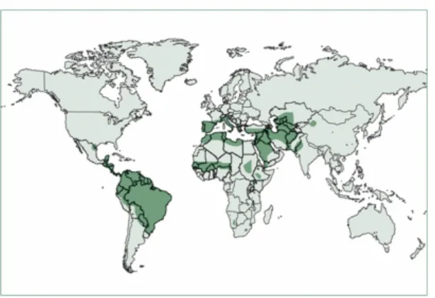
VL, the systemic form of leishmaniasis, is caused by the Leishmania species belonging to the Leishmania donovani complex, namely L. chagasi (New World) L. donovani and L. infantum (Wold World) (Table I). There is growing evidence to suggest that L. chagasi and L. infantum are the same specie (Momen et al., 1987; Rioux et al., 1990; Cupolillo et al., 1994; Mauricio et al., 1999). Indeed, several authors refer to them as synonyms, despite the disagreement of others (Shaw, 1994; Lukes et al., 2007; Chappuis et al., 2007). The transmission of L. infantum and/or L. chagasi is zoonotic, while it is anthroponotic to L. donovani (Table I). While L. donovani infects all age groups, L. infantum infects mainly children and immunosuppressed individuals. More than 90% of VL cases occur in Bangladesh, India, Nepal, Sudan, Ethiopia and Brazil (Figure 6) (Chappuis et al., 2007). Indeed, the estimated number of VL new cases and deaths each year is, respectively, 500000 and 50000 (Desjeux et al., 2004). The increased incidence of the disease has been associated to migration, lack of control measures, and HIV co-infection
Chapter I Leishmania spp. and leishmaniasis
15
(Boelaert et al., 2000; Desjeux, 2001). Cases of HIV/Leishmania co-infection have been reported in 35 countries worldwide. The HIV/Leishmania co-infected individuals present a high risk of developing VL upon Leishmania infection due to their immunosuppressed status (Moreno et al., 2000). Furthermore, they present high parasite loads in organs, low sensitivity to the serological-based diagnostic tests and treatment failure (Murray et al., 1999; Deniau et al., 2003). Several cases of HIV/Leishmania co-infection have been reported in South-Western Europe (Desjeux, 2000).
PKDL, a VL complication that arises after treatment, is very frequent in Sudan and more rarely, in East African countries and Indian subcontinents (Zijlstra et al., 2003). PKDL cases are highly infective, since the nodular lesions contain many parasites, and they might represent a putative reservoir for anthroponotic VL transmission (Addy and Nandy, 1992).
Figure 6. Worldwide distribution of the visceral leishmaniasis. 90% of VL cases occur in Bangladesh, India, Nepal, Sudan, Ethiopia and Brazil (Adapted from Chappuis et al., 2007).
2.2 Diagnosis
The large clinical spectrum of leishmaniasis makes it difficult to diagnose. Furthermore, other pathologies with similar clinical manifestations (e.g. leprosy, skin cancers, tuberculosis, cutaneous mycoses for CL and malaria or schistosomiasis for VL) are common in Leishmania endemic areas (Escobar et al., 1992). Therefore, specific and sensitive tests are very important, not only for a prompt and accurate diagnosis but also for a successful treatment and subsequent disease control.
2.2.1 Microscopy
The visualization of the parasite by microscopy examination of Giemsa-stained: lesion biopsy smears (for CL) or lymph nodes, spleen or bone marrow aspirates (for VL) remains the gold standard confirmatory diagnostic method for leishmaniasis in mainly endemic areas. Occasionally, the culture of biopsy triturates or aspirates is
Chapter I Leishmania spp. and leishmaniasis
16
also performed (Escobar et al., 1992). Even though it is very specific, the sensitivity of this method is highly variable and depends largely on the number and dispersion of parasites in the biopsy, the technical skills of the personnel, the sampling procedure and the sample origin (Herwalt, 1999). Indeed, in the diagnosis of VL, the sensitivity of the microscopy is higher for the spleen (93-99%), when compared to bone marrow (53-86%) or lymph node (53-65%) aspirates (Siddig et al., 1988). However, spleen aspirates require considerable technical expertise, since life threatening haemorrhages can occur (Kager et al., 1983).
2.2.2 PCR
Among the molecular biology methods available to detect Leishmania parasites, the most used are the PCR-based ones. The PCR revealed to be very specific and sensitive in the detection of Leishmania parasites. It has been considered a better approach in samples with low parasite load (MCL) (Garcia et al., 2005) and from less invasive methods such as blood (Cruz et al., 2002) as well as conjunctive (Strauss-Ayali et al., 2004) than the conventional microscopy. The relevance of this technique is extended to the diagnosis of Leishmania in HIV co-infected individuals (Cruz et al., 2002; De Doncker et al., 2005). Furthermore, the possibility of identifying the Leishmania specie and predicting the disease severity and treatment outcome is becoming important in the patients’ clinical management (Murray et al., 2005). Genetic markers of high copy number (e.g. rRNA genes, kinetoplast DNA minicircles, mini-exon genes) (Bensoussan et al., 2006) are usually selected to perform detection, quantification and viability studies, while repeated and polymorphic sequences are targeted to perform species identification (e.g. gp63, hsp70, and cysteine proteinases) (Garcia et al., 2005).
However it must be stressed that the feasibility of PCR diagnosis in endemic countries is still limited, since it requires high equipment and working costs.
2.2.3 Serological tests
Several tests for the detection of anti-Leishmania specific antibodies have been developed. Their applicability in the diagnosis of CL, however, is rare due to the variable specificity and sensitivity (Kar, 1995), and in the diagnosis of VL there are two main limitations. One is related to the persistence of the anti-Leishmania antibodies for long periods of time after the cure (Hailu, 1990;De Almeida Silva et al., 2006) and the other has to do with the detection of anti-Leishmania antibodies in healthy individuals from endemic areas with no story of VL (Sundar et al., 2006). These tests should therefore be used in combination with clinical symptoms for an
Chapter I Leishmania spp. and leishmaniasis
17
accurate diagnosis of VL. The several limitations of the poor Leishmania endemic areas have boosted the development of diagnostic tests that could be used in the field: easy to perform, cheap, reproducible and rapid. Therefore, two serological tests have been adapted and validated to field conditions – the fast agglutination screening test (FAST), a modified version of the direct agglutination test (DAT), and the rK39-immunocromatography or dipstick based test (ICT) (Schoone et al., 2001; Silva et al., 2005; Hailu et al., 2006; Chappuis et al., 2007). While the agglutination tests are based on the use of L. donovani stained promastigotes that agglutinate in the presence of a serum containing anti-Leishmania antibodies (el Harith et al., 1988), the rK-39 dipstick uses a recombinant 39-amino acid repeat that is part of a kinesin-related protein in L. chagasi and which is conserved within the L. donovani complex (Burns et al., 1993). The diagnostic performance of the latter has been evaluated in the several VL endemic areas (Chappuis et al., 2006; Sundar et al., 2007; Ritmeijer et al., 2007; Boelaert et al., 2008).
2.2.4 Antigen detection
Among the limitations of the antibody detection diagnostic-based tests is the impossibility to distinguish between an acute and asymptomatic disease and the antibodies’ cross-reactivity. An antigen-based diagnostic test has been developed for VL. Indeed, it is based on the detection of a heat-stable, low-molecular-weight carbohydrate antigen in the urine of VL patients (Attar et al., 2001; Sarkari et al., 2002). Although, improvements concerning the test sensitivity are required (Chappuis et al., 2006; Sundar et al., 2007).
2.3 Disease control
In general, the control of leishmaniasis is based on case detection followed by treatment, but also on vector and reservoir control, since no effective anti-Leishmania vaccine is available. A brief overview of each approach will be given, but treatment options, due to their relevance in the scope of this thesis, will be dealt with in more detail in the next chapter.
2.3.1 Control of vectors and reservoirs
Since sandflies are susceptible to insecticides, spraying houses with them is the most widely used intervention to control sandflies that are endophagic (mainly stay indoors after feeding) and endophilic (mainly feed indoors). The house spraying


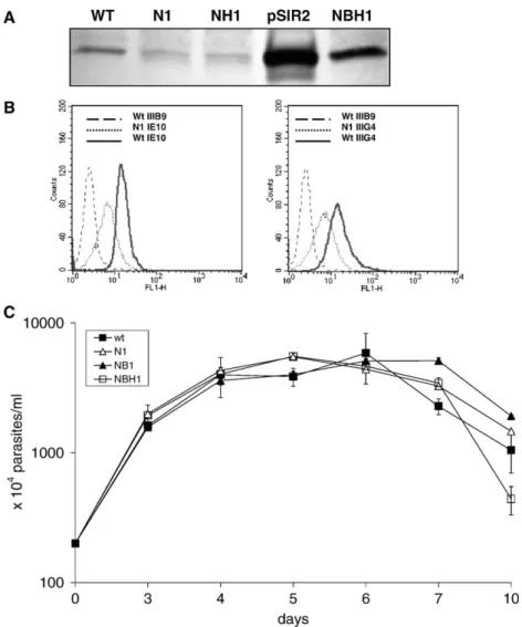
![Fig. 4. Plasmid curing experiments. Both NB1 (LiSIR2/LiSIR2::neo[pXG-BSDLiSIR2]) and NBH1 (LiSIR2::neo/LiSIR2::hyg[pXG-BSDLiSIR2]) clones were maintained in culture over 21 weeks by weekly subpassages without selection pressure](https://thumb-eu.123doks.com/thumbv2/123dok_br/19204402.955270/134.892.226.689.106.507/plasmid-experiments-bsdlisir-bsdlisir-maintained-subpassages-selection-pressure.webp)