Airways: Role in Airway Smooth Muscle Migration and
Chemokine Production
Jingbo Zhang1,4, Lianyu Shan1, Latifa Koussih1, Naresh Singh Redhu1, Andrew J. Halayko1,2, Jamila Chakir3, Abdelilah S. Gounni1*
1Department of Immunology, Faculty of Medicine, University of Manitoba, Winnipeg, Manitoba, Canada,2Department of Physiology, Faculty of Medicine, University of Manitoba, Winnipeg, Manitoba, Canada,3Institut Universitaire de Cardiologie et de Pneumologie de Que´bec, Universite´ Laval, Sainte-Foy, Que´bec, Canada,4Xinqiao Hospital, Third Military Medical University, Chongqing, China
Abstract
Background:Pentraxin 3 (PTX3) is a soluble pattern recognition receptor with non-redundant functions in inflammation and innate immunity. PTX3 is produced by immune and structural cells. However, very little is known about the expression of PTX3 and its role in allergic asthma.
Objectives and Methods:We sought to determine the PTX3 expression in asthmatic airways and its function in human airway smooth muscle cells (HASMC).In vivoPTX3 expression in bronchial biopsies of mild, moderate and severe asthmatics
was analyzed by immunohistochemistry. PTX3 mRNA and protein were measured by real-time RT-PCR and ELISA, respectively. Proliferation and migration were examined using3H-thymidine incorporation, cell count and Boyden chamber assays.
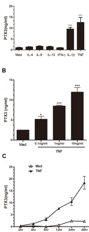
Results:PTX3 immunoreactivity was increased in bronchial tissues of allergic asthmatics compared to healthy controls, and mainly localized in the smooth muscle bundle. PTX3 protein was expressed constitutively by HASMC and was significantly up-regulated by TNF, and IL-1b but not by Th2 (IL-4, IL-9, IL-13), Th1 (IFN-c), or Th-17 (IL-17) cytokines.In vitro, HASMC
released significantly higher levels of PTX3 at the baseline and upon TNF stimulation compared to airway epithelial cells (EC). Moreover, PTX3 induced CCL11/eotaxin-1 release whilst inhibited the fibroblast growth factor-2 (FGF-2)-driven HASMC chemotactic activity.
Conclusions:Our data provide the first evidence that PTX3 expression is increased in asthmatic airways. HASMC can both produce and respond to PTX3. PTX3 is a potent inhibitor of HASMC migration induced by FGF-2 and can upregulate CCL11/ eotaxin-1 release. These results raise the possibility that PTX3 may play a dual role in allergic asthma.
Citation:Zhang J, Shan L, Koussih L, Redhu NS, Halayko AJ, et al. (2012) Pentraxin 3 (PTX3) Expression in Allergic Asthmatic Airways: Role in Airway Smooth Muscle Migration and Chemokine Production. PLoS ONE 7(4): e34965. doi:10.1371/journal.pone.0034965
Editor:Mauricio Rojas, University of Pittsburgh, United States of America ReceivedNovember 10, 2011;AcceptedMarch 8, 2012;PublishedApril 18, 2012
Copyright:ß2012 Zhang et al. This is an open-access article distributed under the terms of the Creative Commons Attribution License, which permits unrestricted use, distribution, and reproduction in any medium, provided the original author and source are credited.
Funding:This work was supported by the Canadian Institutes of Health Research (CIHR) grant and Manitoba Research Chair to ASG. JZ is supported by a postdoctoral fellowship from the Manitoba Health Research Council-Manitoba Institute of Child Health (MHRC-MICH). NSR is supported by the Canadian Thoracic Society-Canada Lung Association (CTS-CLA) graduate studentship. The funders had no role in study design, data collection and analysis, decision to publish, or preparation of the manuscript.
Competing Interests:The authors have declared that no competing interests exist.
* E-mail: [email protected]
Introduction
Asthma is a chronic inflammatory condition of the airways characterized by bronchial hyperresponsiveness, infiltration of inflammatory cells, and airway remodeling. Increased airway smooth muscle mass and deposition of extracellular matrix (ECM) proteins, is an important feature of airway remodeling [1]. Cumulative evidence suggest that airway smooth muscle cells (ASMC) are not only the target for but also a rich source of different pro-asthmatic factors [2]. These studies indeed suggest that ASMC may be classified as ‘inflammatory-like’ cells by virtue of their capacity to produce and respond to multiple pro-inflammatory mediators [2,3,4]. These mediators in turn modulate the ASMC functions such as cell proliferation, migration, and
matrix production, eventually leading to airway remodeling. Importantly, it is now evident that ASMC can directly respond to inhaled environmental factors via mechanisms that are either immune-mediated such as both high- and low-affinity IgE receptors [5,6,7,8,9], or in an immune-independent fashion such as protease activated receptor-2 (PAR2) or calreticulin [10,11], summarized in [12]. Targeting ASM, therefore, presents an attractive treatment regimen for chronic airway inflammatory diseases.
PTX3 interacts with other biologically active molecules, such as fibroblast growth factor-2 (FGF-2) [32] and P-selectin [33].
Considering an ‘inflammatory-like’ phenotype of ASMC [2], we hypothesized that PTX3 may also be involved in airway diseases such as allergic asthma. Thus, we first investigated the expression of PTX3 in bronchial biopsies of asthmatic patients, and then analyzed the production of PTX3 in human primary ASMC and epithelial cells (EC) in vitro. We further investigated the possible effects of recombinant human PTX3 on ASMC functions such as inflammatory mediator release, cell migration, and proliferation.
Results
Expression of PTX3 is increased in bronchial biopsies of allergic asthmatic patients
Bronchial biopsy specimens from healthy controls (n = 10) and subjects with allergic asthma (n = 27; 9 with mild, 10 with moderate, and 8 with severe asthma) were used for immunohis-tochemistry staining using mAb anti-human PTX3. Positive PTX3 immunoreactivity was detected in all cases in the epithelium, bronchial mucosa inflammatory infiltrating cells and ASM cell layers (Figure 1 A, C, E and G). Substitution of the first Ab with matched rat-IgG2b control eliminated the positive signal, demonstrating the specificity of the analysis (Figure 1 B, D, F and H).
Interestingly, since PTX3 was abundantly present at sites of ASM bundle, we performed semi-quantitative assessment through blind grading [34]. PTX3 staining intensity was significantly increased in asthmatic groups compared to healthy controls (Figure 1 I, P,0.001). A significantly higher staining intensity was found in mild (Figure 1 C) (P,0.05), moderate (Figure 1 E) (P,0.01) or severe (Figure 1 G) (P,0.05) asthmatic subjects compared to healthy controls (Figure 1 A). Furthermore, comparing the mean values of PTX3 staining intensity in the subgroups of asthmatic subjects revealed that severe and moderate group had higher intensity than mild asthmatic group without reaching statistical significance (P.0.05).
PTX3 protein is highly expressed by human ASMC (HASMC)in vitro
Lung EC lines have been previously shown to express PTX3 [26]. Similarly, PTX3 immunoreactivity was observed in our study in both ECs and HASMC within bronchial biopsies of asthmatic and healthy donors (Figure 1). We then decided to compare PTX3 release from HASMC (n = 4 donors) with that in human airway ECs (n = 4 donors). Under similar culture conditions and the same cell number, levels of PTX3 released from cultured HASMC was far greater than the airway EC at the baseline (mean value more than 36-fold) or upon TNF stimulation (mean value more than 115-fold) (Figure 2), suggesting that HASMC are one of the main producers of PTX3 in the airways.
we further characterized the dose or time course effect of TNF on PTX3 expression by HASMC. A statistically significant increase in PTX3 protein release from HASMC occurred with 0.1 ng/ml (*P,0.05), 1 ng/ml (***P,0.001), and 10 ng/ml (***P,0.001) concentration of TNF (n = 5) (Figure 3 B). Furthermore, time course assay showed that PTX3 protein release by HASMC was time-dependent and reached a maximum at 48 h in response to TNF (10 ng/ml) stimulation (Figure 3 C).
Recombinant PTX3 induces CCL11/eotaxin-1 but not TGFb1, CXCL8/IL-8, or IL-6 protein release from HASMC
Because the functions of PTX3 are not yet completely defined, we explored whether PTX3 could be an inducer of inflammatory mediators. To test this hypothesis, serum-deprived HASMC were stimulated with a graded concentration of human recombinant PTX3 (10–500 ng/ml). A statistically significant increase in eotaxin-1/CCL11 release from HASMC occurred with 50, 100, and 500 ng/ml PTX3 at 24 h (P,0.05, Figure 4 A). However, PTX3-stimulated HASMC showed no statistical significance in TGFb1 (Figure 4 B), IL-6 (Figure 4 C) or CXCL8/IL-8 release (p.0.05, n = 3; data not shown). Taken together, the above data demonstrate that PTX3 can induce CCL11/eotaxin-1 production in HASMC but fails to affect TGF-b1, IL-6 or CXCL8/IL-8.
Recombinant PTX3 inhibits FGF2-induced chemotaxis but not proliferation in HASMC
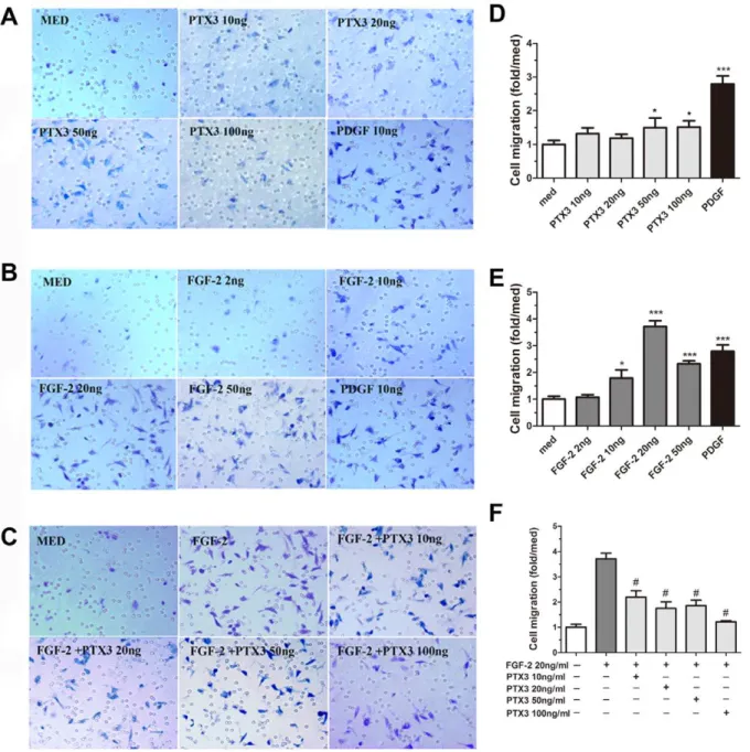
PTX3 was previously shown to play an important role in tissue remodeling and matrix assembly [38]. We therefore investigated its role in HASMC migration as a surrogate marker of airway tissue remodeling. Migration of HASMC was slightly increased by PTX3 alone (50 ng/ml, 1.5060.40- fold and 100 ng/ml, 1.5160.32-fold, P,0.05). Platelet-derived growth factor (PDGF, 10 ng/ml), used as positive control, significantly increased the HASMC chemotaxis (3.0260.65-fold, P,0.001) compared to the medium control (Figure 5 A).
Previous observations have shown the ability of PTX3 to bind FGF-2, thus acting as an FGF-2 antagonist [32,39]. Furthermore, FGF-2 is known to contribute to HASMC proliferation and migration ability that are believed to be major contributors to airway remodeling in asthma [40]. On this basis, we further investigated the capacity of PTX3 to affect FGF-2-dependent HASMC migration. As showed in. Figure 5 B, FGF-2 significantly increased migration of HASMC, with an optimal response at 20 ng/ml (3.7160.71-fold, P,0.001 compared to the medium). Concentration of PTX3 between 10–100 ng/ml significantly inhibited the effect of FGF-2 on HASMC migration (Figure 5 C,#P,0.001 compared to FGF-2 alone (20 ng/ml, n = 3).
stimulated cells, used as positive control (data not shown). Furthermore, FGF-2 stimulated HASMC showed significant proliferation as expected. However, FGF-2-mediated HASMC proliferation was not affected by addition of PTX3 (data not shown).
Taken together, these data demonstrate that PTX3 has the ability to inhibit FGF2-induced migration of HASMC at leastin vitrosuggesting its role as potential protective mechanism against remodelling observed in asthma.
Discussion
The present study is the first description of PTX3 expression in patients with allergic asthma. A significantly higher expression of PTX3 within the ASMC area was observed in mild, moderate, and severe asthmatics compared to healthy donors. PTX3 is abundantly expressed within ASM bundle and epithelial cell layers as well as inflammatory cell infiltrate. Furthermore, at leastin vitro, primary HASMC produced higher levels of PTX3 at the baseline and upon TNF stimulation compared to epithelial cells, suggesting that HASMC are one of the major sources of this mediator in the airways. We also showed that PTX3 can be up-regulated by TNF, but not IFNc, IL-17, IL-4, IL-9, or IL-13 in HASMC. Moreover, we showed that the PTX3 induces CCL11/eotaxin-1 release, but not IL-6, CXCL8/IL-8, or TGF-b and counteracts FGF-2-induced HASMC migration. Taken together, our data suggest a
potential dual role of PTX3 in allergic asthma by enhancing airway inflammation via local CC chemokine expression from HASMC; and down regulating the remodeling by counteracting FGF-induced HASMC migration.
Another study showed an enhanced PTX3 expression in induced sputum of COPD patients compared to controls [46]; whereas the PTX3 knockout mice showed no difference in cigarette smoke-induced pulmonary inflammation, emphysema, and body weight changes compared to the wild-type mice [47]. Therefore, a clear role of PTX3 in the pathogenesis of COPD remains yet to be established.
The reason why PTX3 is highly expressed within the airway of asthmatics remains unknown, but there are at least two possible explanations. First, emerging evidence suggests PTX3 as an important structural component of ECM that plays a crucial role in sustaining its stability [13,14,48]. In particular, PTX3 is involved in the organization of ECM by cross-linking hyaluronan via interaction with TNF-stimulated gene 6 (TSG6) and Inter-a -trypsin inhibitor (IaI) [49,50]. In agreement with the role of PTX3 in matrix organization, our IHC staining showed a significant PTX3 staining within the ASM bundles of asthmatic bronchial tissue compared to healthy donors, which may provide another possible explanation of airway remodeling observed in asthma. Second, PTX3 may potentially contribute to airway inflammation and subsequent remodeling. Furthermore, PTX3 enhanced CCL11/eotaxin-1 release in HASMC, chemokine that plays an important role in eosinophil airway inflammation [51], and remodeling [52].
However, it is worth mentioning that CCL11/eotaxin-1, besides its role in attracting eosinophils, can exert a specific regulatory function of neutrophil recruitmentin vivoas has been shown in a mouse model of endotoxemia [53] via probable down-regulation of CXC chemokine CXCL8/IL-8 [54]. In light of prior evidence and present data, it is tempting to speculate that PTX3-mediated CCL11/eotaxin-1 release may down-regulate exaggerated neu-trophilic inflammation by suppressing CXC chemokine release during acute inflammation, hence creating a negative feedback to establish tissue homeostasis especially in severe asthma that is characterized by more neutrophilic infiltrates [55]. In agreement with this possibility, Mantovani and colleagues reported recently
that PTX3 dampens neutrophil recruitmentin vivovia a P-selectin dependent mechanism [33]; and previous data demonstrated that PTX3 knockout mice develop more myocardial damage associ-ated with more neutrophil infiltration in a model of cardiac ischemia reperfusion injury [56]. These data suggest a potential role of PTX3 in down regulating neutrophilic airway inflamma-tion in allergic asthma. Taken together, it is plausible that on one Figure 2. HASMC produce enhanced PTX3in vitrocompared to
airway EC.(A) 75–80% confluent HASMC or ECs were growth arrested by FBS deprivation for 48 h, and stimulated in fresh FBS-free medium containing TNF (10 ng/ml) or medium alone. Supernatants were collected at 24 h, and PTX3 was quantified using ELISA. #P,0.0001 PTX3 protein release from HASMCs versus from airway ECs by ANOVA. doi:10.1371/journal.pone.0034965.g002
Figure 3. Cytokines induce PTX3 expression in HASMCin vitro. (A) PTX3 protein release from HASMC stimulated with proinflammatory (IL-1b, TNF, 10 ng/ml), Th2 (IL-4, IL-9, 10 ng/ml, or IL-13, 100 ng/ml), or Th1 cytokines (IFN-c, 100 U/ml) for 24 h (n = 3). (B) PTX3 protein release was measured in response to increasing concentrations of TNF at 24 h (n = 5), or (C) at various time-points in presence of TNF (10 ng/ml) (n = 3). *p,0.05, ***p,0.001 compared to medium group.
hand a reduced PTX3 expression in COPD favors a characteristic neutrophilic response [45], whereas enhanced PTX3 expression in asthmatics may embark on an eosinophilic inflammation poten-tially via inducing eotaxin-1/CCL11 expression in airway structural cells. More studies are needed further to investigate these possibilities.
We also found that PTX3 significantly inhibits FGF-2-induced chemotaxis in HASMC. Furthermore, in contrast with previous study in vascular smooth muscle cells [39], we did not find any effect of PTX3 protein on HASMC proliferation, suggesting that PTX3 effect is cell specific. Although PTX3 at higher doses by itself tend to induce ASM migration, it attenuates the FGF-2-induced migration. It may be plausible that the locally-produced PTX3 facilitates immune inflammatory cell recruitment, while counteracts the effects of FGF-2 in inducing migration of ASM itself. Moreover, since PTX3 has been shown to act as an antagonist of FGF-2 by others [32,39], it may be plausible that the inhibitory effect on migration is selective and specific to FGF-2-induced migration. We are exploring these hypotheses further by extensivein vitrostudies and by employing PTX3 knockout mouse model. Besides EC that is the main source of FGF-2 in asthmatic airways, inflammatory cells mobilized into the airways such as eosinophils, macrophages, T-cells, mast cells are also potent producers of FGF-2 [40]. FGF-2 is known to be released directly following allergen-induced degranulation of mast cells which may cause HASMC migration [57]. On this basis, it is enticing to speculate that PTX3 released from HASMC, as well as others sources, may have a regulatory role in smooth muscle migration and thus airway remodeling.
In conclusion, this novel study demonstrated an increased PTX3 expression in bronchial biopsies of allergic asthmatics compared to healthy controls. Considering HASMC as one of the major cell types in the airway, constitutive expression and inducible PTX3 produced from this source may play a dual role in inflammatory and airway remodelling process in allergic asthma.
Some of the results of these studies have been previously presented in American Thoracic Society International Conference and reported in the form of an abstract online.
Materials and Methods
Ethics statement
Human airway smooth muscle cells (HASMC) were obtained from surgical patients following the receipt of written informed
consent in accordance with procedures approved by the Human Research Ethics Board of the University of Manitoba, Winnipeg, Canada. Bronchoscopy to obtain lung tissue (for immunohisto-chemistry) was performed in accordance with procedures approved by the Human Research Ethics Board of Laval University, Quebec, Canada, following the receipt of the subjects’ written informed consent. The current study was approved by the research ethics boards of the University of Manitoba, Winnipeg, and the Laval University, Quebec, Canada.
Reagents
Recombinant human TNF, IL-1b, IL-4, IL-9, IL-13, IFN-c, FGF-2, PTX3 proteins, and ELISA kits for human PTX3 were purchased from R&D Systems (Minneapolis, MN). Monoclonal rat anti-PTX3 (MNB1) was purchased from ALEXIS Biochem-icals (Lausen, Switzerland). All other reagents were procured from Sigma-Aldrich Canada Ltd. (Oakville, ON), unless specified.
Subjects
Asthmatic subjects were fulfilling the American Thoracic Society selection criteria [58]. Mild asthmatics had an FEV1 of .80% predicted, used no inhaled corticosteroids (CS), had symptoms,2 days per week, and rare exacerbations. Moderate asthmatics used inhaled CS on a daily basis and had an FEV1 of .50% predicted and used less than 1000mg fluticasone/day.
Furthermore, moderate asthmatics have a history of emergency department (ED)/urgent care visits or systemic steroid use within the past year. Severe asthmatics needed treatment with high dose of inhaled CS (.1000mg fluticasone/day) or systemic CS. All
asthmatic subjects were atopic with at least one positive response to common allergens on allergy skin prick tests. In the month preceding the study, none of the subjects reported a respiratory infection or an increase in asthma symptoms. Some subjects who had the smoking history had quit at least three months. Healthy subjects were non-atopic non-smokers with no history of asthma, respiratory disease or atopy, except one who had a smoking history and had quit 2.5 years ago. All healthy subjects showed normal pulmonary function testing and a negative methacholine chal-lenge. Subjects’ characteristics are shown in Table 1.
Immunohistochemistry
Paraffin sections were stained with rat anti-human PTX3 mAb or control IgG2b followed by secondary biotin goat anti-rat IgG and streptavidin-alkaline phosphatase developed by fast red as described earlier [59]. Briefly, formalin-fixed tissues were paraffin Figure 4. Recombinant human PTX3 induce cytokine release from HASMC.Eotaxin-1/CCL11 (A), TGF-b1 (B), and IL-6 (C) proteins were measured by ELISA upon 24 h stimulation of serum deprived HASMCs with graded concentrations of recombinant PTX3 (*P,0.05, **P,0.01 and ***P,0.001 compared with medium group).
embedded, and 5-mm-thick sections were prepared, deparaffinized
in xylene, and rehydrated through graded concentrations of alcohol to water and then boiled with microwave for 10 min in sodium citrate buffer (pH, 6.0) for antigen retrieval. Sections were washed and then incubated with blocking solution (5% goat serum, 1% BSA, 0.1% cold fish skin gelatin in PBS) for 60 min at room temperature. Rat anti-human PTX3 mAb or control mouse IgG2b (both at 10mg/ml) were added, and sections were incubated overnight at 4uC. Slides were then washed twice with PBS followed by incubation for 1 h at room temperature with biotin-conjugated goat anti-rat IgG (Jackson ImmunoResearch Laboratories, Inc. West Grove, PA). Slides were then washed extensively with PBS and incubated with streptavidin-alkaline
phosphatase for 30 min at room temperature. After washing with PBS, the slides were developed using Fast Red and counterstained with Mayer’s hematoxylin. Positive cells stained red after development with Fast Red (Sigma-Aldrich Canada Ltd., Oak-ville, ON). Isotype-matched control mAb was used for negative control. Semi-quantitative assessment of specimens were per-formed by three independent observers and a pathologist in a blinded manner as previously described [34]: 0, absent or faint staining of an occasional ASM bundle only; l+, faint staining of several ASM bundle; 2+, moderate intensity staining of most ASM bundle; 3+, intense staining of most ASM bundle and 4+intense staining of all ASM bundle.
Figure 5. PTX3 abrogates FGF-2 induced HASMC migration.Boyden upper chamber-seeded HASMC were exposed to graded concentrations of (A) PTX3, (B) FGF-2, and (C) PTX3 plus FGF-2 (20 ng/ml) in lower compartment for 4 h. The migrated cells were quantified by light microscope (magnification,6200). Migration is expressed as fold increase relative to medium control (D, E, and F). *P,0.05, ***P,0.001vsmedium control. #P,0.001 compared to FGF-2 (20 ng/ml). n = 3.
HASMC preparation
HASMC were obtained as described previously [59,60,61] from macroscopically healthy segments of the trachea after lung resection from surgical patients in accordance with procedures approved by the Human Research Ethics Board of the University of Manitoba, Winnipeg, Canada. Primary HASMC were isolated from explants. HASMC retain smooth muscle-specific actin, SM22, calponin protein expression, and mobilize intracellular Ca2+
in response to acetylcholine [60], and were used at passages 3–5. HASMC were grown on uncoated plastic dishes in complete DMEM (DMEM supplemented with 100mg/ml streptomycin, 100 U/ml penicillin, and 10% fetal bovine serum). Unless otherwise mentioned, cells were grown to a subconfluent (,70%) condition and serum starved to synchronize for 48 h in Ham’s F12 supplemented with 100mg/ml streptomycin, 100 U/ ml penicillin, and 1X ITS (5mg/ml insulin, 5mg/ml transferrin, and 5 ng/ml selenium) before each experiment. Primary bronchial epithelial cells (EC) were isolated from biopsies obtained from healthy controls by using previously described techniques [62,63]. Briefly, epithelial cells were isolated from bronchial biopsies obtained by bronchoscopy, and were characterized by immuno-fluorescence and flow cytometry using anti-cytokeratin antibody from Calbiochem (San Diego, CA). This identification confirmed the purity of the bronchial cell culture as we previously described [62,63]. EC were cultured in DMEM supplemented with 100mg/ ml streptomycin, 100 U/ml penicillin, and 10% fetal bovine serum.
ELISA
PTX3 ELISA was performed according to the protocol provided by the manufacturer (R&D Systems, Minneapolis, MN). Sensitivity was 20 pg/ml for PTX3. IL-6, IL-8, TGFb, and eotaxin-1/CCL11 ELISA was performed in-house using matched Abs from R&D Systems. Sensitivity was IL-6 (7.6 pg/ml), CXCL8/IL-8 (7.6 pg/ml), TGF-b (15 pg/ml), and CCL11/ eotaxin-1 (10 pg/ml).
Proliferation and chemotaxis assay
Cell proliferation was examined by using 3H-thymidine incorporation [64], cell counting, and Alamar blue assays. Cell migration was analyzed according to the previously described using Boyden chamber assay [65]. Briefly, 48 h serum-starved cells were detached from the culture plate using trypsin (0.5 mg/ml)-EDTA (0.2 mg/ml) solution (Invitrogen Canada Inc., Burlington, ON) and resuspended in Ham’s F12 medium containing 100mg/ ml streptomycin, 100 U/ml penicillin, and 1X ITS. A polycar-bonate membrane of 8mm pore size (Neuroprobe, Gaithersburg, MD, USA) was coated with 0.01% collagen type-I in 0.01N HCl solution (Sigma). A 50ml aliquot of HASMC (56104cells/ml) was added to the upper chamber of modified Boyden chamber apparatus (Neuroprobe). In the lower chamber, FGF-2, PDGF, or PTX3 were added as a chemoattractant to the same media as the upper chamber. After 4 h of incubation at 37uC in humidified 5% CO2 incubator, the membranes were peeled-off. Cells on the
upper side of the membrane were scraped off and the cells migrated to the lower side were fixed and stained with HemacolorHstain set (EMD Millipore, Billerica, MA, USA) The number of migrated cells was counted in four-five random fields under620 magnification by phase contrast microscope (Carl Zeiss Canada Ltd., Toronto, ON).
Statistical analysis
Data obtained from experiments performed in triplicate and repeated at least three times was represented as means6SEM. Differences among groups were analyzed using unpaired t-tests or ANOVA together with a post-hoc Bonferroni analysis. Non-parametric data were analyzed using the Kruskal-Wallis test followed by the Mann-Whitney U-test. P values were considered significant at 0.05 levels.
Acknowledgments
We would like to thank the Tissue Bank of the Respiratory Health Network of the Fonds de la Recherche en Sante´ du Quebec. The authors would like Table 1.Clinical characteristics of the subjects.
Category Subcategory Healthy controls Mild asthma Moderate asthma Severe asthma
Number (n) 10 9 10 8
{
Age (years) 2461.5 2461.6 3364.5 3965.9
Sex Male 7 4 2 1
Female 3 5 8 7
Smoking status NS 8 6 9 5
Ex 2 3 1 3
S 0 0 0 0
Atopy Yes 0 9 10 8
No 10 0 0 0
{
FEV1 (%) 98.562.8 96.064.4 86.366.7 78.966.0
{
PC20(mg/ml) 111.4618 1.760.7 0.760.5
-Asthma medication ICS (mg) - - 437.56100.2 1187.56277.2
LABA - - - Yes
{Data are expressed as mean6SEM. NS: Non smoker; Ex: Ex-smoker; S: Smoker. FEV1: forced expiratory volume in 1 second.
LABA: Long actingb2-agonists.
Neurobiol 137: 209–222.
4. Panettieri RA, Jr. (2003) Airway smooth muscle: immunomodulatory cells that modulate airway remodeling? Respir Physiol Neurobiol 137: 277–293. 5. Gounni AS (2006) The high-affinity IgE receptor (FcepsilonRI): a critical
regulator of airway smooth muscle cells? Am J Physiol Lung Cell Mol Physiol 291: L312–321.
6. Gounni AS, Wellemans V, Yang J, Bellesort F, Kassiri K, et al. (2005) Human airway smooth muscle cells express the high affinity receptor for IgE (Fc epsilon RI): a critical role of Fc epsilon RI in human airway smooth muscle cell function. J Immunol 175: 2613–2621.
7. Redhu NS, Saleh A, Shan L, Gerthoffer WT, Kung SK, et al. (2009) Proinflammatory and Th2 cytokines regulate the high affinity IgE receptor (FcepsilonRI) and IgE-dependant activation of human airway smooth muscle cells. PLoS One 4: e6153.
8. Roth M, Tamm M (2010) The effects of omalizumab on IgE-induced cytokine synthesis by asthmatic airway smooth muscle cells. Ann Allergy Asthma Immunol 104: 152–160.
9. Hakonarson H, Grunstein MM (1998) Autologously up-regulated Fc receptor expression and action in airway smooth muscle mediates its altered responsiveness in the atopic asthmatic sensitized state. Proc Natl Acad Sci U S A 95: 5257–5262.
10. Chambers LS, Black JL, Ge Q, Carlin SM, Au WW, et al. (2003) PAR-2 activation, PGE2, and COX-2 in human asthmatic and nonasthmatic airway smooth muscle cells. Am J Physiol Lung Cell Mol Physiol 285: L619–627. 11. Chambers LS, Black JL, Poronnik P, Johnson PR (2001) Functional effects of
protease-activated receptor-2 stimulation on human airway smooth muscle. Am J Physiol Lung Cell Mol Physiol 281: L1369–1378.
12. Roth M, Tamm M (2010) Airway smooth muscle cells directly respond to inhaled environmental factors. Swiss Med Wkly.
13. Manfredi AA, Rovere-Querini P, Bottazzi B, Garlanda C, Mantovani A (2008) Pentraxins, humoral innate immunity and tissue injury. Curr Opin Immunol 20: 538–544.
14. Ortega-Hernandez OD, Bassi N, Shoenfeld Y, Anaya JM (2008) The Long Pentraxin 3 and Its Role in Autoimmunity. Semin Arthritis Rheum. 15. Latini R, Maggioni AP, Peri G, Gonzini L, Lucci D, et al. (2004) Prognostic
significance of the long pentraxin PTX3 in acute myocardial infarction. Circulation 110: 2349–2354.
16. Fazzini F, Peri G, Doni A, Dell’Antonio G, Dal Cin E, et al. (2001) PTX3 in small-vessel vasculitides: an independent indicator of disease activity produced at sites of inflammation. Arthritis Rheum 44: 2841–2850.
17. Luchetti MM, Piccinini G, Mantovani A, Peri G, Matteucci C, et al. (2000) Expression and production of the long pentraxin PTX3 in rheumatoid arthritis (RA). Clin Exp Immunol 119: 196–202.
18. Tong M, Carrero JJ, Qureshi AR, Anderstam B, Heimburger O, et al. (2007) Plasma pentraxin 3 in patients with chronic kidney disease: associations with renal function, protein-energy wasting, cardiovascular disease, and mortality. Clin J Am Soc Nephrol 2: 889–897.
19. Azzurri A, Sow OY, Amedei A, Bah B, Diallo S, et al. (2005) IFN-gamma-inducible protein 10 and pentraxin 3 plasma levels are tools for monitoring inflammation and disease activity in Mycobacterium tuberculosis infection. Microbes Infect 7: 1–8.
20. Mauri T, Coppadoro A, Bellani G, Bombino M, Patroniti N, et al. (2008) Pentraxin 3 in acute respiratory distress syndrome: an early marker of severity. Crit Care Med 36: 2302–2308.
21. Doni A, Peri G, Chieppa M, Allavena P, Pasqualini F, et al. (2003) Production of the soluble pattern recognition receptor PTX3 by myeloid, but not plasmacytoid, dendritic cells. Eur J Immunol 33: 2886–2893.
22. Shelton RC, Liang S, Liang P, Chakrabarti A, Manier DH, et al. (2004) Differential expression of pentraxin 3 in fibroblasts from patients with major depression. Neuropsychopharmacology 29: 126–132.
23. Jaillon S, Peri G, Delneste Y, Fremaux I, Doni A, et al. (2007) The humoral pattern recognition receptor PTX3 is stored in neutrophil granules and localizes in extracellular traps. J Exp Med 204: 793–804.
24. Norata GD, Marchesi P, Pirillo A, Uboldi P, Chiesa G, et al. (2008) Long pentraxin 3, a key component of innate immunity, is modulated by high-density lipoproteins in endothelial cells. Arterioscler Thromb Vasc Biol 28: 925–931.
atherogenic lipoproteins induce expression of pentraxin-3 by human vascular smooth muscle cells. Atherosclerosis 175: 221–228.
28. Bozza S, Bistoni F, Gaziano R, Pitzurra L, Zelante T, et al. (2006) Pentraxin 3 protects from MCMV infection and reactivation through TLR sensing pathways leading to IRF3 activation. Blood 108: 3387–3396.
29. Garlanda C, Hirsch E, Bozza S, Salustri A, De Acetis M, et al. (2002) Non-redundant role of the long pentraxin PTX3 in anti-fungal innate immune response. Nature 420: 182–186.
30. Gaziano R, Bozza S, Bellocchio S, Perruccio K, Montagnoli C, et al. (2004) Anti-Aspergillus fumigatus efficacy of pentraxin 3 alone and in combination with antifungals. Antimicrob Agents Chemother 48: 4414–4421.
31. Salustri A, Garlanda C, Hirsch E, De Acetis M, Maccagno A, et al. (2004) PTX3 plays a key role in the organization of the cumulus oophorus extracellular matrix and in in vivo fertilization. Development 131: 1577–1586.
32. Camozzi M, Rusnati M, Bugatti A, Bottazzi B, Mantovani A, et al. (2006) Identification of an antiangiogenic FGF2-binding site in the N terminus of the soluble pattern recognition receptor PTX3. J Biol Chem 281: 22605–22613. 33. Deban L, Russo RC, Sironi M, Moalli F, Scanziani M, et al. Regulation of
leukocyte recruitment by the long pentraxin PTX3. Nat Immunol 11: 328–334. 34. Fregonese L, Swan FJ, van Schadewijk A, Dolhnikoff M, Santos MA, et al. (2005) Expression of the anaphylatoxin receptors C3aR and C5aR is increased in fatal asthma. J Allergy Clin Immunol 115: 1148–1154.
35. Renauld JC (2001) New insights into the role of cytokines in asthma. J Clin Pathol 54: 577–589.
36. Amrani Y (2007) TNF-alpha and calcium signaling in airway smooth muscle cells: a never-ending story with promising therapeutic relevance. Am J Respir Cell Mol Biol 36: 387–388.
37. Berry MA, Hargadon B, Shelley M, Parker D, Shaw DE, et al. (2006) Evidence of a role of tumor necrosis factor alpha in refractory asthma. N Engl J Med 354: 697–708.
38. Maina V, Cotena A, Doni A, Nebuloni M, Pasqualini F, et al. (2009) Coregulation in human leukocytes of the long pentraxin PTX3 and TSG-6. J Leukoc Biol 86: 123–132.
39. Camozzi M, Zacchigna S, Rusnati M, Coltrini D, Ramirez-Correa G, et al. (2005) Pentraxin 3 inhibits fibroblast growth factor 2-dependent activation of smooth muscle cells in vitro and neointima formation in vivo. Arterioscler Thromb Vasc Biol 25: 1837–1842.
40. Bosse Y, Rola-Pleszczynski M (2008) FGF2 in asthmatic airway-smooth-muscle-cell hyperplasia. Trends Mol Med 14: 3–11.
41. Mauri T, Bellani G, Patroniti N, Coppadoro A, Peri G, et al. Persisting high levels of plasma pentraxin 3 over the first days after severe sepsis and septic shock onset are associated with mortality. Intensive Care Med 36: 621–629. 42. Dias AA, Goodman AR, Dos Santos JL, Gomes RN, Altmeyer A, et al. (2001)
TSG-14 transgenic mice have improved survival to endotoxemia and to CLP-induced sepsis. J Leukoc Biol 69: 928–936.
43. Breviario F, d’Aniello EM, Golay J, Peri G, Bottazzi B, et al. (1992) Interleukin-1-inducible genes in endothelial cells. Cloning of a new gene related to C-reactive protein and serum amyloid P component. J Biol Chem 267: 22190–22197.
44. Koppelman GH, Stine OC, Xu J, Howard TD, Zheng SL, et al. (2002) Genome-wide search for atopy susceptibility genes in Dutch families with asthma. J Allergy Clin Immunol 109: 498–506.
45. Van Pottelberge GR, Bracke KR, Pauwels NS, Vermassen FE, Joos GF, et al. (2011) COPD is associated with reduced pulmonary interstitial expression of Pentraxin-3. Eur Respir J.
46. Pizzichini MM, Kleveston T, Morato EF, Pinheiro JT, Steidle LJ, et al. (2009) Pentraxin 3 (PTX3): A New Marker To Study Airway Inflammation? Am J Respir Crit Care Med April 1: A2532.
47. Pauwels NS, Bracke KR, Maes T, Van Pottelberge GR, Garlanda C, et al. (2010) Cigarette smoke induces PTX3 expression in pulmonary veins of mice in an IL-1 dependent manner. Respir Res 11: 134.
48. Mantovani A, Garlanda C, Doni A, Bottazzi B (2008) Pentraxins in innate immunity: from C-reactive protein to the long pentraxin PTX3. J Clin Immunol 28: 1–13.
50. Varani S, Elvin JA, Yan C, DeMayo J, DeMayo FJ, et al. (2002) Knockout of pentraxin 3, a downstream target of growth differentiation factor-9, causes female subfertility. Mol Endocrinol 16: 1154–1167.
51. Humbles AA, Conroy DM, Marleau S, Rankin SM, Palframan RT, et al. (1997) Kinetics of eotaxin generation and its relationship to eosinophil accumulation in allergic airways disease: analysis in a guinea pig model in vivo. J Exp Med 186: 601–612.
52. Fulkerson PC, Fischetti CA, Rothenberg ME (2006) Eosinophils and CCR3 regulate interleukin-13 transgene-induced pulmonary remodeling. Am J Pathol 169: 2117–2126.
53. Cheng SS, Lukacs NW, Kunkel SL (2002) Eotaxin/CCL11 is a negative regulator of neutrophil recruitment in a murine model of endotoxemia. Exp Mol Pathol 73: 1–8.
54. Cheng SS, Lukacs NW, Kunkel SL (2002) Eotaxin/CCL11 suppresses IL-8/ CXCL8 secretion from human dermal microvascular endothelial cells. J Immunol 168: 2887–2894.
55. Fukakusa M, Bergeron C, Tulic MK, Fiset PO, Al Dewachi O, et al. (2005) Oral corticosteroids decrease eosinophil and CC chemokine expression but increase neutrophil, IL-8, and IFN-gamma-inducible protein 10 expression in asthmatic airway mucosa. J Allergy Clin Immunol 115: 280–286.
56. Salio M, Chimenti S, De Angelis N, Molla F, Maina V, et al. (2008) Cardioprotective function of the long pentraxin PTX3 in acute myocardial infarction. Circulation 117: 1055–1064.
57. Redington AE, Roche WR, Madden J, Frew AJ, Djukanovic R, et al. (2001) Basic fibroblast growth factor in asthma: measurement in bronchoalveolar lavage fluid basally and following allergen challenge. J Allergy Clin Immunol 107: 384–387.
58. (1987) Standards for the diagnosis and care of patients with chronic obstructive pulmonary disease (COPD) and asthma. This official statement of the American Thoracic Society was adopted by the ATS Board of Directors, November 1986. Am Rev Respir Dis 136: 225–244.
59. Shan L, Redhu NS, Saleh A, Halayko AJ, Chakir J, et al. (2010) Thymic stromal lymphopoietin receptor-mediated IL-6 and CC/CXC chemokines expression in human airway smooth muscle cells: role of MAPKs (ERK1/2, p38, and JNK) and STAT3 pathways. J Immunol 184: 7134–7143.
60. Naureckas ET, Ndukwu IM, Halayko AJ, Maxwell C, Hershenson MB, et al. (1999) Bronchoalveolar lavage fluid from asthmatic subjects is mitogenic for human airway smooth muscle. Am J Respir Crit Care Med 160: 2062–2066. 61. Redhu NS, Saleh A, Halayko AJ, Ali AS, Gounni AS (2011) Essential role of
NF-kappaB and AP-1 transcription factors in TNF-alpha-induced TSLP expression in human airway smooth muscle cells. Am J Physiol Lung Cell Mol Physiol 300: L479–485.
62. Chakir J, Page N, Hamid Q, Laviolette M, Boulet LP, et al. (2001) Bronchial mucosa produced by tissue engineering: a new tool to study cellular interactions in asthma. J Allergy Clin Immunol 107: 36–40.
63. Semlali A, Jacques E, Plante S, Biardel S, Milot J, et al. (2008) TGF-beta suppresses EGF-induced MAPK signaling and proliferation in asthmatic epithelial cells. Am J Respir Cell Mol Biol 38: 202–208.
64. Goncharova EA, Lim P, Goncharov DA, Eszterhas A, Panettieri RA, Jr., et al. (2006) Assays for in vitro monitoring of proliferation of human airway smooth muscle (ASM) and human pulmonary arterial vascular smooth muscle (VSM) cells. Nat Protoc 1: 2905–2908.