Rita Joana Rodrigues da Silva Rua Ferreira
Licenciada em Anatomia Patológica, Citológica e Tanatológica
Cilia motility studies in zebrafish embryos
Dissertação para a obtenção do Grau de Mestre em
Genética Molecular e Biomedicina
Orientador: Susana Santos Lopes,
Investigadora Principal, Centro de Estudos de Doenças Crónicas,
Faculdade de Ciências Médicas, Universidade Nova de Lisboa
Co-orientador: Paula Gonçalves, Professora Auxiliar,
Faculdade Ciências e Tecnologia, Universidade Nova de Lisboa
Júri:
Presidente: Prof. Dra Paula Gonçalves Arguente: Dr. Jorge Carneiro
Cilia motility studies in zebrafish embryos
Submitted by
Rita Joana Rodrigues da Silva Rua Ferreira
A thesis submitted in fulfilment of the requirements for the degree of
Masters in Molecular Genetics and Biomedicine
!
Cilia motility studies in zebrafish
Copyright Rita Joana Rodrigues da Silva Rua Ferreira, FCT/UNL, UNL
A Faculdade de Ciências e Tecnologia e a Universidade Nova de Lisboa têm o direito, perpétuo e
sem limites geográficos, de arquivar e publicar esta dissertação através de exemplares impressos
reproduzidos em papel ou de forma digital, ou por qualquer outro meio conhecido ou que venha a ser
inventado, e de a divulgar através de repositórios científicos e de admitir a sua cópia e distribuição
com objectivos educacionais ou de investigação, não comerciais, desde que seja dado crédito ao
To my family,
Acknowledgements
First, I would like to thank Susana Lopes, my supervisor, for being such a good scientist and for
knowing how to motivate her students to accomplish more and more. Our snack breaks were always
a nice place for discussions and new ideas came from that. Thank you for giving me the opportunity
to work in a newly established lab and for encouraging me to search new scientific challenges. For all
this time thank you for being my friend and for being so quickly in correcting my thesis. I will never
forget our support and I hope we will meet again in a near future.
I thank Petra and Bárbara for this last year of laughing and nice work environment. Our lab was
always a funny place to work and learn with our discussions. I will miss our lunch times where
Bárbara has always new amazing things to tell us. She knows a lot about everything. Thank you for
your support during this time. I wish the best for both of you.
I would like to thank Cláudia and Marta for being such a nice lab neighbors. We became friends and
now I am sure I will miss you a lot. Hope you will come to Strasbourg to visit me.
I also thank Maysa Franco and Liliana Vale for taking care of IGC’s Fish Facility. I could not forget
Pedro Almada for all the help in the Spinning Disk. During this year I bugged you a lot and you always
help me. Thanks, thanks, thanks!!!
I would like to thank specially to my family just for being there every time I needed. My mood after
work made me treated you not always the best way but I love you all and without your support nothing
of this would be possible. Thanks Pai João, Mãe Té, Jotapê, Gui, Inês e Catarina. Thanks also to my
grandparents that don’t have a clue on what I am doing so far but supported me all my whole life. I
dedicate this work to all my family.
Another special thanks to my boyfriend Tiago, that always tried to kept my mind out of work. Thank
you for the times we spent watching sport channels, USA Open, Rolando Garros, among others,
because sometimes I really needed your ‘wake-up call’ to see that life is not all work, work and work.
Thank you for your unconditional love!
A special thanks to my best friend Sara that loves and supports me unconditionally although in the
last year she had my company just a few times. Work always kept me distant from our meetings.
Fortunately cell phones exist and you still considered myself as your ‘sister’! Thanks for your support.
I dedicate this work also to you, Sara.
Finally, my deepest gratitude goes to Adán Guerrero for teaching me so many things about science.
Without you this work would not have been possible in the way it was. You are a really good scientist
and teacher and I am sure that your future will be great. I am feeling very lucky for having you as a
Abstract
Motile ciliary dysfunctions cause specific Ciliopathies that affect mainly the respiratory tract,
fertilization and left-right body establishment. The embryonic organ where left-right decisions are first
taken is called the organizer, a ciliated organ where a leftward cilia driven fluid-flow is generated. The
organizer is named node in the mouse and Kupffer’s vesicle (KV) in zebrafish. The correct left-right
axis formation is highly dependent on signaling pathways downstream of such directional fluid-flow.
Motile cilia need to be coordinated and Ciliary Beat Frequency (CBF) is characteristic of different
types of cilia depending on their function. Using zebrafish as a model, our group has been studying
cilia length regulation and motility in wild-type and deltaD-/- mutant embryos. Recently, we showed
that Notch signalling was directly involved in the control of cilia length in the KV cells given that the
deltaD-/- mutant present shorter KV cilia.
The goal of this project was to characterize the CBF of deltaD-/- KV cilia vs. wild-type cilia and reveal
how potential differences in CBF impact on KV fluid flow, using spectral analysis associated with
high-speed videomicroscopy. By decomposing and comparing the obtained CBF with Fast Fourier
Transform, we identified two major populations of motile cilia in wild-type as well as in deltaD-/- mutant
embryos. However, we found the CBF populations had differential relative contributions and different
distributions between wild-type and mutant embryos. Furthermore, by measuring the velocity of native
particles we studied the KV fluid-flow and concluded that the dispersion of the flow velocity was much
wider in the deltaD-/-mutants. On the other hand, based on a gene expression study of motility genes
downstream of DeltaD, we concluded that motility related genes (dnah7, rsph3 and foxj1a) were
deregulated in the mutants.
During this project we generated data that led to new hypotheses that will allow us to test the
causality between the described correlations.
Key-words: Kupffer’s vesicle, motile cilia, cilia beat frequency, fluid-flow, deltaD-/- mutants
Resumo
Cílios primários disfuncionais causam Ciliopatias que afectam principalmente o trato respiratório, a
taxa de fertilização e o estabelecimento do eixo esquerda-direita. O órgão embrionário onde
primeiramente são tomadas decisões relativas ao eixo esquerda-direita é denominado de
‘organizador’. É um órgão ciliado onde um fluído direcionado para a esquerda é gerado pelo
movimento dos cílios. Este ‘organizador’ é denominado de ‘nó’ no rato e de ‘vesícula de Kupffer’ (VK)
no peixe-zebra. A correta formação do eixo direita-esquerda é altamente dependente das vias de
sinalização a jusante do fluxo direcional para a esquerda.
Os cílios móveis necessitam de ser coordenados e a frequência de batimento ciliar (FBC) é
característica de cada tipo de cílio e está dependente da sua função. Usando o peixe-zebra como
modelo, o nosso grupo tem vindo a estudar a regulação do tamanho e da motilidade ciliar em
embriões selvagens e em mutantes deltaD-/-. Recentemente, o nosso grupo mostrou que a
sinalização Notch está diretamente envolvida no controlo do tamanho dos cílios das células da VK,
tendo em conta que os mutantes deltaD-/- apresentam cílios curtos.
O objectivo deste projeto foi a caracterização das FBC dos mutantes deltaD-/- em comparação com
embriões wild-type, e revelar qual o impacto de potenciais diferenças das FBC no fluxo do fluído da
VK. Realizámos uma análise espectral dos cílios individualmente, através da utilização de
vídeo-microscopia de alta velocidade. Por decomposição e comparação das FBC obtidas com a
transformada rápida de Fourier, identificámos duas populações de cílios móveis tanto em embriões
selvagens como nos mutantes deltaD-/-. Para além disso, medimos a velocidade através da filmagem
de partículas naturalmente existentes na VK, estudámos o fluxo do fluído e concluímos que existia
uma maior dispersão dos valores de velocidade das partículas nos mutantes. Por outro lado, com
base num estudo de expressão genética realizado em genes relacionados com a motilidade ciliar a
jusante do DeltaD, observámos que três desses genes (dnah7, rsph3 and foxj1a) apresentavam uma
expressão desregulada nos mutantes deltaD-/-.
Até agora, mostrámos que não só os cílios são mais curtos na VK dos mutantes deltaD-/-, como a sua
motilidade é diferente. A expressão de genes de motilidade também está desregulada.
Durante este projecto obtivemos dados que nos direccionaram para novas hipóteses, que nos
permitirão testar a causalidade entre as correlações descritas.
Palavras chave: Vesícula de Kupffer, cílios móveis, frequência de batimento ciliar, fluxo, mutantes
-/-General index
Acknowledgements ix
Abstract xi
Resumo xiii
General index xv
Figures index xix
Tables index xxi
1. Introduction 1
1.1. Cilia and flagella 3
1.1.1. Cilia localization 5
1.2. Ciliopathies 7
1.2.1. Primary ciliary dyskinesia 8
1.3. Role of cilia in te left-right patterning 9
1.3.1. The two left-right models 9
1.3.2. Left-right signalin cascade 11
1.4. Embryonic left-right organizers 11
1.4.1. Mouse node 11
1.4.2. Zebrafish Kupffer’s vesicle 14
1.5. Motile cilia generated flow in zebrafish 17
1.5.1. Motile cilia beat pattern 17
1.5.2. Motile Cilia Beat Frequency 17
1.5.3. Fluid hydrodynamics 18
1.5.4. Directional flow 19
1.5.5. Interplay between models and experiments 21
2. Materials and Methods 23
2.1. Recording Cilia Beat Frequencies in the Kupffer’s vesicle 25
2.1.1. Zebrafish mating 25
2.1.2. Mouting zebrafish live embryos for recording Cilia Beat Frequency in the Kupffer’s
vesicle 25
2.1.3. Microscope setup 26
2.1.4. Recording Kupffer’s vesicle cilia – image frames acquisition 27
2.1.5. Image processing and kymograph design 28
2.1.6. Cilia Beat Frequency spectral analysis 30
2.1.7. Statistical analysis of Cilia Beat Frequency results 31
2.2. Cilia localization/distribution inside Kupffer’s vesicle 31
2.3. High-speed videomicroscopy for measuring the Kupffer’s vesicle fluid flow 32
2.4. Molecular study of motility genes downstream of DeltaD 33
2.4.1. Whole-mount in situ Hybridization 33
2.4.2. Quantitative real-time Polymerase Chain Reaction 34
2.4.3. Cloning of dynein axonemal heavt chain 7 (dnah7) 36
2.4.3.1. PCR amplification 36
2.4.3.2. dnah7 cloning into pGEM®-T easy vector system 37
2.4.3.3. mRNA in vitro transcription and probe production 38
3. Results 41
3.1. Recording Cilia Beat Frequencies in the Kupffer’s vesicle 43
3.1.1. Cilia Beat Frequency analysis in wild-type identifies two cilia populations 43
3.1.2. deltaD-/- mutants have a wider range of Cilia Beat Frequencies 46
3.2. Cilia localization/distribution in the Kupffer’s vesicle 50
3.3. High-speed videomicroscopy for measuring the Kupffer’s vesicle fluid flow 51
3.3.1. Cilia Beat Frequency variations see to affect KV vesicle fluid flow 51
3.4. Molecular study of motility genes downstream of DeltaD 52
3.4.1.3. rsph3 gene expression 54
3.4.2. Motile-related genes were up-regulated in deltaD-/- mutants by qPCR 56
4. Discussion 59
References 65
Annex I 73
Annex II 77
Figure 1.1 – Schematics of a 9+2 configuration cilia 3
Figure 1.2 – The green alga Chlamydomonas reinhardtii with two motile cilia/flagella 4
Figure 1.3 – The ciliated Tetrahymena 4
Figure 1.4 - Motile and primary cilia in diverse organisms and cell types 5
Figure 1.5 – Schematic representation of “9+2”and “9+0” axoneme cross-sections and IFT 6
Figure 1.6 - Organs affected in human Ciliopathies 7
Figure 1.7 – Body plan and axes in zebrafish: anterior-posterior, dorsal-ventral, left-right 9
Figure 1.8 – Schematic representation of the two L-R models 10
Figure 1.9 – Scanning Electron micrographs of a mouse node 12
Figure 1.10 - Model of the generation of nodal flow 12
Figure 1.11 – Trajectory of Node Cilia Movement 13
Figure 1.12 – Cilium in wild-type mouse node 13
Figure 1.13 – DIC images of the zebrafish KV 14
Figure 1.14 – Immunofluorescence experiment for visualizing KV cilia 15
Figure 1.15 – Typical three-dimensional (3-D) reconstructions of confocal data sets
of KV cilia at the 9th somite stage projected in the Cave 16
Figure 1.16 – Mouse, medaka and zebrafish left-right organizers 16
Figure 1.17 - Fluid dynamics at low Reynolds number 19
Figure 1.18 - Pumping flow with motile cilia at low Reynolds numbers 20
Figure 2.1 – Mounting of zebrafish live embryos for recording CBF in the KV 25
Figure 2.2 – DIC image of a zebrafish KV from a live embryo filmed from the dorsal side 26
Figure 2.3 – ROI representation 27
Figure 2.4 – Motile cilia after image processing 28
Figure 2.5 – ImageJ kymograph design 29
Figure 2.6 – Kymograph with the traced line represented 29
Figure 2.7 - Fast Fourier transform (FFT) spectral analysis 30
Figure 2.10 – Example of a plate for qPCR 36
Figure 2.11 – Scheme of pGEM®-T easy vector system 38
Figure 3.1 – WT CBF population 43
Figure 3.2 – WT cilia population with single CBFs 44
Figure 3.3 - WT cilia population with double CBFs 45
Figure 3.4 - Classification of double CBFs into dominant and secondary frequencies
of the WT double cilia population 46
Figure 3.5 – deltaD-/- mutants CBF population 47
Figure 3.6 - deltaD-/- cilia population with single CBFs 47
Figure 3.7 - deltaD-/- cilia population with double CBFs 48
Figure 3.8 - Classification of double CBFs into dominant and secondary frequencies
of the deltaD-/- double cilia population 49
Figure 3.9 – Spatial distribution of the two motile cilia populations in the KV 50
Figure 3.10 - Comparison of KV fluid flow velocity between WT and deltaD-/- mutant embryos 51
Figure 3.11 – WISH experiment for ntl gene in WT 8th somite-stage embryos 52
Figure 3.12 – WISH experiment for dnah7 in WT (upper panel) and deltaD-/- mutants
at 8th somite-stage embryos (lower panel) 53
Figure 3.13 – WISH experiment for foxj1a in WT (upper panel) and deltaD-/- mutants
at bud-stage and 8th somite-stage embryos (lower panel) 54
Figure 3.14 – WISH experiment for rsph3 in Wild-type (upper panel) and deltaD-/- mutants
at 8th somite-stage embryos (lower panel) 55
Figure 3.15 – Gene expression analysis of cilia motility-related genes by qPCR in WT
Table 1.1 – Cilia properties in various embryonic models 18
Table 2.1 – WISH experiment 34
Table 2.2 – qPCR analysis 35
Table 2.3 – Reaction mixture for the dnah7 DNA ligation with pGEM®-T easy vector system 37
Table 2.4 – Reaction mixture for dnah7 insert release with Not I 38
Table 2.5 – Reaction mixture for theplasmid digestion with SacI 39
Table 2.6 – Reaction mixture for theplasmid digestion with SacII 39
Table 2.7 – Reaction mixture for in vitro transcription and RNA probe production 40
Table 3.1 – Summary statistics over variability in CBF 49
CHAPTER 1
1.1 Cilia and Flagella
Cilia and flagella are ubiquitous antenna-like organelles encountered from protists to mammals that
protrude out of nearly all vertebrate cells into the local environment (Supatto & Vermot 2011; Fliegauf
et al. 2007).
The distinction between cilia and flagella is mostly historical, as both organelles display a common
architecture (Theegarten & Ebsen 2011; Afzelius 1995). Cilia were probably first observed and their
motile function appreciated by Antony van Leeuwenhoek in 1674-75, although the term for these
organelles was probably first used by Otto Friedrich Müller in 1786 (reviewed in Bloodgood 2010).
Cilia and flagella number, length and disposition can be highly variable from one cell type to another.
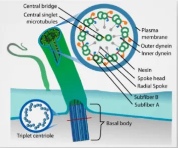
They elongate from a basal body, made of nine triplets of microtubules surrounding a cartwheel, a
hub-like central axis with radiating arm-like extensions located at the proximal tip of the basal body
(Vincensini et al. 2011). The core structure of cilia and flagella is the axoneme, a cylinder composed
of nine doublets of microtubules (Vincensini et al. 2011) - Figure 1.1.
Cilia and flagella play important roles in cellular motility, signal transduction, embryonic development,
and human disease (Nicastro et al. 2011), and can be found in several distinct organs and tissues
(Figure 1.4).
Figure 1.1 – Schematics of a 9+2 configuration cilia. Representation of a basal body and a cross section of a 9+2 axoneme. Adapted by Bloodgood et al. (Bloodgood 2010).
The most striking function of cilia and flagella is their involvement in cell motility. In protists, flagella
propel the organism in the aqueous environment, a feature also displayed by gametes in many
species (Vincensini et al. 2011; Guerrero et al. 2011).
Many major discoveries about cilia have been made in ‘simple’ organisms such as the nematode
Paramecium and Tetrahymena (Figure 1.3), or the kinetoplastid Trypanosoma, that are more
amenable to experimentation and genetic manipulation than vertebrate models − mouse, zebrafish
and Xenopus (Vincensini et al. 2011). Ciliates such as Paramecium and Tetrahymena are solid models for cilia studies, especially for the understanding of basal body and centriole assembly
(Vincensini et al. 2011).
Figure 1.2 – The green alga Chlamydomonas reinhardtii with two motile cilia/flagella. The green alga Chlamydomonas possesses two flagella (∼12 µm in length) that protrude from the apical end of the cell. The flagella beat in a breaststroke fashion and play an important role in response to light and in gamete adhesion. Chlamydomonas displays numerous practical and technical advantages for the study of the eukaryotic flagellum. Chlamydomonas cells exhibit complex swimming behaviours in response to various light stimuli, allowing for the dissection of flagellar beating regulatory pathways (Vincensini et al. 2011). Adapted from (Vincensini et al. 2011).
!
Figure 1.3 – The ciliated Tetrahymena. The 50µm long Tetrahymena
cell contains approximately 750 basal bodies. The huge number of cilia and basal bodies is a great advantage for biochemical studies of basal bodies, RNAi (RNA interference) studies, GFP tagging, genetic knockins and knockouts, and ultrastructural studies (Vincensini et al. 2011).
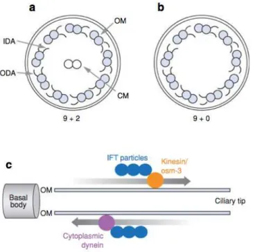
The synthesis of structural and functional components of cilia occurs in the cytoplasm and a
specialized system termed intraflagellar transport (IFT), is responsible for moving cargo (IFT particles)
toward the axonemal tip or away from it (anterograde and retrograde transport, respectively - Figure
1.5c). Pioneering work in the green alga Chlamydomonas reinhardtii, led to the discovery and
exhaustive characterization of a large number of genes involved in flagellum motility and construction,
such as axonemal dynein motors (reviewed in (Vincensini et al. 2011)) or the actors of the
intraflagellar transport (IFT) machinery, including motors (Pazour et al. 2000; Vincensini et al. 2011;
Dentler et al. 2009) and particle componentes (Piperno & Mead 1997; Cole et al. 1998), reviewed in
1.1.1 Cilia Classification
Cilia are classified according to their internal molecular arrangement and their ability to move (Supatto
& Vermot 2011). Two cilia types can be defined: primary and motile cilia.
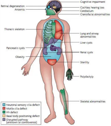
Figure 1.4 - Motile and primary cilia in diverse organisms and cell types. (a) The protozoan Paramecium is covered with motile cilia that enable it to swimming. (b) Motile cilia in the mammalian trachea. (c) Primary cilia in the renal tubules epithelia. (d) Electron micrograph of a mouse pyramidal neuron displaying a primary cilium. MC and DC denote the mother and daughter centrioles, respectively. (e) Primary cilia in the epithelial cells surrounding the lumen of pancreatic ducts. (f ) Micrograph of a primary cilium emerging from a human odontoblast. Figure from (Badano et al. 2006).
The primary cilium is generally shorter and immotile, and is present as a solitary structure on most
cells including epithelial cells, fibroblasts or neurons (Vincensini et al. 2011; Supatto & Vermot 2011).
They are related with sensory and signalling functions. According to internal arrangement, primary
cilia have a 9+0 axoneme (no central microtubule pair) and lack of dynein arms (Vincensini et al.
2011) - Figure 1.5b.
They were long thought to be vestigial, but were recently found to act as a complex signalling centre
(Singla & Reiter 2006). Primary cilia participate in numerous biological processes ranging from
chemo- and mechano-sensation to the transduction of an expanding list of signalling cascades that
are essential to regulate cellular and tissue homoeostasis (Vincensini et al. 2011; Singla & Reiter
2006). Their functional importance is highlighted by the growing list of diseases caused by defects at
the level of the primary cilium.
In contrast, motile cilia are found by the hundreds at the surface of epithelial cells lining the ventricles
of the brain, the respiratory tract and the oviduct, and they beat in a synchronized manner to direct
structure (Figure 1.1 and 1.5a) and are involved in ciliary beating. A 9+2 structure means the
axoneme of motile cilia is composed of nine peripheral doublet microtubules with attached outer and
inner dynein arms (ODA and IDA, respectively) and radial spokes, surrounding a central complex
(CC) with two central microtubules and the central sheath. ODAs and IDAs (axonemal dyneins*)
generate the force necessary for motility. The motor activity of dyneins is also regulated by several
other componentes (Supatto & Vermot 2011; Theegarten & Ebsen 2011; Schwabe et al. 2008): (i) the
radial spokes, which are T-shaped projections originating from each peripheral doublet and directed
towards the central pair, (ii) the projections associated with the central pair, and (iii) the dynein
regulatory complex, which is constituted by the nexin links. The basis for axonemal movement is the
sliding of microtubules relative to one another (Supatto & Vermot 2011; Theegarten & Ebsen 2011;
Schwabe et al. 2008). Dynein-generated forces that cause sliding between pairs of doublet
microtubules produce axonemal bending for axonemal microtubules are anchored to the cell via the
basal bodies, microtubule sliding causes the axoneme to bend (Supatto & Vermot 2011; Theegarten
& Ebsen 2011; Schwabe et al. 2008).
Figure 1.5 – Schematic representation of “9+2”and “9+0” axoneme cross sections and intraflagellar transport. (a, b)
Simplified diagram of the ultrastructure of motile and primary cilia. The axoneme of (a) 9+2 cilia is composed of nine outer doublets of microtubules (OM) surrounding a central pair (CM), whereas in (b) 9+0 the latter is not present. Inner (IDA) and outer (ODA) dynein arms are responsible for generating force for movement and project from one outer doublet to the next. (c) Along the outer microtubule doublets (OM) and under the ciliary membrane, IFT particles are transported towards the ciliary tip (anterograde) by kinesin and back to the basal body and the cell (retrograde) by the molecular motor cytoplasmic dynein. Figure and legend from (Badano et al. 2006).
!
!!!!!!!!!!!!!!!!!!!!!!!!!!!!!!!!!!!!!!!!!!!!!!!!!!!!!!!!!!!!!
*1.2 Ciliopathies and laterality defects
Given the multiple functions of cilia in development, cilia defects cause multiple human diseases
known as Ciliopathies (Marshall 2008; Fliegauf et al. 2007; Goetz & Anderson 2010), like polycystic
kidney disease (PKD) and primary ciliary dyskinesia (PCD). The role of motile cilia in a number of
physiological processes has been long recognized and thus the consequences of motile cilia
dysfunction have four major manifestations in mammals: early embryonic death due to failure of
embryonic turning; respiratory dysfunction; reproductive sterility; and hydrocephalus (Badano et al.
2006) (Figure 1.6). Ciliary dysfunctions involving motile cilia also result in body laterality defects
(McGrath 2003), like situs inversus (Afzelius 1995).
Not all Ciliopathies are related to motile cilia. Some are based in defects occurred in primary cilia, like
PKD (polycystic kidney disease), BBS (Bardet−Biedl syndrome), Joubert Syndrome, orofacial digital
syndrome, Alström syndrome or Meckel Gruber syndrome (Fliegauf et al. 2007; Vincensini et al.
2011). The link between the primary cilium and genetic diseases was first established by the study of
Pazour et al. (Pazour et al. 2000) who discovered that IFT88, a gene essential for IFT and flagellum
construction in Chlamydomonas reinhardtii was mutated in a mouse model for PKD. This mouse
model has been instrumental in the revelation of the essential role of the primary cilium in the
development of several tissues in vertebrates (Pazour et al. 2000; Vincensini et al. 2011).
1.2.1 Primary ciliary dyskinesia
Primary ciliary dyskinesia (PCD) is a rare, usually inherited, autosomal disease with a recessive
pattern. Estimation of prevalence is difficult and given between 1:10,000 to 40,000 live births (Afzelius
1995; Afzelius 2004; Theegarten & Ebsen 2011). Includes a group of diseases that present alterations
in the motility of respiratory cilia. The coexistence of PCD and situs inversus† is called Kartagener
syndrome and has a frequency between 40% and 50% among patients with PCD (Afzelius 1995).
Symptoms vary according to the age in which diagnosis is made, but commonly PCD is characterized
by the presence of simultaneous chronic infections of the upper and lower respiratory tracts, including
the middle ear, starting from the time of birth. Early diagnosis is very important because it is possible
to institute a number of respiratory measures that help to prevent the irreversible lung damage
(Ellerman & Bisgaard 1997). Diagnosis of PCD requires the presence of the characteristic clinical
phenotype and either specific ultrastructural ciliary defects identified by TEM or/and by abnormal
ciliary function (Bush et al. 2007; Theegarten & Ebsen 2011; Armengot et al. 2012).
Although PCD is a uniform clinical entity, studies conducted with electron microscopy have shown
that there are different subgroups within it. Axonemal dynein absence affects 70%-80% of patients
(Afzelius 2004; Jorissen et al. 1997). However, there are cases of patients with PCD and situs
inversus (Kartagener syndrome) with a completely normal ciliary ultrastructure (Afzelius 2004; Theegarten & Ebsen 2011; Merveille et al. 2010). On the other hand, it has been observed that there
may be ciliary abnormalities in the context of infection and inflammation (secondary ciliary
dyskinesia). For these reasons, the diagnostic validity of the ultrastructural study is limited (Bush et al.
2007), and has been replaced by high-speed videomicroscopy (Armengot et al. 2012; Green et al.
1993). Normal cilia beat frequency (CBF) is about 11-18 Hz, and any mean frequency less than 10 Hz
is considered abnormal (Green et al. 1993). According to Jackson et al. (Jackson et al. 2012)
investigation, high-speed videomicroscopy for studying CBF and motility pattern have to be done
under 37°C since different temperatures may increase the risk of PCD misdiagnosis. At present,
guidelines and algorithms have been developed to standardize diagnostic procedures (Bush et al.
2007) in order to ameliorate the PCD diagnostics.
Studies carried out in the green alga Chlamydomonas reinhardtii established the link between cilia
and several genetic diseases (Vincensini et al. 2011). Loss-of-function mutations in the human
ortholog of Chlamydomonas reinhardtii ODA7 disrupt dynein arm assembly and cause PCD (reviewed
in Vincensini et al. 2011).
!!!!!!!!!!!!!!!!!!!!!!!!!!!!!!!!!!!!!!!!!!!!!!!!!!!!!!!!!!!!!
†1.3 Role of cilia in the left-right patterning
The establishment of left-right (L-R) asymmetries in vertebrates occurs during early stages of
embryonic development through a complex process involving epigenetic and genetic mechanisms
(Lopes et al. 2010). Three consecutive axes are established during early development; they are the
dorsal-ventral, anterior-posterior and left-right axes (Figure 1.7) (Hirokawa et al. 2009).
Figure 1.7 – Body plan and axes in zebrafish: anterior-posterior, dorsal-ventral, left-right.
Adapted from (A. A. Smith et al. 2012)
The embryonic node, also known as the L-R organizer of the vertebrate embryo, is a ciliated structure
where motile cilia generate an important fluid-flow named ‘Nodal flow’. The Nodal flow is thought to
generate the first cues to establish the L-R axis of symmetry (Nonaka et al. 1998), either a chemical
or/and a mechanosensory signalling pathway that will culminate with correct positioning of internal
organs. Sometimes this process fails and gives rise to laterality defects or heterotaxy (Nonaka et al.
1998; McGrath 2003) like in Kartagener syndrome.
1.3.1 The two left-right models
The mechanism by which L-R is transferred to the LPM remains a major unanswered question in L-R
patterning (Hamada 2008; Marshall & Nonaka 2006). There are two prevailing models: one arguing
that a morphogen becomes concentrated on the left side of the node in response to flow - ‘morphogen
model’ (Okada et al. 2005; Nonaka et al. 1998; Tanaka et al. 2005) (Figure 1.8A); and other
suggesting that ‘two different cilia types’ in the node perform different functions (McGrath et al. 2003;
Tabin & Vogan 2003) (Figure 1.8B).
During the development of L-R theories, Tanaka and colleagues (Tanaka et al. 2005) argued that
lipid-bound vesicles containing morphogens are carried leftwards, breaking on the left side of the
node and thereby releasing their cargoes asymmetrically. This study showed that fibroblast growth
factor (FGF) signaling triggers the secretion of little membrane-vesicles (0.3–5"µm) termed ‘nodal
vesicular parcels’ (NVPs), that carry Sonic hedgehog (Shh) and retinoic acid (RA) inside the mouse
node (Tanaka et al. 2005). Based on Tanaka’s findings, Cartwright and colleagues (Cartwright et al.
2006) modeled the movement of NVPs across the mouse node and demonstrated that the flow
should indeed cause them to accumulate on the left side of the node, as required for symmetry
that the morphogens can not be delivered to the surrounding cells by their mechanical rupture either
by the cilia or the flow. They postulate that if there is rupture it must be induced by a biochemical
mechanism not yet discovered (Cartwright et al. 2006). Both studies contributed for the ‘morphogen
model’.
The second model is called ‘two-cilia model’ and is based on the discovery that in the embryonic
mouse node, there are motile cilia, identifiable with left-right dynein (Lrd) (McGrath et al. 2003). In addition, a population of non-motile cilia was reported and suggested to be capable of sensing the
mechanical stress of the stronger leftward flow (mechanosensory cilia) (McGrath et al. 2003; Tabin &
Vogan 2003). It has been proposed that the mechanosensory cilia trigger a left-sided calcium signal
through the action of polycystic kidney disease 2 (Pkd2) channels localized on their axonemes. Pkd2
is known to form a functional calcium channel that is thought to sense the bending of primary cilia
induced by urine flow in the renal tubule, thus functioning as a mechanosensor (Nauli et al. 2003).
Polycystic kidney disease 1 - like 1 (Pkd1l1) is the functional partner of Pkd2 in L-R patterning being conserved between medaka and mouse (Field et al. 2011). Pkd1l1-Pkd2 colocalize on motile cilia in
the medaka KV where all cilia are thought to be motile (Kamura et al. 2011).
Currently, a new idea in the field is emerging: all cilia (motile and immotile) may have sensory
functions. Motile cilia of the mammalian respiratory epithelium have been reported to exhibit both
mechanosensitivity and chemosensitivity (Bloodgood 2010). The mucociliary clearance in the airways
is a known example of mechanosensation where rises in viscosity are sensed by the motile cilia that
lead to an increase in cytosolic calcium which in turn is associated with increased CBF to accelerate
mucus clearance (reviewed in Bloodgood 2010). Recently, it was reported an unexpected
demonstration of chemoreception by the localization of different members of the bitter taste-receptor
family to motile cilia of airway epithelial cells (Shah et al. 2009). Indeed, bitter compounds induce a
rise in intracellular-calcium concentration in ciliated cells as well as an increase in CBF (Shah et al.
2009).
To date, both LR models are equally plausible because the molecular sensors of nodal flow are still
unknown.
Figure 1.8 – Schematic representation of the two L-R models. (A) ‘morphogen model’ - NVPs appear to be released from dynamic microvilli, transported to the left side by the nodal flow, and fragmented with the aid of cilia at the left periphery of the node. (B) ‘two cilia model’ - non-motile cilia are capable of sensing the mechanical stress of the leftward flow generated by motile cilia. Adapted from Tanaka et al. 2005) and (Tabin & Vogan 2003).
A
1.3.2 Left-right signaling cascade
The establishment of L-R patterning comprises three key steps (Shiratori & Hamada 2006): (i) the first
step is the symmetry-breaking event in the embryonic node, a leftward extracellular fluid flow that is
generated by the rotational movement of monocilia; (ii) the second step is the transduction of this
asymmetric information to the left LPM, where genes such as Nodal are asymmetrically expressed.
However, the mechanism that produces asymmetrical expression of Nodal and Cerl2 on the left and
right sides of the mouse node, and its potential link to the leftward nodal flow, is not fully understood
(Lopes et al. 2010); (iii) in the third step, these asymmetric signals drive the morphogenesis of various
asymmetric visceral organs (Shiratori & Hamada 2006).
Ciliary motility and the resulting nodal flow have also been shown to be required for L-R axis
formation at the posterior notochord in rabbit, the gastrocoel roof plate in Xenopus laevis, and in
Kupffer’s vesicle in zebrafish and medaka fish, finally resulting in the asymmetrical activation of the
Nodal signaling cascade in the left LPM (Okada et al. 2005; Essner 2005; Hojo et al. 2007;
Schweickert et al. 2007).
To date, the relationship between the fluid flow inside KV and the asymmetric function of charon in
medaka (Hojo et al. 2007) and zebrafish (Lopes et al. 2010), and its homologue coco in Xenopus
have been studied (Schweickert et al. 2007).
In summary, nodal flow is highly conserved and is important for L-R patterning in mouse, Xenopus
and fish.
1.4 Embryonic Left-Right organizers
1.4.1 Mouse node
Several studies indicate that the mouse node plays an important role in the establishment of L-R
asymmetry (Nonaka et al. 1998; Okada et al. 2005), and that is the reason why this organ is also
called mouse L-R organizer. The mouse node (Figure 1.9) is found at the rostral end of the primitive
streak. It consists dorsally of epiblast and ventrally of the most caudal aspect of the notochordal plate
(Brennan et al. 2002). The node gives rise to midline structures such as the notochord and floor plate
that act as a midline barrier necessary for maintaining correct laterality (reviewed in Brennan et al.
Figure 1.9 – Scanning Electron micrographs of a mouse node. Low-magnification view of a mouse embryo at 7.5 days post-coitum. Reichert’s membrane was removed. Higher-magnification view of the mouse node, where is visible nodal cilia and nodal pit cells. Adapted from (Okada et al. 2005).
!
Ultrastructural studies have shown that monocilia present on cells of the exposed ventral surface of
the node generate a net leftward flow of extra-embryonic fluid known as the nodal flow (Sulik et al.
1994; Nonaka et al. 1998), as a result of a clockwise rotation of polarized, posteriorly tilted cilia
(Shiratori & Hamada 2006; Nonaka et al. 2005) (Figure 1.10 and 1.11). These motile cilia bend like
ropes being whirled in circles (Cartwright et al. 2006). In mouse, posterior tilting of cilia occur
passively, since the apical surface of ventral node cells has a convex curvature and, because cilia
localize to the posterior side of the plasma membrane, this results in a tilt (reviewed in Brennan et al.
2002).
The precise structure of the motile cilia in the mouse node is still a point of discussion. Motile cilia,
such as those of the human respiratory tract or oviduct, generally have a 9+2 configuration in the
ciliary axoneme (Figure 1.1 and 1.5a). In contrast, mouse node cilia generally have a 9+0 microtubule
arrangement, i.e., they are devoid of central microtubule pair, and thus usually thought to be immotile
(Figure 1.5b), which is clearly not the case. Transmission EM of node cilia showed a 9+0
configuration (Sulik et al. 1994; Essner 2005).
At present it is thought that 9+0 node cilia are motile and produce a vortical motion in which the cilium
maintains a straight extended orientation but the distal end moves in a circle around the axis of
rotation - in contrast to the whip-like back – and - forth motion characteristic of 9+2 cilia (Nonaka et al.
1998). However, two recent studies have identified 9+2 cilia in the mouse node (Figure 1.12), perhaps
due to improved fixation conditions (Caspary et al. 2007). Thus, additional studies are needed to
define the structure and spatial organization of motile cilia in mouse node.
Figure 1.11 – Trajectory of Node Cilia Movement. (A) Trace of node cilia in enhanced DIC images after background subtraction. Positions of root are indicated in black, and tip in blue, green, and orange. Most cilia have a pattern consistent with the projection of a tilted cone (blue and green) whereas some cilia move in a D-shape (orange). A, P, L, and R refer to anterior, posterior, left, and right sides of the node, respectively. The direction of cilia rotation was clockwise (arrows); (B) Relationship between essentially rotatory movement of cilia and their projected images at various tilt angles.
Adapted from (Nonaka et al. 2005)
!
!
! ! ! !!
!
Several transcription factors are required for the formation of normal node cilia, like Foxj1 that is
specifically required for the formation of motile cilia in many tissues, and Rfx3 which acts as
transcription factor that binds to X-box motifs present in the regulatory regions of many ciliary genes
(Lee & Anderson 2008). In the absence of murine Foxj1, cilia of normal length are present on the
node but appear to be immotile, and situs is randomized as a result (Chen et al. 1998; Brody et al.
2000; M. M. Zhang et al. 2004). Rfx3 mutants form nodal cilia but they grow slowly and are shorter
than the wild-type cilia at the two-somite stage (when nodal flow is required)(Bonnafe et al. 2004).
Rfx3 mutants show randomized L-R situs, suggesting that cilia must have a specific length to generate effective nodal flow (Bonnafe et al. 2004).
1.4.2 Zebrafish Kupffer’s vesicle
Kupffer’s vesicle (KV) is the equivalent to the mouse node ciliated organ of asymmetry in the
zebrafish embryo that initiates L-R development of the brain, heart and gut (Essner 2005). First
described in 1868 by Kupffer, KV is a conserved structure among teleost fishes, like Fundulus
heteroclitus (Brummett & Dumont 1978). In zebrafish, KV is formed from a group of approximately
two-dozen cells, known as dorsal forerunner cells (DFCs‡) that migrate at the leading edge of the
embryonic shield (the zebrafish equivalent of the mouse node that gives rise to the notochord and
floorplate) during gastrulation. At the end of gastrulation, DFCs migrate deep into the embryo and
organize to form KV (reviewed in (Essner 2005)). During subsequent somite stages, KV constitute a
small but distinctive epithelial closed vesicle containing fluid, located mid-ventrally posterior to the yolk
cell or its extension, and transiently present during most of the segmentation period (Kimmel et al.
1995; Kramer-Zucker 2005).
! ! ! ! ! !
Figure 1.13 – DIC images of the zebrafish KV. (A and C) KV dorsal view in a 10th
somite zebrafish embryo; (B) KV lateral view in a 10th
somite zebrafish embryo.
!!!!!!!!!!!!!!!!!!!!!!!!!!!!!!!!!!!!!!!!!!!!!!!!!!!!!!!!!!!!!
‡ In contrast to other cells in this region, DFCs do not involute during gastrulation, but remain at the leading edge of epibolic movements.
Figure 1.14 – Immunofluorescence experiment for visualizing KV cilia. Imaging experiment using a sox17:GFP transgenic zebrafish line, showing KV monocilia labelled with an antibody anti- acetylated alpha-tubulin (in red).
The epithelium of the KV is made of 9+2 (Ferrante et al. 2009; Kramer-Zucker 2005; Kreiling et al.
2007) monociliated cells (Figure 1.14) that generate a counterclockwise fluid flow. This flow triggers
asymmetric calcium response on the left side of the cavity (Francescatto et al. 2010; Sarmah et al.
2007) and is involved in establishing and maintaining the L-R asymmetry of the body axis (Essner
2005; Kramer-Zucker 2005) as described in this Chapter. Even though, the presence of sensory
immotile cilia is still a matter of debate in the fish (Borovina et al. 2010; Okabe et al. 2008).
Currently, it is assumed that KV is analogous to the mouse node in terms of L-R patterning (Essner
2005). While the ciliated surface of the mouse node is relatively flat, KV is a hollow sphere containing
cilia, projecting both from dorsal roof and ventral floor (Kreiling et al. 2007; Amack et al. 2007).
Kramer-Zucker et al. (Kramer-Zucker 2005) reported that cilia rotate counterclockwise when viewed
from the apical side in the KV, which is opposite to what is observed in the mouse node. Kreiling et al.
(Kreiling et al. 2007) found by 3D images analysis that cilia are concentrated in the dorsal-anterior
region of the vesicle, suggesting that these cilia cause the dominant counterclockwise flow in KV
(Figure 1.15). Okabe at al. (Okabe et al. 2008) hypothesized that the net flow in zebrafish KV is
analogous to flow in the mouse node and medaka fish in terms of L-R patterning even though the
ciliated cell structures appear to differ in architecture. Furthermore, Okabe et al. (Okabe et al. 2008)
SEM and video microscopy studies demonstrate that numerous cilia within the vesicle are tilted to the
posterior direction. Although the posterior tilt of the cilia in the mouse node is thought to be due to a
shift of the base of the cilia to the posterior, the same can not be confirmed in KV (Okabe et al. 2008).
It is more likely that the combination of cilia bending and cell orientation of KV causes cilia to be
pointed towards the posterior (Okabe et al. 2008).
!
!
!
!
!
Figure 1.15 – Typical three-dimensional (3-D) reconstructions of confocal data sets of KV cilia at the 9th somite stage projected in the
Cave. Distribution of KV cilia (green); anterior (a) is on top, posterior (p) is on the bottom. Some of the anterior cilia have been marked (red cones). The asymmetric distribution of cilia can be seen as a clustering of cilia on the anterior surface of KV. Adapted from (Kreiling et al. 2007).
Some differences were found between mouse node and KV from medaka and zebrafish (Figure 1.16).
According to Kamura et al. (Kamura et al. 2011), all medaka KV cilia are motile, whereas in the
mouse node McGrath et al. (McGrath et al. 2003) showed the presence of two populations of cilia,
one motile and other immotile. Furthermore, Caspary et al. (Caspary et al. 2007) showed cilia from
the mouse node with central pair, adding the presence of another type of cilia to the granted
hypothesis of the 9+0 cilia (Nonaka et al. 2005) (as described previously – Figure 1.12). In zebrafish,
immotile and motile cilia were seen in the KV, although is still a matter of debate (Borovina et al.
2010; Okabe et al. 2008).
However, the fluid dynamics inside KV is not completely understood and several groups are trying to
figure it out (Supatto & Vermot 2011; Kreiling et al. 2007; Okabe et al. 2008; A. A. Smith et al. 2012;
Cartwright et al. 2008). The diversity of cilia motility and fluid mechanics in the embryo is starting to be
studied in vivo (Supatto & Vermot 2011). The number of flows that can be generated by such simple
structure is limited, but the combination of cilia beating pattern and particular topology of the
environment can participate to build up complex flows.
1.5 Motile cilia generated flow in zebrafish
1.5.1 Motile cilia beat pattern
A key parameter dictating cilia-mediated hydrodynamics is the type of beat they generate (Figure
1.18). The beat pattern seems related with the internal organization of the cilia but many unresolved
questions remain concerning the correlation of structure and cilia beat in different developing organs.
The motility depends on the presence of dynein arms that are attached to the microtubules, the
dynein regulatory complex (DRC) and on the presence of radial spokes (reviewed in Vincensini et al.
2011). Vertebrates 9+2 cilia can significantly bend during its motion with effective and recovery
strokes, while 9+0 cilia have an almost perfect circular motion (Nonaka et al. 1998; Nonaka et al.
2005). In protists, the presence of the central pair of microtubules is critical in shaping the overall
tridimensional motion of the beating cilium. The so-called central pair hypothesis constitutes an
attractive view of the structural basis of cilia beat§ (Castleman et al. 2009). In humans and zebrafish,
mutants for the radial spoke heads affect cilia motion (Castleman et al. 2009) and the DRC is critical
for proper cilia motility in zebrafish (Colantonio et al. 2009).
In terms of motility, little is known about potential factors that could control the direction and frequency
of beating cilia in the zebrafish embryo. The relationship between the beat pattern and the directional
flow will be discussed further in this section.
1.5.2 Motile Cilia Beat Frequency
In cases of Ciliopathies such as PCD, cilia beat frequency (CBF) is an important feature for the
definitive diagnose (Armengot et al. 2012; Shoemark et al. 2012; Jackson et al. 2012). Cilia from
nasal brushings and biopsy material are screened using high-speed videomicroscopy, as a way of
studying CBF (Armengot et al. 2012; Shoemark et al. 2012; Jackson et al. 2012). CBF values under
10 Hz are considered abnormal (Green et al. 1993).
Several studies have been done in order to better characterize the CBF of ciliated organs in distinct
animal organisms (Table 1.1). Some methodologies and techniques used for human CBF analysis
were originated and validated in animal in vivo studies. Nevertheless, CBF should be even more
characterized in model organisms, especially when related with L-R problems (Kunimoto et al. 2012;
Lechtreck et al. 2008; Norris & Grimes 2012; Lucas et al. 2011).
!!!!!!!!!!!!!!!!!!!!!!!!!!!!!!!!!!!!!!!!!!!!!!!!!!!!!!!!!!!!!
§Table 1.1 – Cilia properties in various embryonic models. Zebrafish, mouse, medaka and Xenopus are used as ciliated models. Cilia ultrastructure, CBF, length and rotation direction, among other ciliary characteristics are reviewed in this table, for distinct organs. Figure from (Supatto & Vermot 2011).
1.5.3 Fluid hydrodynamics
Essentially, the main function of motile cilia is to generate fluid flow at the micrometer scale. However,
fluid dynamics are governed by laws that are not trivial at this scale and the resulting fluid flow
presents features that are not intuitive when used to human scale (Supatto & Vermot 2011).
In fluid mechanics, the Reynolds number (Re) is a dimensionless number that characterizes the
nature of a fluid flow and the relative contribution of inertia and viscous dissipation. The cilia-driven
flow involved in zebrafish development exhibits characteristic scales, L < 100µm and U < 100 µm s-1
(L = length; U = typical velocity) (Supatto & Vermot 2011). Using the kinetic viscosity of water (ν≈ 106
µm2 s-1), the resulting Re is < 10-2. The flow generated by beating cilia is characterized by a low Re
(Re << 1) environment**, and it is governed by Stokes equations - being referred as ‘creeping flow’ or
‘Stokes flow’ (Supatto & Vermot 2011).
From a modeling perspective, the low value of the Re enables the simplification of the Navier–Stokes
flow equation, the general model governing fluid dynamics (Supatto & Vermot 2011). If Re << 1, the
inertial forces can be neglected compared to the viscous forces and the unresolved Navier–Stokes
flow equation can be approximated by the linear Stokes flow equation (Figure 1.17). This equation is
the starting point of any modeling of cilia-driven flows (Cartwright et al. 2006; D. J. Smith et al. 2008;
D. J. Smith et al. 2010; A. A. Smith et al. 2012; Supatto et al. 2008). Thus, the flow generated by an
ensemble of cilia can be approximated as the sum of the flow induced by each single cilium. In
addition, the linearity and the time reversibility of ‘Stokes flow’ equation provides fundamental
properties of the flow that can be generated by motile cilia (Supatto & Vermot 2011).
!!!!!!!!!!!!!!!!!!!!!!!!!!!!!!!!!!!!!!!!!!!!!!!!!!!!!!!!!!!!!
**At the human scale, a swimmer (L ≈ 1m and U ≈ 1ms-1) should swim in a fluid that is 108 times more viscous than water to experience such fluid behavior.
!
Figure 1.17 - Fluid dynamics at low Reynolds number. The velocity field
u of a fluid can be generally described by the Navier–Stokes flow equation. Each term of this equation scales as a force per unit volume: finertia includes
the time-dependent and the nonlinear intertial components (blue), fpressure is
the force generated by a pressure gradient (green), fviscosity is the viscous
dissipation term (orange), and fext corresponds to external forces applied to
the liquid (such as force generated by a motile cilium). The Reynolds number (Re) compares finertia with fviscosity to check if one of these terms can be
neglected in the Navier–Stokes equation. Re << 1 means that viscous forces dominate inertia. In this case, the inertial term can be neglected to obtain the ‘Stokes flow’ equation. Figure and legend from(Supatto & Vermot 2011).
1.5.4 Directional flow
Once cilia beat in their fluidic environment, how can they generate a directional flow? Based on
‘Stokes flow’ equation, the first challenge to the generation of a directional flow is the difficulty to
obtain a net flow (Supatto & Vermot 2011). The absence of inertia results in a velocity that is
proportional to the force applied to the fluid†† (Supatto & Vermot 2011; A. A. Smith et al. 2012). Time
reversibility of the ‘Stokes flow’ equation means that the time asymmetry is not sufficient to create a
net flow (reversing time does not affect the equation), as described by (Supatto & Vermot 2011; A. A.
Smith et al. 2012). According to both studies (Supatto & Vermot 2011; A. A. Smith et al. 2012), in
order to produce a directional flow at low Re, a beating cilium needs an asymmetry is space or shape.
Models predict that a directional flow cannot be produced by either a stiff cilium rotating in free space
(Buceta et al. 2005) or a distribution of cilia arranged on a spatially asymmetric pattern (Cartwright et
al. 2006), as reviewed in (Supatto & Vermot 2011). Presumably due to the cilia ultrastructure, length,
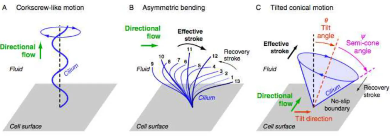
and/or orientation, three types of spatially asymmetric cilia beating patterns have been proposed
theoretically and observed experimentally in different organs of developing embryos (Figure 1.18): the
‘corkscrew-like motion’, the ‘asymmetric bending’, and the ‘tilted conical’ motion (Supatto & Vermot
2011).
!!!!!!!!!!!!!!!!!!!!!!!!!!!!!!!!!!!!!!!!!!!!!!!!!!!!!!!!!!!!!
Figure 1.18 - Pumping flow with motile cilia at low Reynolds numbers: three types of spatially asymmetric beating patterns observed experimentally: (A) helical motion or corkscrew-like motion which pump fluids along the cilium, (B) asymmetric motion which is related to an asymmetric bending of the cilium during its movement, (C) cylindrical rotation with a tilted cilium. In (B) and (C), the effective stroke corresponds to the cilium momentum where fluid is moved efficiently in the direction of motion, whereas poor transport occurs during the cilia recovery phase (Supatto & Vermot 2011).
The ‘corkscrew-like motion’ (Figure 1.18A) is well known and is used by flagella to propel bacteria, but
is not common in cell epithelium. Nevertheless, such cilia motion pattern has been observed during
zebrafish kidney development (Kramer-Zucker 2005). In the ‘corkscrew-like motion’, the direction of
the net flow generated is parallel to the rotation axis of the cilium, and it depends on the rotation
direction and the helix orientation (Supatto & Vermot 2011).
In the ‘asymmetric bending’ (Figure 1.18B), the cilium changes its shape while rotating. During one
beating cycle, its bending is not the same on one way compared to the other, producing an effective
and a recovery stroke (Supatto & Vermot 2011). The flow direction is perpendicular to the rotation
axis of the cilium and depends on the bending asymmetry. Flow direction does not directly depend on
the cilium direction of rotation, but on the orientation of the effective stokes (Supatto & Vermot 2011).
Such beating pattern has been observed in the left–right organizer in xenopus (Schweickert et al.,
2007).
At last, the ‘Tilted conical’ motion (Figure 1.18C) is characterized by having a directional flow
generated by cilia that exhibits a symmetrical circular motion without asymmetric shape as in the two
previous cases. This type of cilia motion has been observed experimentally in mouse, rabbit, and fish
L-R organizer (Okada et al. 2005). Recent models proposed that the spatial symmetry can be broken
without the bending of the cilium. In this case, the cilium interaction with the cell surface plays a
critical role (Cartwright et al. 2006; D. J. Smith et al. 2008; D. J. Smith et al. 2010; A. A. Smith et al.
2012; Vilfan et al. 2012): if the velocity of the fluid at the cell surface match the velocity of the surface
itself (no-slip boundary condition (Supatto & Vermot 2011)), the rotation forms an effective stroke
when the cilium is far from the surface, and a recovery stroke when it is close to the surface. Thus,
perpendicular to the direction of the cilium tilt and parallel to the surface. The model predictions of a
cilium tilt have been experimentally confirmed in vivo in mouse (Okada et al. 2005) (Figure 1.10) and
in zebrafish (Supatto et al. 2008). Interestingly, among the three asymmetric beating patterns
generated by biological cilia, the tilted conical motion is the easiest to reproduce artificially as it does
not require controlling the bending of the cilium. For this reason, the generation of directional flow at
low Re in microfluidic devices can be obtained using stiff artificial cilia beating with tilted conical
motion (Nonaka et al. 2005; Vilfan et al. 2012).
Among the asymmetric cilia beating patterns, it is interesting to mention that only the ‘tilted conical’
motion allows breaking the L-R symmetry without preexisting L-R asymmetry at the scale of the cilium
(Supatto & Vermot 2011). This pattern allows translating an anterior–posterior or dorsal-ventral
asymmetry into a L-R asymmetry (Supatto & Vermot 2011). This would not be true for ‘corkscrew-like’
motion or ‘asymmetric bending’: a net flow in the L-R direction would require having a preexisting L-R
asymmetry at the level of the cilium (Supatto & Vermot 2011).
1.5.5 Interplay between models and experiments
Efforts have been made to address the hydrodynamics involved in the L-R symmetry breaking during
early vertebrate development. Through the modeling of the leftward directional flow in the L-R
organizer, some key predictions have been explored.
In recent years, several models and simulations have been developed based on mouse node
experimental data (section 5.1 of this Chapter). In these theoretical studies, the cilium is modeled
either as an infinitesimal sphere rotating in its place (Cartwright et al. 2006), a small sphere moving on
a fixed trajectory in the vicinity of a planar surface (Vilfan et al. 2012) or a slender body (D. J. Smith et
al. 2008). Whatever the complexity of the model used, they are a great advantage to understand how
biological processes occur, by explaining new observations, raising specific predictions, and
suggesting the next set of experiments.
How the spatial asymmetry allows the generation of a directional flow within the L-R organizer was
the first question that theorists wanted to address. The proposition that cilia were beating with a tilted
conical motion‡‡ was first suggested by Cartwright et al. (Cartwright et al. 2006) and further
investigated by demonstrating the importance of the cell surface and the no-slip boundary condition to
obtain efficient and recovery strokes (D. J. Smith et al. 2008; Vilfan et al. 2012). It is noted that the
beating pattern of KV’s cilia is different from the mouse nodal cilia since they do not share the same
ultrastructure. More sophisticated models are now taking into account the geometry of the L-R
organizer and the spatial distribution and the density of cilia in mice. Kreiling et al. (Kreiling et al.
!!!!!!!!!!!!!!!!!!!!!!!!!!!!!!!!!!!!!!!!!!!!!!!!!!!!!!!!!!!!!
‡‡2007) showed that 80% of the cilia are located on the dorsal side of KV, revealing that the structure of
KV could be more similar to the ventral node in mouse embryos than previously believed.
While many features are common between mice and fish, each species remain distinct and might
have developed different ways to break the embryonic symmetry using the same basic ciliary
machinery (Supatto & Vermot 2011). Thus, it is crucial to improve the characterization of the L-R
organizers, especially the differences between the different animal organisms, in order to establish
more accurate L-R theoretical models.
1.6 Project goals
The main goal of this Master project was the characterization of cilia motility through the study of cilia
beat frequencies (CBFs) in the Kupffer’s vesicle (KV) of zebrafish embryos. It was our objective to
understand how could potential changes in CBFs impact on KV fluid flow, important for the
establishment of the left-right axis of asymmetry.
The first part of the project consisted in studying CBF in the Kupffer’s vesicle monociliated cells using
spectral analysis. It was our objective to film KV cilia, using high-speed videomicroscopy, and apply a
designed script, that allowed the extraction and decomposition of CBF results from the movies. The
second part of this project was to study the cilia-generated fluid flow inside the zebrafish KV by
tracking native particles and calculating their velocity in a non-invasive assay. Through the study of
CBF and KV fluid flow mean velocity, we aimed to investigate if there were different populations of
cilia based on CBF criteria and to classify the range of fluid flow velocities operating in this organ.
A tissue-specific screen previously done in our lab was the foundation for the third part of my Master
project. With this screen we found several differential expressed genes in deltaD-/- mutants, compared
to wild-type embryos. Having identified some motile cilia-related genes, we proposed a validation
study of gene expression using quantitative real time polymerase chain reaction (qPCR) and
whole-mount in situ hybridyzation (WISH). The three motile cilia-related genes we proposed to validate were
dnha7§§, rsph3*** and foxj1a†††. It was our ultimate goal, to test causality between CBF patterns and gene expression.
With this Master project we wanted to test if deltaD mutants had a different CBF evaluation and
whether that could partially explain the defects in L-R patterning reported (Lopes et al. 2010).
!!!!!!!!!!!!!!!!!!!!!!!!!!!!!!!!!!!!!!!!!!!!!!!!!!!!!!!!!!!!!
§§dnha7 (Dynein Heavy Chain 7) was one of such differentially expressed genes. The expression of dnha7
mRNA is induced during ciliated cell differentiation, being present in normal motile cilia. Zhang et al (2002) identified DNAH7 as an inner dynein arm component of human cilia important for its motility. Cilia from PCD patients have an absence of DNAH7.
*** Initially identified in Chlamydomonas,
rsph3 (Radial Spoke Protein Homolog 3)81is 1 of more than 20 identified
radial spoke structural components of motile cilia and is required for axonemal sliding and flagellar motility. Radial spoke proteins are thought to be important in transducing signals from the inner pair of microtubules to the outer doublets in the flagellar axoneme, regulating dynein-mediated axonemal sliding and subsequent flagellar motility. †††
foxj1a gene is considered the master motile ciliogenic transcription factor 82, and as showed by Lopes et al.27