Dirceu Henrique Paulo Mabunda
maio de 2014
Electroconvulsive therapy in psychotic
patients: interface between perceived stress,
anxiety and depression and the clinical
outcome
Electroconvulsoterapia em pacientes
psicóticos: interface entre o stress percebido,
ansiedade e depressão e resultado clínico
UMinho|20 14 Dir ceu Henriq ue P aulo Mabunda Electroconvulsive t herap y in psy cho
tic patients: inter
face be
tw
een perceived s
tress, anxie
ty and depression and t
he clinical outcome
Universidade do Minho
Escola de Ciências da Saúde
Trabalho efetuado sob a orientação do
Prof. Doutor João Miguel Seiça Bessa Peixoto
e co-orientação do
Professor Doutor António Pacheco Palha
Dirceu Henrique Paulo Mabunda
maio de 2014
Dissertação de Mestrado
Mestrado em Ciências da Saúde
Electroconvulsive therapy in psychotic
patients: interface between perceived stress,
anxiety and depression and the clinical
outcome
Electroconvulsoterapia em pacientes
psicóticos: interface entre o stress percebido,
ansiedade e depressão e resultado clínico
Universidade do Minho
Escola de Ciências da Saúde
iii
«As for a future life, every man must judge for himself between conflicting vague probabilities.» Charles Darwin
v
AGRADECIMENTOS
Ao Prof DoutorJoão Bessa por me ter orientado ao longo deste ano. Pela sua disponibilidade, boa disposição e pela sinceridade que muitas vezes ouvi«Qual é a tua ideia.O que pensas fazer? Está bem assim. É esta a tua ideia. Perfeito». Muito Obrigado!
Ao Professor Doutor António Pacheco Palha pelo suporte, sem mãos a medir, sem o qual não seria possível alcançar os actuais resultados. Pela sinceridade, objectividade e flexibilidade- um verdadeiro mestre. O meu muito obrigado!
À Professora Doutora Joana Palha por ter criado condições para que pudessemos frequentar o curso nesta escola, com ela tivemos o primeiro contacto nesse sentido. Muito Obrigado
Às irmãs hospitaleiras e todos funcionários da Casa de Saúde do Bom Jesus, especialmente ao Dr António Guimarães pelos ensinamentos e disponibilidade em apoiar a minha formação. Ao Dr Jorge Mota pelo apoio incondicional prestado para a materialização deste projecto.Aos funcionários do Hospital de Magalhães Lemos- unidade de electroconvulsoterapia pela hospitalidade e apoio incondicional prestado durante a realização do estudo naquela unidade hospitalar.À Prof Doutora Margarida Correia-Neves pelo apoio incondicional que prestou nos primeiros dias da nossa estadia no minho e pelo contínuo suporte nas actividades académicas. Ao Prof. Doutor João Cerqueira pelos ensinamentos e pela disponibilidade em ensinar. O meu muito obrigado Ao grupo das neurociências por todo o apoio prestado, em especial ao José Carlos Nunes Portugal pelo apoio prestado nos momentos dificeis nesta academia- muito obrigado por me ter ajudado a compreender e a adaptar-me ao ritmo da escola, à Sofia Neves e à Mónica Morais.Ao Ministério da Saúde de Moçambique, em especial a Dra Lídia Gouveia pelo apoio e pela contribuição ímpar para a materialização da minha formação nesta escola. O meu muito obrigado.
Agradecimento especial à minha esposa Anatércia, às minhas filhas Olívia e Maira( que Deus te proteja no eterno descanso) pela compreensão e suporte para materialização do sonho de estudar para além das terras do índico.Aos meus pais, Maria Olívia da Costa e Paulo Vasco Mabunda. Em especial aos meus irmãos por sempre me incentivarem a continuar na caminhada em busca do conhecimento.Aos meus colegas,Aida, Flávio, Sámia e Wilza pelos momentos que partilhamos dentro e fora da academia diariamente. O meu muito obrigado.
vii
Electroconvulsive therapy in psychotic patients: interface between perceived stress, anxiety and depression and the clinical outcome
ABSTRACT
Electroconvulsive therapy (ECT) continues to be considered an effective treatment of psychotic patients with hallucinatory and delusional symptoms resistant to antipsychotic medication. Several studies have evaluated the effectiveness of ECT in major depression as well as the effect of cortisol in the evolution and prognosis of those patients. However, studies that evaluate the evolution of the delusional and hallucinatory schizophrenic patients, psychotic depression and schizoaffective psychotic patients treated with ECT and its correlation with the salivary cortisol are scarce. Moreover, stress plays a significant role in modulation of mental disorders, and the hypothalamic-pituitary-adrenal (HPA) axis regulates the biological mechanisms of stress.
In this project we have assessed the clinical evolution of patients in three diagnostic groups, namely patients psychotic depression, schizoaffective disorder and schizophrenia before and after ECT. The Positive and Negative Syndrome Scale (PANSS) was used to evaluate psychotic symptoms, the Perceived Stress scale (PSS) to evaluate perceived stress, the Hospital Anxiety and Depression Scale (HADS) to evaluate anxiety and depression and salivary cortisol levels to evaluate the HPA axis function. The results of our study demonstrate that ECT is effective and a valuable therapeutic option which could be useful as adjunctive therapy in psychotic patients refractory to antipsychotics agents. There was a noticeable improvement in the PANSS, HADS and PSS scores after ECT in all psychotic patients in our study. Moreover, this study shows the effectiveness of ECT in chronic psychotic patients, while most studies have studied first psychotic episodes. The decrease of salivary cortisol after ECT in our study suggests that it may impact in the HPA axis, improving its function. However, the mechanism by which it can improve HPA axis function remains unclear. Finally, the correlations between variables revealed that the HADS score may predict the rate of response to ECT.
In conclusion, the present study has confirmed the role of ECT in the treatment of psychotic disorders. Importantly, a relation between HPA axis function and ECT was revealed specifically in patients with schizophrenia and psychotic depression but not in schizoaffective disorder, suggesting a distinct involvement of this key neurobiological factor.
ix
Eletroconvulsoterapia em pacientes psicóticos: interface entre o stress percebido, ansiedade e depressão e resultado clínico
RESUMO
A eletroconvulsoterapia (ECT) continua a ser considerada um tratamento eficaz para pacientes psicóticos com sintomatologia delirante e alucinatória resistente à medicação antipsicótica. Vários estudos têm avaliado a eficácia da ECT na depressão major, bem como o efeito do cortisol na evolução e prognóstico desses pacientes. No entanto, estudos que avaliam a evolução em pacientes com esquizofrenia , depressão psicótica e psicose esquizoafetiva tratados com ECT e sua correlação com o cortisol salivar são escassos. Além disso, o stress desempenha um papel significativo na modulação de perturbações mentais sendo regulado pelo eixo hipotálamo-hipófise-adrenal (HPA).Neste projeto, avaliamos a evolução clínica dos pacientes em três grupos diagnósticos, nomeadamente pacientes com depressão psicótica, perturbação esquizoafetiva e esquizofrenia, antes e depois da ECT. A escala de sintomas positivos e negativos (PANSS) foi utilizada para avaliar sintomas psicóticos, a escala de stress percebido (PSS) para avaliar a percepção de stress, a escala de ansiedade e depressão hospitalar (HADS) para avaliar a ansiedade e a depressão e os níveis de cortisol salivar para avaliar a função do eixo HPA.
Os resultados do nosso estudo demonstram que a ECT é eficaz e uma valiosa opção terapêutica que pode ser útil como terapia adjuvante em pacientes psicóticos refratários a agentes antipsicóticos. Observou-se uma melhoria significativa nas pontuações PANSS, HADS e PSS após ECT em todos os grupos de pacientes psicóticos no nosso estudo. Além disso, este estudo demonstrou a eficácia da ECT em pacientes psicóticos crónicos, enquanto a maioria dos trabalhos têm estudado primeiros episódios psicóticos. A diminuição do cortisol salivar após ECT no nosso estudo sugere que esta tem um impacto sobre o eixo HPA, melhorando a sua função. No entanto, o mecanismo pelo qual ela modula a função do eixo HPA permanece desconhecido. Finalmente, as correlações entre as variáveis revelaram que os niveis de HADS podem predizer a taxa de resposta à ECT.
Em conclusão, o presente estudo confirmou o papel da ECT no tratamento de perturbações psicóticas. De realçar, a relação entre a função do eixo HPA e ECT foi revelada especificamente em pacientes com esquizofrenia e depressão psicótica, mas não na perturbação esquizoafetiva, sugerindo um envolvimento distinto deste factor neurobiológico.
xi Index DECLARAÇÃO ... II AGRADECIMENTOS ... V ABSTRACT ... VII RESUMO ... IX 1. INTRODUCTION ... 1 1.1. Psychotic disorders ... 3
1.1.1. Schizophrenia: from the origin to nowadays ... 4
1.1.2. Dopaminergic Pathways: ... 5
1.1.3. Glutamatergic Pathways: ... 5
1.1.4. Etiology of Psychosis ... 6
1.1.5. Antipsychotics ... 7
1.2. Electroconvulsive Therapy: ... 8
1.2.1. Mechanism of action of ECT: ... 8
1.2.2. ECT Technique: ... 9
1.2.3. ECT effectiveness:... 10
1.2.4. Indications of Electroconvulsive Therapy ... 10
1.3. Stress and psychosis: ... 11
1.3.1. The role of HPA-Axis ... 11
1.4. Final considerations ... 12
2. OBJECTIVES ... 15
3. MATERIAL AND METHODS ... 19
3.1. Sample: ... 21
3.1.1. Inclusion criteria: ... 21
3.1.2. Exclusion criteria: ... 21
xii 3.3. Experimental Design ... 23 3.4. Variables assessment ... 23 3.4.1. Clinical Variables: ... 24 3.5. Cortisol Sampling: ... 24 3.5.1. Principle of assay ... 24 3.5.2. Test protocol ... 25
3.6. The Positive and Negative Syndrome Scale Assessment(PANSS) ... 26
3.7. Perceived Stress Scale Assessment ... 26
3.8. Hospital Anxiety and Depression Scale Assessment ... 26
3.9. Statistical Analysis ... 27
4. RESULTS ... 29
4.1. Clinical diagnostic and socio-demographic characteristics: ... 31
4.1.1. Response to ECT among psychotic patients ... 31
4.1.2. Effects of ECT on Perceived stress ... 33
4.1.3. Effects of ect on Anxiety and depression scores ... 34
4.1.4. effects of ect in salivary Cortisol ... 35
4.2. Differences in clinical outcome between gender ... 36
4.2.1. Improvement of negative, positive and general symptoms after ECT ... 38
4.2.2. Anxiety and depression among psychotics patients ... 39
4.2.3. Perceived Stress ... 41
4.2.4. Cortisol among psychotics patients undergoing ECT ... 41
4.3. Correlation between Variables ... 42
4.3.1. Correlation between variables among Schizophrenic patients ... 43
4.3.2. Correlation between variables in Psychotic Depression patients group ... 47
4.3.3. Correlation between variables in Schizoaffective psychotic patients group ... 50
4.4. Model regression to predict clinical outcome In psychotic patients undergoing ect ... 51
4.4.1. Anxiety and Depression before ect as a predictor of response to ect in psychotic patients. . 51
4.4.2. Perceived Stress before ect as a good predictor of response to ect in psychotic patients .... 52
xiii
5.1. Clinical and socio-demographic characteristics of patients... 57
5.2. Response to ECT in psychotic patients... 57
5.3. Psychometric measurements of clinical improvement after ECT ... 57
5.4. the effect of ect in the HPA- axis ... 58
5.5. Correlations between cortisol and clinical variables ... 58
5.6. Correlations between variables,and regression model ... 58
5.7. Conclusion: ... 61
5.8. Limitations and future directions: ... 63
6. REFERENCES ... 65
7. ATTACHMENTS ... 78
Tables index Table 1. Procedure of ELISA ... 25
Table 2. Effectiveness of ECT on PANSS... 32
Table 3. Descriptive statistic of perceived stress ... 33
Table 4. Descriptive statistic of HADS ... 34
Table 5.Descriptive statistic of salivary cortisol ... 35
Table 6. Descriptive statistic of differences between gender ... 36
Table 7. Independent t-test differences between gender ... 36
Table 8. General correlations ... 42
Table 9. Correlations in schizophrenic patients ... 43
Table 10. Correlation in psychotic depression patients ... 47
Table 11. Correlation in schizoaffective patients ... 50
Table 12. HADS model regression ... 51
Table 13. ANOVA HADS... 51
Table 14. Coefficients of regression ... 52
Table 15. PSS Model ... 52
Table 16.ANOVA PSS ... 52
Table 17. Coefficient of regression model ... 53
Figure 1.Schematic representantion of biphasic brief pulse. ... 10
Figure 2 Schematic representation of electrode placement.. ... 23
xiv
Figure 4. Average change in PANSS scores after ECT in the different experimental groups ... 31
Figure 5. Effects of ECT on PANSS. ... 32
Figure 7. Effectiviness of ECT in Anxiety and Depression. ... 34
Figure 8. Effects of ECT on salivary cortisol levels among psychotic patients ... 35
9. Differences between genders in BMI ... 37
10. Differences between genders on PANSS. ... 37
Figure 11 Measure of PANSS before and after ECT . ... 39
Figure 12 Measure of Anxiety and Depression . ... 40
Figure 13Perceived stress among patients undergoing ECT ... 41
Figure 14 Cortisol in three psychotic patients ... 42
.Figure 15.Correlation between salivary cortisol and perceived stress before ... 44
Figure 16.Correlation between salivary cortisol after ECT and HADS score ... 45
Figure 17.Correlation between perceived stress and anxiety and depression in schizophrenic. ... 46
Figure 18. Correlation between perceived stress after ECT and negative symptoms ... 46
Figure 19.Correlation between perceived stress after ECT and anxiety and depression before ECT ... 46
Figure 20. Correlation between negative symptoms after ECT and depression after ECT ... 47
Figure 21. Correlation between salivary cortisol and anxiety score after ECT ... 48
Figure 22. Correlation between perceived stress and other variables. ... 49
xv ABBREVIATIONS LIST
ECT- electroconvulsive therapy HIV- human immunodeficiency virus GABA- gamma amino butyric acid NMDAR- N-methyl-D-Aspartate receptor GluN1- Ionotropic glutamate receptors
GluA1- Glutamate receptor 1, family of alpha-amino-3-hydroxy-5-methyl-4-isoxazole propionate (AMPA)
D1- dopamine 1 like receptor GlyT1- glycine transporter PFC- prefrontal cortex
COMT- catechol-o-methyl-transferase gene
5-HTTLPR- serotonin-transporter-linked polymorphic region SERT- serotonin transporter
DAT- dopamine transporter
fMRI -functional magnetic resonance image DTI- Diffusion tensor imaging
PET- Positron emission tomography
HPA-axis- hypothalamus-pituitary-adrenal axis SSRI- Selective serotonin re-uptake inhibitors
xvi D2- dopamine 2 receptor
5-HT1A - 5-hydroxytryptamine type receptor 1A 5-HT2- 5-hydroxytryptamine type receptor 2 BF- bifrontal electrode placement
BL- bilateral electrode placement E-field- electrical field
IST- initial seizure threshold BMI- body mass index
MAP- Medial Arterial Pressure GRs- glucocorticoid receptors MRs- mineralocorticoid receptors PSS- perceived stress scale
HADS- Hospital Anxiety Depression Scale PANSS- positive and negative syndrome scale
DSMV-TR- Diagnostic and Statistical Manual of Mental Disorders- text revised
UPY- unity pack year(number of cigarette smoked per day x number of years smoking/20) RIA- radioimmunoassay
ELISA- enzyme linked immunosorbent assay ECLIA- automated electrochemiluminescent assays
LCMSMS- liquid chromatographic methods coupled with mass spectrometry IBL- international laboratory producer of cortisol kits
xvii
TMB solution- substrate solution, contains: TMB, buffer, stabilizers TMB STOP- solution contain: 1M H2SO4( Sulphuric acid )
1
3 1.1. Psychotic disorders
Psychotic disorders are psychiatric conditions characterized by specific psychopathological phenomena in the domains of thought and perception. In the domain of thought, changes can occur in thought content with the development of delusional ideas and in thought form manifested by disorganization of speech. In the perception domain, the expression of perceptions in the absence of external stimuli that are experienced in the external subjective space and cannot be controlled by the patient are defined as hallucinations which in association with delusions, constitute the backbone for he definition of psychosis. Furthermore, the lack of insight concerning the psychopathological phenomena described is important not only for diagnosis but also for treatment adherence.
In the diagnostic category of psychotic disorders, schizophrenia is the most common and well characterized disorder. Other psychotic disorders include paranoid disorder, substance-induced psychosis, schizoaffective disorder or shared psychotic disorder. Importantly, psychotic symptoms are often present in mania, depression and cognitive disorders (Thara, Taj, & Tirupati, 2008). Psychotic symptoms such as aggressive behavior, apathy and hallucinations have been recognized in Dementia, Parkinson’s Disease and other degenerative disorders such as Huntington´s disease, motoneuron disease (Jellinger, 2012) and in infectious disorders such as HIV (Mohraz et al., 2014) and Syphilis (Allen et al., 2014).Patients with bipolar disorder may present high rates of psychotic symptoms associated with the core symptoms (Canuso, Bossie, Zhu, Youssef, & Dunner, 2008), which is associated with worse outcome of the disease (Mazzarini et al., 2010). Interestingly, coping strategies have been shown to contribute to better outcomes in patients with psychotics symptoms (Wigman et al., 2014). Finally, the duration of untreated psychosis is directly correlated with poor outcomes and with the severity of positive and negative symptoms (Tang et al., 2014), a fact that emphasizes the importance of treatment effectiveness and adherence.
4
1.1.1. SCHIZOPHRENIA: FROM THE ORIGIN TO NOWADAYS
Emil Kraeplin used the term ‘dementia praecox’ to describe the sub-acute development (Palha & Esteves, 1997) of a condition of mental weakness occuring at a youthful age. Kraeplin also distinguished dementia praecox from manic-depressive psychosis. In 1911, Bleuler renamed dementia praecox as schizophrenia, and thought that it resulted from splitting the psychic functions mostly cognitive and affective (Hunter & Woodruff, 2005). Bleuler described the four ‘A’s ‘ that were thought to be the core features of schizophrenia: autism, ambivalence, abnormal affect and loosening of associations (Tsoi, 2008).
Before the early descriptions by Kraeplin and Bleuler, negative symptoms were recognized as a one of principal features of the phenomenology of psychosis. Nowadays, negative symptoms such as blunted affect, apathy, alogy and anhedonia are recognized as a separate symptom domain and play important role as a mediator between cognition and functional outcome (Foussias, Agid, Fervaha, & Remington, 2013). Negative symptoms may be present in a wide range psychiatric disorders such as schizoaffective disorders, schizotypal personality disorder, major depression and have been linked with functional disability. Curiously, recent studies suggest that anhedonia may be considered as a different phenomena from negative and cognitive symptoms in schizophrenia(Huxley & Fonseca, 2014).
Schizophrenia is commonly diagnosed in young adulthood. At the time of first episode of psychosis, positive symptoms are generally improved by available antipsychotics, although negative symptoms and neurocognitive dysfunction seem to improve weakly with current psychopharmacological approaches. Recent studies suggests GABAergic-Glutamatergic balance via N-methyl-D-Aspartate receptor modulators, dopaminergic signaling and possibly, oxytocinergic and cannabinoidergic neurotransmission as potential targets to new pharmacological approaches (Millan, Fone, Steckler, & Horan, 2014). Furthermore, recent genomic studies suggests that inherited factors may be related with the abnormalities in white matter that are commonly described in schizophrenia (Duncan et al., 2014).
5
1.1.2.
DOPAMINERGIC PATHWAYS:
Four distinct dopamine pathways act in synchronization in the brain: nigrostriatal, mesolimbic, mesocortical and tubero-infundibular (Reynolds, 2005). This complex dopaminergic system exerts different actions within several systems involved in motor function, motivation and reward attention, learning and memory ,and disruptions that can appear within this systems might lead to psychiatric disorders (Goto & Grace, 2007).In schizophrenic patients, studies have shown excessive dopamine in the striatum with concomitant depletion of dopamine in prefrontal cortex (Laruelle, 2014).
A recent study in anhedonic animals has shown that stress causes structural changes in mesolimbic pathways through neuronal morphological alterations in the nucleus accumbens, and that those changes are reversible after treatment with antidepressants (Bessa et al., 2013). Furthermore, recent data suggests that dopamine may play an important role in hippocampal neurogenesis via D1 like receptor (Takamura et al., 2014).
1.1.3.
GLUTAMATERGIC PATHWAYS:
The serendipitous observation that the NMDA receptor (NMDAR) antagonists phencyclidine and ketamine, are able to induce psychotic symptoms opened a window to the hypothesis that glutamatergic signaling may provide an important therapeutical target in schizophrenia. Recent studies suggest that selective activation of metabotropic glutamate receptors mGlu5 and mGlu2 and muscarinic receptors, as well as blockade of glycine transporter GlyT1 may elicit antipsychotic effects in schizophrenia (Field, Walker, & Conn, 2011). Another study also suggests a role of D-serine in improved outcomes in schizophrenia patients (Labrie, Wong, & Roder, 2012). NMDAR´s are located in sub cortical regions on GABAergic neurons, and glutamatergic neurons in the PFC involved in the thalamocortical glutamatergic pathway (Vinson & Conn, 2012). NMDAR agonist drugs have shown to improve negative symptoms (Laruelle, 2014), positive symptoms, depressive symptoms and cognitive symptoms (Lin, Lane, & Tsai, 2012).
6
1.1.4.
ETIOLOGY OF PSYCHOSIS
Recent studies have shown the role of genetic and epigenetic regulation in the etiology of psychosis namely in schizophrenia and bipolar disorder, with the identification of a common risk allele in chromosome 16p11.2 (Steinberg et al., 2014), Single Nucleotide Polymorphism (Bramon et al., 2014) and a role of low COMT hemizygosity (Gothelf et al., 2014). Furthermore, an increased risk of psychotic symptoms has been identified in patients with S-allele of 5-HTTLPR that predispose resistance to antidepressants (Stamm et al., 2013).Furthermore, post-mortem brain analysis in schizophrenia and depression subjects have shown alteration in histone methylation (Peter & Akbarian, 2011). Other studies have suggested a role of polymorphisms of SERT and DAT in resistance to treatment of psychosis (Bilic, Jukic, Vilibic, Savic, & Bozina, 2014). Genetic data analysis with bioinformatics tools and correlation with functional findings may advance our understanding of neurobiological mechanisms (Bray, Leweke, Kapur, & Meyer-Lindenberg, 2010).Finally, contradictory results have been published about the presence of neuronal surface antibodies in patients with primary psychiatric disorders (Coutinho, Harrison, & Vincent, 2014).
By using fMRI, DTI, cognitive data and biological markers researchers have demonstrate that it is possible to identify individual who have had a first episode psychosis, differentiating then this individuals with those with ultra high risk. This approach might be useful as diagnostic tool in clinical settings and health care services (Pettersson-Yeo et al., 2013).
Younger patients with psychotic depression and anxiety symptoms have shown improvement in psychotic symptoms when medicated with SSRI than older patients with the same clinical symptoms (S. J. C. Davies et al., 2014). Another study suggests that the deregulation of the HPA-axis in individuals with high risk of psychosis may be associated with depression and perceived stress in the psychotic episode (Thompson et al., 2007). Different early developmental pathways contribute for developing psychosis later in life in children with multiple developmental disorders (Sprong et al., 2008). Subjective reported negative attitude of others towards oneself was associated to vulnerability to psychosis (R K R Salokangas et al., 2012), and has been suggested has an early indicator of psychotic development (Raimo K R Salokangas et al., 2009).Another risk factor for psychotic depression is a familiar story of mental disorder, maternal
7
mental disorder increases risk off severe depression than a paternal disorder (Ostergaard, Waltoft, Mortensen, & Mors, 2013).
1.1.5.
ANTIPSYCHOTICS
Classical antipsychotic drugs like Haloperidol are potent dopamine D2 receptors antagonists (Karam et al., 2010) while the more recent atypical antipsychotics like risperidone have less potentD2 receptor antagonism but also modulate different neurotransmitter receptors such as serotonin, noradrenalin and acetylcholine. Most of excitatory synapses in the central nervous system are formed onto dendritic spines (Glausier & Lewis, 2013).A neurochemical model for schizophrenia is the NMDA receptor hypo function hypothesis (Abi-Dargham & Laruelle, 2005), which proposes that NMDA receptor dysfunction may result in both positive and negative symptoms, as well as neurocognitive deficits (Davis, Horan, & Marder, 2013). Epigenetic modifications such as methylation or acetylation may also prove relevant (Ross, Margolis, Reading, Pletnikov, & Coyle, 2006). Studies have also suggested the use of drugs that modulate metabotropic glutamate receptors in the treatment of early phase of psychosis (Paz, Tardito, Atzori, & Tseng, 2008).
All antipsychotic medications are D2receptors antagonists, except aripiprazole (Brosda, Jantschak, & Pertz, 2014) which exhibits partial agonist effect at D2 receptor and demonstrated effectiveness for both positive and negative symptoms, improving cognitive and memory via action on 5-HT1A receptors (S. Leucht et al., 2012). Atypical antipsychotics exhibit efficacy in treatment of positive symptoms with low extrapiramidal side effects, due to its core features of 5-HT2 antagonism and D2 antagonism (Hood, Orr, & Nutt, 2007). Quetiapine, iloperidone and malperone, increase dopamine and acetylcholine release in pre frontal cortex through partial agonism on 5-HT1A receptor (Ichikawa, Li, Dai, & Meltzer, 2002). Recent studies suggests important role of clozapine (Gemperle, Enz, Pozza, Lthi, & Olpe, 2003) in long-term potentiation and demonstrate an correlation between volume of basal ganglia and treatment with risperidone (Hutcheson, Clark, Bolding, White, & Lahti, 2014).
Studies suggests that haloperidol affects neuroplasticity playing an important role in synapse formation and rearrangement (Konradi & Heckers, 2001). Recent data confirms that
8
antipsychotics differ from each other in terms of efficacy (Samara, Cao, Helfer, Davis, & Leucht, 2014), while other study suggests that depot antipsychotic are clinically superior compared to oral in outpatients by reducing relapses rates (C. Leucht et al., 2011). Despite recent advances in neuroscience and psychopharmacology, antipsychotic treatment does not improve outcome in about 30 % of psychotic patients (Nguimfack, 2004). In these refractory cases ,electroconvulsive therapy (ECT)remains the last and most effective therapeutical approach (Fablet-Vergnaux, Loirat, & Vanelle, 2003).
1.2. Electroconvulsive Therapy:
Electroconvulsive therapy (ECT) is a non-pharmacological biological treatment consisting of the successive application of an electrical current in the human brain, for the treatment of a mental disorder (Thirthalli, Prasad, & Gangadhar, 2012).
Giovanni Aldini was the first to use electricity to treat melancholia in 1801, and at that time the electricity was used as a punishment method. In 1937, Professor Lucio Bini due to serendipity reported the use of electricity to safely induce seizures in dogs. Hugo Cerletti, chairman of Bini, developed the task to improve the use of electric stimulation for human approaches. That discovery constituted a therapeutic revolution, and in august of 1939, at the 3rd International
Neurological Congress in Copenhagen, Professor Bini reported the first use of electrical stimulation in psychotic patients for therapeutical purposes (Faedda et al., 2010). The recognition of the therapeutic effect of complete seizures belongs to von Meduna, although the concept of induce seizure through chemical means had been used since the 16th century (Payne
& Prudic, 2011).
1.2.1. MECHANISM OF ACTION OF ECT:
The mechanism of action of ECT remains unknown, despite the major advances in the field of neuroscience. Studies have shown a reduction of 5-HT1A receptor binding in hippocampus,
9
amygdala, cingulate and orbitofrontal cortices of depressed patients after ECT (Lanzenberger et al., 2013) and effectiveness in delusional depression, psychosis and catatonic conditions, suggesting that ECT shares similar molecular mechanisms with antidepressant treatments (Gersner, Toth, Isserles, & Zangen, 2010). An anorexigenic effect of ECT mediated by the ventromedial hypothalamus has also been demonstrated (Segi-Nishida et al., 2013). ECT also stimulates proliferation of endothelial cells in the hypothalamus, improving hypothalamic dysfunction associated with mood disorder and psychosis (Jansson, Hellsten, & Tingström, 2006).A recent study reveals important effects of ECT in the limbic system, demonstrating an increase of amygdala and hippocampus volumes after ECT (Tendolkar et al., 2013).
Recent studies demonstrated that ECT generates different pattern of electrical field in portions of the brain. Bifrontal ECT( BF) generates higher E-field in prefrontal structures compared to bilateral and right unilateral ECT. On the other hand, BL ECT produces higher E-field in thalamus and hypothalamus this pattern exert an role in superior antidepressant efficacy of BL ECT (Lee et al., 2012). The initial seizure threshold (IST) should be defined based on all parameters, not only in the summary metric (Peterchev & Lisanby, 2010). IST of 68,2 mC is considered within the stimulus dose range of ‘fixed dose’ method (van Waarde, Verwey, & van der Mast, 2009). Clinical symptoms were resolved with ECT in an PET study of cerebral glucose consumption with shifts in the balance of corticolimbic function (Suwa et al., 2012).
1.2.2. ECT TECHNIQUE:
In ECT practice three electrode placement are commonly used: bitemporal, bifrontal and right unilateral configurations. A recent review shows that bilateral electrode placement is the more efficient placement method (Fink, 2014).
Neurons depolarize in a width in the order of 0,1 to 0,2 milliseconds (ms).Pulse width lower than 0,5 milliseconds is considered ultra brief-pulse ECT, pulse width 0,5-2,0 milliseconds range are considered brief pulse ECT (Bai, Loo, Al Abed, & Dokos, 2012).,Excessive pulse width should be avoided, as this energy administered during refractory period may cause cognitive impairments (Sackeim, Prudic, & Lisanby, 2008). Studies have shown no significant differences in efficacy between ECT administered twice or three tome per week (Siskind, Charlson, Saraf, Scheurer, & Lie, 2012)(Siskind et al., 2012).
10
Figure 1.Schematic representation of biphasic brief pulse Adapted from (Bai et al., 2012).
1.2.3. ECT EFFECTIVENESS:
Schizophrenic patients show higher short-term improvement in symptomatic and cognitive outcomes in BF ECT than in BT ECT (Phutane et al., 2012).A case-control study demonstrated security and safety of ECT in first-episode psychosis, with quickly clinical improvement and shorter hospitalization (Zhang et al., 2012). Nevertheless, a controversy remains about the impact of ECT in neuroendocrine and immunological patterns. While some studies show that acute ECT does not alter neuroendocrine-immune function (Fernandes et al., 2009)(Fluitman et al., 2011), others show an alteration in neuroendocrine function (Haghighi et al., 2013)(Stelzhammer et al., 2013).A recent study revealed that patients with higher body mass index increase more the systolic blood pressure (SBP) after ECT, and this elevation in SBP after ECT was linearly related to BMI (Takagi, Iwata, & Nakagawa, 2012). Bilateral ECT in schizophrenia results in hemodynamic changes in PFC with left dominant asymmetric alteration (Fujita et al., 2011). Finally, ECT can be considered a secure and safe adjunctive treatment according to a recent study that shows an efficacy in preventing recurrence of depression episodes without cognitive impairment over several years (Elias, Chathanchirayil, Bhat, & Prudic, 2014).
1.2.4. INDICATIONS OF ELECTROCONVULSIVE THERAPY
ECT is effective in the treatment of different psychiatric disorders, including major depression, mania, schizophrenia and schizoaffective disorder (Baghai & Moller, 2008). It is also indicated in the treatment of psychiatric disorders in pregnancy (Bulbul et al., 2013) by presenting a minimal
11
risk to the fetus and pregnant women (Anderson & Reti, 2009). Schizophrenic patients who do not respond to pharmacological treatment (Lévy-Rueff, Gourevitch, Lôo, Olié, & Amado, 2010) show improvement of psychotic symptomatology with ECT (Shimizu et al., 2007)(Zervas, Theleritis, & Soldatos, 2012). Recent studies have shown that ECT is more effective than repetitive transcranial magnetic stimulation in the treatment of maintenance in schizophrenics with hallucinatory activity (Matheson, Green, Loo, & Carr, 2010), resistant psychotic depression and schizoaffective disorder (Ren et al., 2014).
1.3. Stress and psychosis:
1.3.1. THE ROLE OF HPA-AXIS
In acute stressful conditions, adaptative mechanisms mediated by the HPA-axis and facilitation of the ventral route to the amygdala enhances emotional arousal. On the other hand, chronic stress has negative effects by reducing the surface expression of glutamate receptors in prefrontal cortex (GluN1 , GluA1), leading to a disruption of excitatory synaptic transmission and neuroplasticity. Genetic background which can serve as a resilience factor or vulnerability factor plays an important role in psychopathology development (Timmermans, Xiong, Hoogenraad, & Krugers, 2013). Stress can be present before the first psychotic episode and confer vulnerability to the patient(van Venrooij et al., 2012) and can be a trigger of psychotic symptoms (Tso, Grove, & Taylor, 2012).
Salivary cortisol is one of the most reliable indicators of the levels of activation of the hypothalamic-pituitary-adrenal and is better to shown the neuroendocrine effects of ECT as its correlation with plasma cortisol is about 0,7 (Obayashi, 2013) and does not suffer rapid variations (Corcoran et al., 2003). Previous studies have shown that in patients with schizophrenia, negative symptoms were not associated with low levels of self-reported stress (A. S. Cohen, Docherty, Nienow, & Dinzeo, 2003) and that the acute reaction stress is delayed or reduced in these patients (K Brenner et al., 2009).
12
The deregulation of the hypothalamic-pituitary-adrenal axis (HPA) plays an important role in the psychosis onset and course of schizophrenia (van Winkel, Stefanis, & Myin-Germeys, 2008). Cortisol is a corticosteroid hormone that acts in stress response and neuronal networks (Corcoran et al., 2003). The bind of cortisol to glucocorticoid receptors (GRs) and mineralocorticoid receptors (MRs) can induce changes in neuronal function. Mineralocorticoids receptors have higher affinity for cortisol than glucocorticoids receptors. The hippocampus contains MRs and GRs, these receptors also are expressed in cortical layers and various brain regions. The persistent elevated levels of cortisol can be neurotoxic, as previous studies have shown reduction of hippocampal volume (Walker, Mittal, & Tessner, 2008), and cognitive impairments with memory deficits. A recent study has shown low biological stress response in medicated patients, indicating that impairments in stress mechanisms may be considered an endophenotype or vulnerability factor in schizophrenia (van Venrooij et al., 2012). Stress activated complex neuronal circuitry, and the current research are centered on genetics factors that may explain differences in stress response (Sousa & Almeida, 2012).
A study in patients with major depression showed that the improvement of the clinical picture after ECT was higher in patients who had higher levels of cortisol after dexamethasone suppression (Vukadin, Birkenhäger, Wierdsma, Groenland, & van den Broek, 2011). Studies have shown that the levels of the neurosteroid dehidroenpiandrosterone sulphate, are predictors of response to ECT in psychotic patients (Maayan et al., 2000). However, the relationship between cortisol levels and the therapeutic response to ECT has not yet been studied in schizophrenia (Kronfol, Hamdan-Allen, Goel, & Hill, 1991). Neuroleptic treatment, in particular atypical antipsychotics, has shown to be effective in restoring some of the cognitive dysfunction and negative symptoms in psychotics patients. However, the effects of ECT is not well studied in these symptoms (van Venrooij et al., 2012).
1.4. Final considerations
Electroconvulsive therapy is still the most effective treatment for patients psychotic disorders that are resistant to the various psychopharmacological approaches. The mechanism of ECT action remains unknown, despite the advances and new approaches reached in the last decades with animal models and neuroimages studies. Thus, understanding how ECT acts to relief and
13
improve psychotics symptoms such as delusions and hallucinations might be essential to improve current psychopharmacological tools, and develop new drug target.
15
17
General: Using an observational study in clinical settings, we were able to better understand the relations between stress, anxiety and depression amongst patients with psychotic symptoms with indication to perform ECT.
Specifics:
The present work aim to:
1- Evaluate the evolution of positive and negative symptoms in psychotic patients undergoing ECT.
2- Assess clinically perceived stress, anxiety and depression in these patients, with the use of PSS scale and HADS scale.
3- Correlate salivary cortisol levels with disease activity and progression, evaluated in terms of clinical evolution after ECT.
19
21 3.1. SAMPLE:
All procedures were conducted according to the ethical principles, with local hospital authorizations and patients included in study assigned informed consent. A total of 36 inpatients were included in the study, from Casa de Saúde do Bom Jesus in Braga and from Hospital de Magalhães Lemos in Porto from October 2013 to April 2014. All participants had formal indication to perform ECT, and were divided according to three psychiatric diagnosis (DSMIV-TR): Psychotic depression, Schizoaffective Disorder and Schizophrenia.
3.1.1. INCLUSION CRITERIA:
-Diagnosis of Schizophrenia, Psychotic depression or Schizoaffective disorder according to DSMV-TR.
-Clinical Criteria to carry out ECT -Age between 20 and above. -Sign informed consent
3.1.2. EXCLUSION CRITERIA:
- Infection at the time of study,
- Substance abuse or dependence( alcohol, cannabis) - Organic impairment, such as : Dementia,
- significant cardiovascular, respiratory conditions. -Epilepsy or other neurological conditions,
22 3.2. TASKS:
Task 1 – Evaluation of the effect of ECT in the evolution of delusional symptoms
The presence, intensity, frequency, quality and characteristics of the hallucinatory and delusional symptoms of patients who accepted to participate in the study and fulfill the inclusion criteria were assessed retrospectively using the clinical process prior to treatment, and compared with similar data collected prospectively through a weekly evaluation on four weeks after the end of treatment. In addition the Positive and Negative Syndrome Scale (PANSS)were also applied to each patient, before the treatment and after the end of the treatment, comparing the results in both times to each individual (Rami et al., 2008). Social demographic data were also collected, which were related with PANSS scores for each patient.
Task 2 -Cortisol in patients undergoing ECT, relation with stress, anxiety and depression.
To evaluate the state of stress exposure salivary cortisolwas measured (Karene Brenner, St-Hilaire, Liu, Laplante, & King, 2011). The samples were collected with Salivette devices in fasting (at 09:00 a.m) in the week preceding the implementation of ECT and in the week after the end of treatment. Samples were subsequently centrifuged and frozen at -86oC until the time of analysis
with a commercial ELISA kit for cortisol. Cortisol levels were analyzed and correlated with the PSS score and HADS score applied in the collection time.
Task 3- ECT techniques
ECT procedures were conducted by a multidisciplinary team composed of an anesthesiologist, psychiatrist and nurse. Bitemporal electrode placement ECT was performed in all patients for being effective and with fewer side effects (Phutane et al., 2012). The number of ECT treatment sessions ranged from 6 -10, twice or three time per week, using a modified protocol with propofol for anesthetic and suxamethonium to induce muscle relaxation.
23
Figure 2 Schematic representation of electrode placement.(A)- position of electrodes for bitemporal ECT. Electrodes are positioned symmetrically on either side of the forehead just 3cm above midpoint of line running from the outer cantus of the eye to the external auditory meatus.
3.3. EXPERIMENTAL DESIGN
Figure 3Schematic representation of experimental design
3.4. VARIABLES ASSESSMENT
Alcoholic habits were measured by quantity of alcohol intake per day, and converted to unit g/l. Smoke habits, measured by unity pack year (UPY), were calculated with the formula: UPY=number of cigarette smoked per day x number of years smoking/20. The weight and height
24
were transformed to Body Mass Index by the following formula: BMI=weight/( height)2 , weight(
kilograms) and height (meters). The BMI is the most accurate (Melmer et al., 2012) to predict anthropometric parameters, glucose, lipidic homeostase and blood pressure . The cardiac rate, systolic and diastolic blood pressure were assessed. Patients with schizophrenia have shown low cortisol reaction when submitted to psychological stressors, but the heart rate and blood pressure increases in these patients(K Brenner et al., 2009).
3.4.1. CLINICAL VARIABLES:
The clinical file of the patients was used to collect the following variables: years passed since the first diagnoses of psychosis and the last outbreak of disease. Psychiatric diagnosis according to Diagnostic and Statistical Manual of Mental Disorders-Text Revised (DSMIV-TR). Symptoms before the ECT and symptoms after ECT. Psychological follow up, psychiatric medication in course.
3.5. CORTISOL SAMPLING:
Patients were trained (not to eat and do not ingest liquid 30 minutes prior to harvest) to take samples of saliva (Restituto et al., 2008) at 09:00 o'clock in the morning, and each one received a Salivette® (Sarstedt, Numbrecht, Germany). The samples were centrifuged at 2000rpm for 2 minutes, and stored at -86 °C in aliquots until analysis. Salivary cortisol follows the diurnal plasma and serum rhythm cortisol and reflect biological free-cortisol. To collect the sample, an absorbent swab was applied in the mouth for 30 seconds to3 minutes until saturation and then transferred into the tube. Several assay techniques(Inder, Dimeski, & Russell, 2012) to measure salivary cortisol are available: radioimmunoassay (RIA), enzyme linked immunosorbent assay (ELISA), automated electrochemiluminescent assays (ECLIA) and liquid chromatographic methods coupled with mass spectrometry (LCMSMS). The advantage of ELISA is that it is a more convenient method, with low cost, specificity and does not require expertise. The disadvantages are that it is less sensitive than LCMSMS and the possible cross-reactivity with other steroids.
3.5.1. PRINCIPLE OF ASSAY
In competitive assay labeled antigen binds to antibody-binding sites unoccupied by sample antigen. The addiction of sample antigen in the system leads to a reduction in the number of the
25
free binding sites(C. Davies, 2013). Three factors contribute to sensitivity of the assay: the equilibrium constant, the signal measurement and the level of non-specific binding.
3.5.2. TEST PROTOCOL
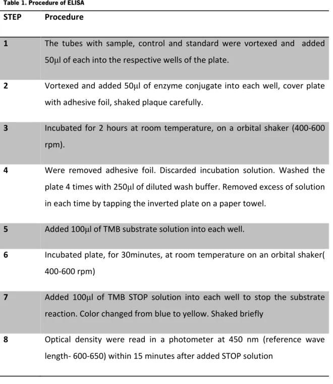
Cortisol ELISA kits ( RE52611) produced by IBL-international GMBH were used. The reportable range for salivar cortisol is 0,015 - 4 μg/dl cortisol.
Table 1. Procedure of ELISA
STEP Procedure
1 The tubes with sample, control and standard were vortexed and added 50μl of each into the respective wells of the plate.
2 Vortexed and added 50μl of enzyme conjugate into each well, cover plate with adhesive foil, shaked plaque carefully.
3 Incubated for 2 hours at room temperature, on a orbital shaker (400-600 rpm).
4 Were removed adhesive foil. Discarded incubation solution. Washed the plate 4 times with 250μl of diluted wash buffer. Removed excess of solution in each time by tapping the inverted plate on a paper towel.
5 Added 100μl of TMB substrate solution into each well.
6 Incubated plate, for 30minutes, at room temperature on an orbital shaker( 400-600 rpm)
7 Added 100μl of TMB STOP solution into each well to stop the substrate reaction. Color changed from blue to yellow. Shaked briefly
8 Optical density were read in a photometer at 450 nm (reference wave length- 600-650) within 15 minutes after added STOP solution
26
3.6. THE POSITIVE AND NEGATIVE SYNDROME SCALE ASSESSMENT(PANSS)
PANSS was assessed in two time points, before the first ECT , and one week after the last ECT, to measure the positive and negative symptoms. PANSS is a good instrument to assess both negative and positive symptoms (S. Leucht et al., 2005) and general psychopathology. It includes psychometric items with higher reliability (Peralta & Cuesta, 1994), validity and evaluates the pharmacological effect of treatment (Kay, Fiszbein, & Opler, 1987)sensitivity. A recent study (Santor, Ascher-Svanum, Lindenmayer, & Obenchain, 2007) has shown that the positive and negative subscales are more sensitive to change, the PANSS items are good to assess severity of disease.
3.7. PERCEIVED STRESS SCALE ASSESSMENT
PSS was assessed before the first ECT, and one week after the last ECT. The scores were correlated with the salivary cortisol levels, heart rate, mean blood pressure and symptoms. The PSS is an objective measure of self report and important tool to assess the stress levels in patients (S. Cohen et al,, 1983). The main advantage is that it allows to measure the risk of disease, with events that occurred during the last month and minimize bias of subjective measures (S. Cohen et al., 1983). A study has shown that patients with negative symptoms of schizophrenia have low capacity to experience stress (A. S. Cohen et al., 2003), and suggests that stress may not cause relapses of negative symptoms. Other study showed that patients with high PSS score have lower levels of cortisol after awaking than patients with low PSS score(Maldonado et al., 2008).
3.8. HOSPITAL ANXIETY AND DEPRESSION SCALE ASSESSMENT
The Portuguese version (Pais-Ribeiro et al., 2007) of the Hospital anxiety and Depression Scale (HADS).was administered to each patient who agreed to participate in the study. HADS self-assessment with fourteen items measures symptoms of anxiety and depression of the past 7 days, with the score of each item ranging from 0 to 3. Scores above 8 indicate severe depression or anxiety. This scale is important for research purposes(Herrmann, 1997) as it characterizes the prevalence and intensity of depression and anxiety. HADS is limited to 14 items which makes it easy to use. We used 8 for both anxiety and depression as a cutoff point.
27 3.9. STATISTICAL ANALYSIS
All statistical analysis was conducted in the SPSS software package version 22( IBM corporation, New York). Normality of data sets was assessed using the Kolmogorov-Smirnov test, Kurtosis and Skweness. As our data met normality, parametric tests were used throughout the analysis. Comparisons means between and within groups of diagnosis(Psychotic Depression, Schizophrenia and Schizoaffective Disorder) were performed using the ANOVA REPEATED MEASURE analysis. Pearson correlations were computed between continuous variables. The demographic, clinical, psychometric and laboratory were reported with use of descriptive statistics (frequencies, averages and standard deviation).
29
31
4.1. CLINICAL DIAGNOSTIC AND SOCIO-DEMOGRAPHIC CHARACTERISTICS:
The clinical and socio-demographic profile of the subjects was assessed with the use of descriptive statistics. During the study period, 36 patients were included, in which 17 patients were diagnosed with Schizophrenia (47,2%), 10 with Psychotic Depression ( 27,8%) and 9 with Schizoaffective Psychosis ( 25%). The diagnosis was established according to the Diagnostic and Statistical Manual for Mental Disorders-TR (DSMIV-TR) by a senior psychiatrist. All patients were physically healthy with no infection or history of drug or alcohol abuse. The mean age of the patients was 49,22 years (sd=15,130). The duration of the psychiatric disorders was 14,67 years (sd=12,191). The mean education level of our patients was 7,64 years (sd=3,972).
Twenty three patients were female (63,9%) and thirteen were male (36,1%) and all of them were medicated with antipsychotics. Our sample displayed a mean body mass index (BMI) of 26,5710 kg/m2 (sd=4,00862) and mean arterial pressure (MAP) of 92,5648mmHg (sd=10,93264) before
ECT. No significant differences were observed in these measures after ECT.
4.1.1. RESPONSE TO ECT AMONG PSYCHOTIC PATIENTS
P e rc e n ta g e o f P A N S S a ft e r E C T
Figure 4. Average change in PANSS scores after ECT in the different experimental groups
The analysis of the clinical response to ECT revealed a decrease in the PANSS scores in the three experimental groups (Figure 4). However, no significant differences were observed between psychotic patients in the average change in PANSS scores, demonstrating the efficacy of this treatment independently of the diagnosis.
32
Table 2. Effectiveness of ECT on PANSS
Descriptive statistics of differences of PANSS between psychotics patients undergoing ECT
Psychotic Disorder Mean Std.
Deviation N PANSSbefore Schizophrenia 127,35 16,733 17 Schizoafective_p 124,56 17,016 9 Psychotic_Depres 104,30 9,334 10 Total 120,25 17,848 36 PANSSafter Schizophrenia 55,35 16,175 17 Schizoafective_p 61,44 13,408 9 Psychotic_Depres 52,10 11,666 10 Total 55,97 14,417 36
The descriptive statistics of the differences in PANSS scores (table 2) reveal that the mean score of PANSS was lower after ECT in all three psychotic patients’ groups. To further analyze the differences between the three psychotic patients’ groups, Anova Repeated Measures was performed with Tukey post-hoc test. Our data reveale statistically signifcant differences between three groups in PANSS scores, HADS scores and Perceived Stress.
*
**
****
33
The repeated measures ANOVA analysis revealed a significant global effect of ECT on the PANSS scores F(2,33)=6,948, p=0,003. The Tukey post-hoc test revealed a significant difference in PANSS scores between schizophrenic patients and psychotic depression one week after ECT (p=0,045) but no significant differences between schizophrenic patients and schizoaffective patients (p=0,951) and psychotic depression and schizoaffective patients (p=0,051)(Figure 5).
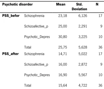
4.1.2. EFFECTS OF ECT ON PERCEIVED STRESS
Table 3. Descriptive statistic of perceived stress
Psychotic disorder Mean Std.
Deviation N PSS_befor Schizophrenia 23,18 6,126 17 Schizoafective_p 25,00 2,291 9 Psychotic_Depres 30,80 3,225 10 Total 25,75 5,628 36 PSS_after Schizophrenia 14,71 5,022 17 Schizoafective_p 16,00 2,872 9 Psychotic_Depres 16,90 5,567 10 Total 15,64 4,722 36
The descriptive statistics of the differences in PSS scores (table 3) reveal that the mean score of PSS was lower after ECT in all three psychotic patients’ groups.
**
*
** **
34
The repeated measures ANOVA analysis revealed a significant global effect of ECT on the PSS scores F(2,33)=4,841 p=0,014. The Tukey post-hoc test revealed a significant difference in PSS scores between schizophrenic patients and psychotic depression one week after ECT (p=0,015) but no significant differences between schizophrenic patients and schizoaffective patients (p=0,639) and psychotic depression and schizoaffective patients (p=0,202)(Figure 6).
4.1.3. EFFECTS OF ECT ON ANXIETY AND DEPRESSION SCORES
Table 4. Descriptive statistic of HADS
Psychotic Disorder Mean Std.
Deviation N HADS_before Schizophrenia 21,41 8,675 17 Schizoafective_p 25,56 6,560 9 Psychotic_Depres 33,30 5,034 10 Total 25,75 8,729 36 HADS_after Schizophrenia 11,53 5,076 17 Schizoafective_p 12,11 6,234 9 Psychotic_Depres 14,50 4,327 10 Total 12,50 5,207 36
The descriptive statistics of the differences in HADS scores (table 4) reveal that the mean score of HADS was lower after ECT in all three psychotic patients’ groups.
*** *** ***
*
35
The repeated measures ANOVA analysis revealed a significant global effect of ECT on the HADS F(2,33)=6,575 p=0,004. The Tukey post-hoc test revealed a significant difference in HADS scores between schizophrenic patients and psychotic depression one week after ECT (p=0,006) but no significant differences between schizophrenic patients and schizoaffective patients (p=0,563) and psychotic depression and schizoaffective patients (p=0,132) (Figure 7).
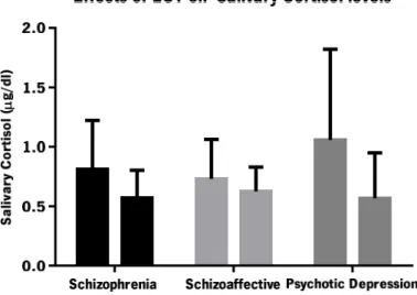
4.1.4. EFFECTS OF ECT IN SALIVARY CORTISOL
Table 5.Descriptive statistic of salivary cortisol
DSMIV_tr Mean Std. Deviation N Cortisol_Before Schizophrenia ,81050 ,411265 16 Schizoafective_p ,73025 ,331653 8 Psychotic_Depres 1,05780 ,763147 10 Total ,86435 ,525503 34 Cortisol_After Schizophrenia ,57069 ,232323 16 Schizoafective_p ,62525 ,203408 8 Psychotic_Depres ,56820 ,380697 10 Total ,58279 ,270941 34
The descriptive statistics of the differences in salivary cortisol levels (table 5) reveal that the mean levels of cortisol after were lower in the schizophrenia and psychotic depression groups.
Figure 8. Effects of ECT on salivary cortisol levels among psychotic patients
The repeated measures ANOVA analysis revealed that there were no significant global effects of ECT on the salivary cortisol levels F(2,31)=2,180 p=0,130 (Figure 8).
36
4.2. DIFFERENCES IN CLINICAL OUTCOME BETWEEN GENDER
Table 6. Descriptive statistic of differences between gender
gender N Mean Std. Deviation Std. Error Mean age Male 13 41,23 16,300 4,521 Female 23 53,74 12,657 2,639 PANSSafter Male 13 49,31 9,160 2,540 Female 23 59,74 15,615 3,256 PSS_befor Male 13 23,46 7,102 1,970 Female 23 27,04 4,248 ,886 PSS_after Male 13 13,31 5,023 1,393 Female 23 16,96 4,084 ,852 cortisol_level1 Male 13 ,82962 ,597452 ,165703 Female 22 ,86536 ,487868 ,104014 cortisol_level2 Male 12 ,65025 ,380993 ,109983 Female 22 ,54600 ,187896 ,040059 BMI_Bef Male 13 23,6545 2,40731 ,66767 Female 23 28,2194 3,81263 ,79499 BMI_Af Male 13 23,6136 2,44965 ,67941 Female 23 28,1596 3,80261 ,79290
N-sample size. Std- standard deviation. PANSS- positive and negative symptoms scale. PSS- Perceived Stress Scale. BMI- body mass index.
The results of the descriptive statistics (Table 6) reveal that females display higher mean ranges of age, PANSS After ECT, PSS before ECT, PSS after ECT, cortisol levels after and before ECT, BMI after and BMI before ECT.
Table 7. Independent t-test differences between gender
Variable measured t-test for Equality of Means
t df Sig. (2-tailed) Mean Difference Std. Error Difference 95% Confidence Interval of the Difference Lower Upper age Equal variances assumed -2,566 34 ,015 -12,508 4,876 -22,417 -2,600 PANSSafter Equal variances assumed -2,196 34 ,035 -10,431 4,750 -20,084 -,778 PSS_befor Equal variances assumed -1,901 34 ,066 -3,582 1,884 -7,410 ,247 PSS_after Equal variances assumed -2,369 34 ,024 -3,649 1,540 -6,779 -,519 cortisol_level1 Equal variances assumed -,193 33 ,848 -,035748 ,185527 -,413207 ,341710
37 cortisol_level2 Equal variances assumed 1,075 32 ,291 ,104250 ,097005 -,093343 ,301843 BMI_Bef Equal variances assumed -3,888 34 ,000 -4,56494 1,17419 -6,95119 -2,17870 BMI_Af Equal variances assumed -3,868 34 ,000 -4,54600 1,17538 -6,93466 -2,15734 Significant level. p<0,05
There were statistically significant differences between males and females in the following variables: age t(34)=-2,566 p=0,015 medium effect size r=0,4, PANSS after ECT t(34)=-2,196 p=0,035 medium effect size r=0,35, PSS after ECT t(34)=-2,369 p=0,024 medium effect size r=0,38, body mass index( BMI) before ECT t(34)=-3,888 p<0,001 large effect size r=0,55 and BMI after ECT t(34)=-3,868 p<0,001. There were no statistically significant difference between male and female in the following variables: PSS before ECT t(34)=-1,901 p=0,66, Salivary cortisol before ECT t(33)=-0,193 p=0,848 and salivary sortisol after ECT t(32)=0,291 p=0,291.
Mal e
Fem ale
****
9. Differences between genders in BMI
Differences between gender on PANSS and Perceived Stress scores
Gender Mal e Fem ale 0 20 40 60 80 PANSS after Perceived Stress before Perceived Stress after
*
* *
38
4.2.1. IMPROVEMENT OF NEGATIVE, POSITIVE AND GENERAL SYMPTOMS AFTER ECT
The first goal of our work was to evaluate the evolution of positive and negative symptoms in psychotic patients undergoing ECT. For this aim, patients were evaluated by a psychiatrist with the use of PANSS scale before the first ECT, and one week after the last ECT. As expected, the patients shown decrease in PANSS scores which is in line with the improvement of positive, negative and general symptoms. The psychotic depression group were those whom shown more clinical improvement after ECT.
A) B) **** **** **** **** Schizophrenic patients S c o re s PAN SS_b efor e PAN SS_a fter Pos itive _bef or Pos itive _aft er Neg ativ e_be fore Neg ativ e_af ter Gen eral _bef or Gen eral _afte r 0 50 100 150 200 **** **** **** ****
39 C)
****
**** ****
****
Figure 11 Measure of PANSS before and after ECT in three different psychotic inpatients groups: Schizophrenic patients ( A ), Psychotic Depression ( B ) and Schizoaffective Psychosis ( C ). In all of them the analysis was performed using dependent t-Test. Bars mean group scores before and after ECT ( t-test, **** p<0,001).
4.2.2. ANXIETY AND DEPRESSION AMONG PSYCHOTICS PATIENTS
Results of Hospital Anxiety and Depression scale revealed decrease of anxiety and depression scores in all three patients groups a week after ECT. With the cut-off point of 8, Psychotic depression shown the less reduction of anxiety score, despite the fact that all of them improves anxiety after ECT.
40 A) HADS _bef HADs -Afte r Anxi et_b ef anxi ety_ afte r Dep_ bef dep_ afte r S c o re *** *** *** B) HADS _bef HADs -Afte r Anxi et_b ef anxi ety_ afte r Dep_ bef dep_ afte r *** *** *** HADS _bef HADs -Afte r Anxi et_b ef anxi ety_ afte r Dep_ bef dep_ afte r **** **** **** C)
Figure 12 Measure of Anxiety and Depression perceived before and after ECT in three different psychotic inpatients groups: (A) Schizoaffective Psychosis ( n=9 ), (B)Schizophrenic patients ( n=17 ) and (C)Psychotic
41
Depression ( 10 ). In all of them the analysis was performed using dependent t-Test. Bars - mean of group scores before and after ECT (dependent t-test, **** p<0,001, *** p<0,01).
4.2.3. PERCEIVED STRESS
Our data reveals an improvement of perceived stress after ECT in all psychotic patients’ groups. Psychotic depression patients displayed the higher improvement in perceived stress taking into account that they displayed higher levels of perceived stress before treatment (Figure 13).
Perceived Stress Psychotic patients undergoing ECT
Score of Perceived Stress
0 10 20 30 40 Dep-Bef Dep_Aft Schizoaf_B Schizoaf_A Schizophr_B Schizophr_A *** *** ***
Figure 13Perceived stress among patients undergoing ECT in three group’s diagnosis, after and before ECT.
***p<0,001.
4.2.4. CORTISOL AMONG PSYCHOTICS PATIENTS UNDERGOING ECT
The specific analysis of the variations of the salivary cortisol levels in each experimental group using the dependent t-test revealed that patients with schizophrenia and psychotic depression displayed a significant decrease in this measure after ECT. No significant differences were observed in schizoaffective psychotic patients. (Figure 14)
42
Cortisol in psychotic patients
Sciz ophr enic _bef Sczo phre nic_ aft Psyc hotic _Dep _bef ore Psyc hotic _Dep _afte r Scizo affe ctive _bef Schi zoaf fect ive_a fter 0.0 0.5 1.0 1.5 2.0 * *
Figure 14 Cortisol in three psychotic patients groups before and after ECT. *p<0,05.
4.3. CORRELATION BETWEEN VARIABLES
Table 8. General correlations
ag e Ed. Lev PANS S_B PANS S_A PSS _B PSS _A HAD S_B HAD S_A Cortis ol_B Cortis ol_A Duration_ disord Perc ent of resp Age r -,544 ** ,050 ,309 ,49 0** ,39 5* ,612** ,531** ,238 ,014 ,645** ,314 Ed-Lev r -,54 4** -,026 -,139 -,28 7 ,05 2 -,270 -,354* -,081 ,047 -,397* -,142 PANSS_B r ,05 0 -,026 ,557** -,25 6 ,13 3 -,157 -,053 ,089 ,112 ,019 -,039 PANSS_A r ,30 9 -,139 ,557** ,20 4 ,44 6** ,239 ,377* ,115 -,021 ,036 ,797** PSS_B r ,49 0** -,287 -,256 ,204 ,54 1** ,727** ,529** ,135 -,227 ,298 ,433** PSS_A r ,39 5* ,052 ,133 ,446** ,54 1** ,507** ,471** ,299 ,203 ,112 ,462** HADS_B r ,61 2** -,270 -,157 ,239 ,72 7** ,50 7** ,584** ,062 -,150 ,184 ,391* HADS_A r ,53 1** -,354 * -,053 ,377* ,52 9** ,47 1** ,584** ,102 -,037 ,099 ,475** Cortisol_ B r ,23 8 -,081 ,089 ,115 ,13 5 ,29 9 ,062 ,102 ,615** ,137 ,083 Cortisol_ A r ,01 4 ,047 ,112 -,021 -,22 7 ,20 3 -,150 -,037 ,615** -,037 -,076 Duration_ disord r ,64 5** -,397 * ,019 ,036 ,29 8 ,11 2 ,184 ,099 ,137 -,037 ,028 Percent of resp r ,31 4 -,142 -,039 ,797** ,43 3** ,46 2** ,391* ,475** ,083 -,076 ,028