Marisa Conceição Lima Gomes
SUSCEPTIBILITY OF CANDIDA BIOFILMS TO
PHOTODYNAMIC TREATMENT
Marisa Conceição Lima Gomes
SUSCEPTIBILITY OF CANDIDA BIOFILMS TO
PHOTODYNAMIC TREATMENT
2015
Dissertação de Mestrado
Mestrado em Bioengenharia
Trabalho realizado sob a orientação da
Professora Doutora Mariana Contente Rangel Henriques
Professora Doutora Isabel Maria Pires Belo
DECLARAÇÃO
Nome: Marisa Conceição Lima Gomes
Endereço eletrónico: pg22726@alunos.uminho.pt Número do Bilhete de Identidade: 13930625
Título da Dissertação: Susceptibility of Candida Biofilms to Photodynamic Treatment Orientadoras:
Professora Doutora Mariana Contente Rangel Henriques Professora Doutora Isabel Maria Pires Belo
Ano de conclusão: 2015
Designação do Mestrado: Mestrado em Bioengenharia
É AUTORIZADA A REPRODUÇÃO INTEGRAL DESTA DISSERTAÇÃO APENAS PARA EFEITOS DE INVESTIGAÇÃO, MEDIANTE DECLARAÇÃO ESCRITA DO INTERESSADO, QUE A TAL SE COMPROMETE;
Universidade do Minho, 30 de Janeiro de 2015
AKNOWLEDGEMENTS
I would like to express my deepest gratitude to my supervisors, Dr.ª Mariana Henriques and Dr.ª Isabel Belo, for their sympathy, availability, knowledge, dedication, advices and suggestions. Thank you for all the words of support and understanding!
To Dr. Paulo Coutinho, I want to express my gratefulness for all the support, knowledge and suggestions, and for allowing me to use the irradiation system. I also want to thank Dr.ª Maria Sameiro Gonçalves and Bachu Rama Raju for their availability and for providing the benzophenoxazine compound.
I am truly grateful to Carlos Tiago Alves, for all the teaching, patience, availability, and support. I also want to express my sincere gratefulness to Marlene Lopes, for her availability, guidance, and support throughout the experimental work. I am very appreciative of the time and effort that both of you devoted to guide me in a wide variety of subjects!
To my friends Ana and Vânia, who are always there to cheer me up. Thank you for all your support, encouragement and amusing moments!
Para finalizar, gostaria de agradecer à minha família, em especial à minha mãe, que ao longo da minha formação académica sempre me apoiou e incentivou. Valter, obrigada pelo conforto que sempre encontrei junto de ti e pelo teu apoio incondicional. Obrigada por tudo!
ABSTRACT
In recent decades, the incidence of fungal infections caused by Candida spp. has significantly increased. C. albicans is the most important pathogen within this genus, since it is the most commonly isolated. However, other Candida species have gained importance over the last years, namely C. glabrata and C. parapsilosis. The rise of strains highly resistant to conventional antifungal treatments has increased the research for development of new effective antifungal therapies. Antimicrobial Photodynamic Therapy (APDT) has been proposed as a therapy for a large variety of localized infections.
The aim of this work was to study the antifungal photodynamic effect of a new benzophenoxazine compound – FSc – on C. glabrata, C. parapsilosis and C. albicans biofilms. Biofilms were treated with different FSc concentrations (50 µM – 500 µM) for 18 h and irradiated with a xenon arc lamp (600 ± 2 nm) at various light doses (8 J cm-2 – 50 J cm-2). The efficiency of photodynamic treatment
mediated by two widely used dyes – Nile blue chloride and Protoporphyrin IX – on C. albicans biofilms was also evaluated. Biofilms were incubated with 300 µM of each dye for 18 h and irradiated with a light dose of 36 J cm-2. In order to understand the oxidative stress response of Candida species to
APDT, suspensions of C. albicans, C. glabrata and C. parapsilosis were incubated with 2 mM and 20 mM of two oxidant agents, hydrogen peroxide and paraquat, for 3 h. The effect of these oxidants on both cell viability and induction of antioxidant enzymes was evaluated.
The photoinactivation of Candida biofilms with either FSc or PpIX as a photosensitizer was able to reduce the cell viability. On the other hand, Nile blue-mediated APDT assays showed no effect on cell viability. In general, photoinactivation of Candida cells was dependent on the dye concentration and light dose delivered. Nevertheless, irradiation with increasing light doses only resulted in a slight decrease of biofilm viability. Although the results of this study are encouraging, further investigations are needed to optimize protocols for maximal cell inactivation.
Candida species showed high levels of resistance to the two oxidant agents, with the exception of C. glabrata that presented a greater sensitivity to paraquat. Generally, all Candida strains responded to hydrogen peroxide and paraquat exposure by increasing their content of catalase and superoxide dismutase, respectively. In summary, the data suggest that Candida species have different levels of resistance to oxidant agents and they use different defense mechanisms against these substances, showing differences in the induction of antioxidant enzymes.
RESUMO
Nas últimas décadas, a incidência de infeções fúngicas provocadas por espécies do género Candida tem aumentado significativamente. C. albicans é considerada a espécie mais patogénica dentro deste género, uma vez que é a mais comummente isolada. No entanto, nos últimos anos, outras espécies de Candida têm ganho importância, nomeadamente C. glabrata e C. parapsilosis. O aumento de estirpes altamente resistentes aos tratamentos antifúngicos convencionais tem aumentado a necessidade de desenvolvimento de novas terapias antifúngicas eficazes. A Terapia Fotodinâmica Antimicrobiana (TFDA) tem sido proposta como um tratamento para uma grande variedade de infeções localizadas.
Este trabalho teve como objetivo estudar o efeito fotodinâmico de um corante pertencente ao grupo das benzofenoxazinas – FSc – em biofilmes de C. glabrata,C. parapsilosis e C. albicans. Os biofilmes foram incubados com diferentes concentrações de FSc (50 µM – 500 µM), durante 18 h, e irradiados com várias doses de luz (8 J cm-2 – 50 J cm-2), usando uma lâmpada de arco de xénon (600 ± 2 nm). Foi ainda avaliada
a eficiência da TFDA com dois corantes amplamente utilizados – Azul do nilo e Protoporfirina IX – em biofilmes de C. albicans. Estes foram incubados com 300 µM de cada corante, durante 18 h, e irradiados com uma dose de luz de 36 J cm-2. De forma a compreender a resposta das espécies de Candida ao stresse oxidativo
induzido pela TFDA, suspensões de C. albicans, C. glabrata e C. parapsilosis foram incubadas com 2 mM e 20 mM de dois agentes oxidantes, peróxido de hidrogénio e paraquato, durante 3 h. Determinou-se o efeito destes oxidantes na viabilidade celular e na indução de enzimas antioxidantes.
Ao contrário da TFDA com Azul do nilo, a fotoinativação de biofilmes de Candida com os compostos FSc ou PpIX foi capaz de reduzir a viabilidade celular. De um modo geral, a inativação das células de Candida foi dependente da concentração de corante e da dose de luz aplicada. No entanto, a irradiação com doses crescentes de luz resultou apenas numa ligeira diminuição da viabilidade celular. Embora os resultados deste estudo sejam encorajadores, investigações posteriores são necessárias para otimizar os protocolos de modo a induzir uma maior inativação celular.
As espécies de Candida apresentaram uma grande resistência aos dois agentes oxidantes, à exceção de C. glabrata que mostrou ser mais sensível ao paraquato. De um modo geral, as espécies de Candida
responderam à exposição ao peróxido de hidrogénio e ao paraquato com a indução de catalase e de superóxido dismutase, respetivamente. Em suma, os resultados indicam que as espécies de Candida
possuem distintos níveis de resistência aos agentes oxidantes, utilizando diferentes mecanismos de defesa contra esses mesmos compostos.
TABLE OF CONTENTS AKNOWLEDGEMENTS... iii ABSTRACT ...v RESUMO ... vii TABLE OF CONTENTS ... ix LIST OF FIGURES ... xi
LIST OF TABLES ... xiii
LIST OF ABBREVIATIONS ... xv
PREAMBLE ... xvii
1. CHAPTER I - INTRODUCTION ... 1
1.1. Candida species ... 3
1.1.1. Cell Biology ... 3
1.1.2. Biofilm forming ability ... 5
1.1.3. Conventional antifungals ... 6
1.2. Antimicrobial Photodynamic therapy (APDT) ... 7
1.2.1. Photophysical and photochemical PDT mechanisms ... 8
1.2.2. Mechanism of microbial inactivation ... 11
1.2.3. Photosensitizers ... 11
1.2.3.1. Properties of photosensitizers ... 12
1.2.3.2. The most commonly used antimicrobial photosensitizers ... 13
1.2.3.3. Benzo[a]phenoxazines ... 15
1.2.3.4. New frontiers in APDT ... 16
1.2.4. Light sources and delivery ... 17
1.2.4.1. The concept of irradiance and fluence ... 17
1.2.4.2. The most commonly used light sources ... 18
1.2.5. Clinical applications ... 20
1.2.7. Mechanisms of resistance to APDT ... 21
1.2.8. Oxidative stress ... 23
2. CHAPTER II - MATERIALS AND METHODS ... 25
2.1. Organisms and culture media ... 27
2.2. Antimicrobial photodynamic therapy ... 27
2.2.1. Photosensitizers ... 27
2.2.2. Biofilms formation ... 28
2.2.3. Dark toxicity ... 29
2.2.4. Photodynamic therapy against Candida biofilms ... 29
2.2.5. Determination of biofilm viability: quantification of CFUs ... 30
2.3. Oxidative stress response ... 31
2.3.1. Operating Conditions ... 31
2.3.2. Analytical Methods ... 31
2.3.3. Determination of minimum inhibitory concentrations of PQ and H2O2 ... 33
2.4. Statistical Analysis ... 34
3. CHAPTER III - RESULTS AND DISCUSSION ... 35
3.1. Antimicrobial photodynamic therapy ... 37
3.2. Oxidative stress response ... 45
4. CHAPTER IV - CONCLUDING REMARKS AND FUTURE PERSPECTIVES ... 51
LIST OF FIGURES
CHAPTER I - INTRODUCTION
Figure 1. Stages in the formation of a Candida spp. biofilm. . ... 6
Figure 2. Photophysical and photochemical mechanisms of PDT. ... 9
Figure 3. Comparison of lifetime, diffusion distance and reactivity of different ROS. ... 10
Figure 4. Basic chemical structures of the main groups of antimicrobial PSs.. ... 14
Figure 5. Chemical structure of phenoxazines and benzophenoxazines.. ... 16
CHAPTER II - MATERIALS AND METHODS Figure 6. Chemical structure of FSc (A), Nile blue chloride (B) and PpIX (C) dyes. ... 27
CHAPTER III - RESULTS AND DISCUSSION Figure 7. Logarithm of the number of C. glabrata ATCC 2001 and C. parapsilosis ATCC 22019 biofilm cells per cm2 after 18 h of dark incubation with different concentrations of FSc. Values are average ± standard deviation of at least four independent experiments. * and ** indicates p <0.05 and p <0.01, and consequently statistically different from the respective control, 0 µM. ... 37
Figure 8. Logarithm of the number of C. parapsilosis ATCC 22019 biofilm cells per cm2 after 18 h of dark incubation with different concentrations of FSc followed by irradiation with various light doses. Values are average ± standard deviation of at least two independent experiments. N – Not determined. *, ** and *** indicates p <0.05, p <0.01 and p <0.001, and consequently statistically different from the control, 0 µM (at the same light fluence). ... 38
Figure 9. Logarithm of the number of C. glabrata ATCC 2001 biofilm cells per cm2 after 18 h of dark incubation with 75 µM of FSc followed by irradiation with various light doses. Values are average ± standard deviation of at least two independent experiments. N – Not determined. * and ** indicates p <0.05 and p <0.01, and consequently statistically different from the control, 0 µM (at the same light fluence). ... 40 Figure 10. Logarithm of the number of C. albicans ATCC 90028 biofilm cells per cm2 after 18 h of
average ± standard deviation of at least two independent experiments. * indicates p <0.05; *** and **** indicates p <0.001 and p <0.0001, and consequently statistically different from the control, 0 µM (at the same light fluence). ... 40 Figure 11. Logarithm of the number of C. albicans ATCC 90028 biofilm cells per cm2 after 18 h of
dark incubation with 300 µM of Nile blue chloride (A) and PpIX (B) followed by irradiation with a light dose of 36 J cm-2. Values are average ± standard deviation of at least two independent experiments.
** indicates p <0.01; * and *** indicates p <0.05 and p <0.001, and consequently statistically different from the control, 0 µM (at the same light fluence). ... 42 Figure 12. Ratio of viable cells (ratio of cell viability between each treatment and the respective control) (A); CAT specific activity (B) and SOD specific activity (C) of C. albicans ATCC 90028, C. glabrata ATCC 2001 and C. parapsilosis ATCC 22019 exposed to 2 mM of PQ or H2O2 for 3 h. Values
are average ± standard deviation of at least three replicate measurements. ... 45 Figure 13. Ratio of viable cells (ratio of cell viability between each treatment and the respective control) (A); CAT specific activity (B) and SOD specific activity (C) of C. albicans ATCC 90028, C. glabrata ATCC 2001 and C. parapsilosis ATCC 22019 exposed to 20 mM of PQ or H2O2 for 3 h.
Values are average ± standard deviation of at least three replicate measurements. ... 47 Figure 14. Growth inhibition (%) of cell suspensions of C. albicans ATCC 90028, C. glabrata ATCC 2001 and C. parapsilosis ATCC 22019 (adjusted to 106 cells mL-1), after incubation with different
concentrations of PQ (A) and H2O2 (B) during 24 h. Values are average ± standard deviation of two
LIST OF TABLES
CHAPTER I – INTRODUCTION
Table 1. Morphological characteristics of the most relevant Candida species. ... 4 Table 2. Lasers most commonly used in PDT. ... 18 Table 3. Lamps most commonly used in PDT. ... 19
CHAPTER II - MATERIALS AND METHODS
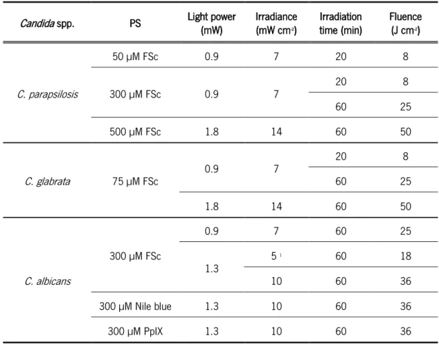
Table 4. Parameters used in the APDT assays against Candida biofilms. ... 30
CHAPTER III - RESULTS AND DISCUSSION
Table 5. MIC90 of PQ and H2O2 (mM) of C. albicans ATCC 90028, C. glabrata ATCC 2001 and C.
LIST OF ABBREVIATIONS
ABC ATP-binding Cassette Abs Absorbance
ALA 5-Aminolevulinic Acid ANOVA Analysis of Variance
APDT Antimicrobial Photodynamic Therapy ATCC American Type Culture Collection ATP Adenosine Triphosphate
BSA Bovine Serum Albumin CAT Catalase
CFU Colony Forming Units CHROMagar Chromogenic Media Agar DNA Deoxyribonucleic Acid ECM Extracellular Matrix
EDTA Ethylenediamine Tetraacetic Acid g Gravity constant (m s-2)
GNP Gold Nanoparticles
HIV Human Immunodeficiency Virus HSP Heat Shock Proteins
Log Logarithm
M Molarity (mol L-1; mmol L-1; µmol L-1)
MB Methylene Blue
MFS Major Facilitator Superfamily MIC Minimum Inhibitory Concentration
MOPS 3-(N-Morpholino)propanesulfonic acid, 4-Morpholinepropanesulfonic acid NCAC Non-C. albicans Candida
NMB New Methylene Blue p Significance value
PBS Phosphate Buffered Saline PDT Photodynamic Therapy
PpIX Protoporphyrin IX PQ Paraquat
PS Photosensitizer
ROS Reactive Oxygen Species rpm Revolutions per minute
RPMI Roswell Park Memorial Institute SDA Sabouraud Dextrose Agar SDB Sabouraud Dextrose Broth SOD Superoxide Dismutase TB Toluidine Blue
TBO Ortho-toluidine Blue
% Percentage
PREAMBLE
Over the last 30 years, the incidence of fungal infections in humans has significantly increased due to several factors, including the use of invasive procedures, prosthetic devices, application of immunosuppressive medications and broad-spectrum antibiotics, as well as the increased incidence of neutropenia and human immunodeficiency virus (HIV) infections [1, 2]. Candida species are among the major human fungal pathogens and have become the second leading cause of invasive fungal infections, being responsible for 70-90 % of all cases [3]. Moreover, systemic candidiasis was observed in 20-40 % of patients with cancer and in approximately 25 % of patients who received bone marrow transplants [4]. Most cases of candidiasis have been attributed to C. albicans, but recently, non-C. albicans Candida (NCAC) spp., such as C. glabrata and C. parapsilosis, have been identified as common pathogens [5]. The worldwide increase in antimicrobial resistance, mainly due to inappropriate prescription of antibiotics and mechanisms developed by microbial cells to increase their resistance to external insults [6] has resulted in the search for alternative antimicrobial therapies [7, 8].
Photodynamic therapy (PDT) initially developed to combat cancerous lesions, has been employed in the treatment of infectious diseases. In fact, it has been found an effective antimicrobial action of PDT against a wide variety of microorganisms, including bacteria and fungi, even in antibiotic resistant strains [9–11], viruses and protozoa [12–17]. Antimicrobial Photodynamic Therapy (APDT) employs a non-toxic dye, termed photosensitizer (PS), and low intensity visible light which, in the presence of oxygen, combine to produce cytotoxic oxygen species leading to cell death.
The development of new classes of PSs, the optimization of dosimetry and further enhancement in technology may drastically change the currently achieved APDT clinical outcome. Thus, this dissertation aimed to evaluate the inactivation of C. glabrata, C. parapsilosis and C. albicans biofilms by APDT through the use of a new benzophenoxazine compound. Dye concentration and light dose were optimized in order to improve the levels of cell inactivation. In addition, the antimicrobial activity of APDT mediated by two commonly used dyes, Nile blue chloride and Protoporphyrin IX, against C. albicans biofilms was also evaluated. With the intent to understand how cells respond to oxidative stress, the main cause of cell death during APDT, cell viability and antioxidant enzymes activity of the three Candida species after exposure to hydrogen peroxide and paraquat (oxidant agents) were
analyzed. It was also determined the minimum inhibitory concentration of both oxidant agents for the three Candida species under study.
The present dissertation is organized into four chapters. Chapter 1 is a brief review of the theoretical basis associated to this work, including cell biology of Candida spp. and their biofilm forming ability; the principles of APDT; the main PSs and light sources used; APDT applications and mechanisms of resistance; and the effect of oxidative stress on cells. Chapter 2 includes the materials and methods used in the experimental work. On chapter 3, all the results obtained, as well as its discussion are presented. Finally, chapter 4 highlights the main conclusions obtained from the realized work and presents some suggestions for future works.
1.
CHAPTER I
INTRODUCTION
INTRODUCTION | 3
1.1.
Candida
speciesThe Candida genus comprises a heterogeneous group of 150 species [18] that are common commensal inhabitant on human mucosal surfaces and skin in an asymptomatic manner. Nevertheless, when that outer layer of protection is compromised, Candida spp. can cause local infection (candidosis). In immunocompromised individuals it can infect deeper layers and if it becomes systemic (candidemia), can lead to death [19, 20]. Candida spp. are most frequently isolated from the oral cavity,gastrointestinal and genital tracts, and can be detected in approximately 31 to 55 % of healthy individuals [21].
C. albicans is the most important pathogen within the Candida genus [22], representing over 80 % of all yeasts isolated in clinical samples [18]. This is the most common fungus affecting oral cavity and immunocompromised patients, namely HIV infected patients and those with oncologic malignancies [7]. However, there are many other Candida species that have gained importance over the last years, because they are also known to be etiological agents of human infections [5], including: C. glabrata, C. parapsilosis, C. tropicalis, C. krusei, C. kefyr, C. rugosa and C. dubliniensis [23]. C. lusitanae, C. orthopsilosis, C. guilliermondii and C. metapsilosis are also pathogenic species but less relevant in comparison with the previous ones [5].The apparent increased occurrence of non-Candida albicans Candida (NCAC) spp. may be related with their inherent higher level of resistance to different antifungal agents compared to C. albicans, and the improvement of diagnostic methods [24].
Some of these species may co-exist competitively and symbiotically in dual biofilms. This is the case of C. albicans and C. glabrata [25] that are the most frequent combination between Candida spp. and have been found in approximately 70 % of the patients with oral candidosis [26]. In addition to the epithelial and mucosal surfaces, Candida can colonize numerous medical devices (vascular catheter, joint prosthesis, hemodialysis fistulas, and peritoneal dialysis catheters).
1.1.1. Cell Biology
Candida species are taxonomically classified in the Ascomycota phylum, and are formed by structures called blastoconidia, which may be round or elongated. They are surrounded by a rigid cell wall that is composed of a mixture of glucan, mannan, chitin and lipoproteins, separated from
4 | INTRODUCTION
the plasma membrane by a periplasmic space [2]. Moreover, some Candida species may also produce a filamentous type of growth such as true hyphae or more frequently, pseudohyphae. Pseudohyphae are formed when the production of blastoconidia continues without separation of the conidia from each other, resulting in filaments with constrictions at the cell-cell junctions. There is no internal cross walls associated with this growth form. In comparison, true hyphae result of a ‘germ tube’ projection which can elongate and then branch with defined septa that divide the hyphae into separated cells [18].
The parameters that lead to germination are not yet well understood. However, in vitro studies demonstrated that this behavior is induced by cultivation in serum, complex or chemicallydefined media; temperatures of 37 ºC and pH of around neutrality. On the other hand, yeast growth is stimulated by temperatures at 25 ºC, acid pH and glucose as a sole carbon source [27].
Macroscopically, Candida spp. colonies on the routinely used Sabouraud dextrose agar (SDA) range in color from cream to yellowish. Depending on the species, their texture may be smooth, pasty, glistening or dry, wrinkled and dull [28]. The use of a differential chromogenic medium such as CHROMagar allows distinguishing between different Candida spp. colonies by color, as a result of distinct biochemical reaction. Table 1 summarizes some of the properties that allow the identification of the most relevant Candida species.
Table 1. Morphological characteristics of the most relevant Candida species. Adapted from [24].
Species Yeast size (µm)
Hyphae/
Pseudohyphae Germ tube
CHROM-agar reaction
C. albicans 4-6 x 6-10 +/+ + Blue-green
C. glabrata 1-4 -/- - White, Pink-purple
C. parapsilosis 2.5-4 x 2.5-9 -/+ - White
As regards the biochemical reactions, it is known that C. albicans can ferment and/or assimilate numerous sugars, with the exception of sucrose; C. glabrata ferments and assimilates only glucose and trehalose; and C. parapsilosis is unable to ferment maltose [21, 29].
INTRODUCTION | 5
1.1.2. Biofilm forming ability
Candida species have an important role in the course of an infection. Their pathogenicity is related with a number of virulence factors, including (i) the ability to express specific host recognition and adhesion biomolecules; (ii) biofilm formation; (iii) secretion of tissue-damaging hydrolytic enzymes (proteases, phospholipases and lipases, and haemolysin) that are responsible for tissue invasion and destruction, as well as for changes on host immunity response [5, 24]; (iv) their versatility and ability to survive in several anatomical sites [27]; and (v) the morphological transitions between yeast and filamentous form (hyphae and pseudohyphae), which plays an important function in tissue invasion and resistance to phagocytosis [30, 31].
The most important virulence factor of Candida spp. is the ability to form biofilm on different surfaces, because biofilm cells are not only more resistant to antimicrobial agents than their free-living counterparts, but also appear to be able to resist the host's defense mechanisms [32, 33]. Both in the environment and during the course of infection, organisms frequently exist in the form of biofilms rather than independent entities. Biofilms are the main cause of microbial infections in humans, being involved in approximately 80 % of all infections [34]. Indeed, it was reported that Candida strains with a high ability to form biofilms are usually more virulent than others [35].
Biofilms are organized microbial communities characterized by sessile cells embedded in a self-created protective extracellular matrix (ECM) composed by diverse polymeric substances such as cell wall glycoproteins and polysaccharides. This structure promotes biofilm adhesion and formation, keeps its integrity, protects cells from phagocytosis, and limits the diffusion of drugs [36, 37]. Biofilms have a distinct phenotype from those of their planktonic counterparts, which is related to the transcribed genes and growth rate [38, 39]. Within biofilms, cell express genes implicated, for example, in the adhesion to host surfaces (such as agglutinin-like gene) that are not expressed in planktonic cells [40]. Furthermore, microbial cells within biofilm are under the control of signaling molecules and therefore have a collective behavior. This improves not only the access to nutrients and niches, but also their collective defense against other organisms [41].
The primary step in the biofilm formation is adhesion between Candida species and host cells/abiotic surfaces (Figure 1). Attachment of Candida species cells to materials is mediated by non-specific interactions, such as hydrophobic and electrostatic forces, as well as by specific adhesin-ligand bonds [42–44]. It is important to note that cell hydrophobicity and charge depend on cell growth
6 | INTRODUCTION
morphology and cell surface structure [45]. Attached cells proliferate to form microcolonies and start to deposit the ECM. Then, biofilm goes through a maturation phase until form a thick layer of matrix involving a complex network of yeasts cells, hyphae and pseudohyphae. At a certain time, cells are detached from biofilm, being able to form new biofilms in the surrounding environment.
Figure 1. Stages in the formation of a Candida spp. biofilm. Adapted from [46].
There are too many reasons for the resistance of C. albicans biofilms to antifungal agents: (i) extracellular matrix acts as a diffusion barrier, inhibiting antifungal diffusion to the inner layers of the biofilm; (ii) slow growth rate (due to the limited availability of nutrients, particularly at the base of the biofilm) leads to slow or insufficient drug uptake; (iii) the presence of a small number of “persister” cells that present a high level of drug resistance; (iv) the presence of efflux drug pumps; and (v) alterations in membrane sterol composition [33, 47–49].
It is known that the formation of mature biofilms and consequent production of ECM is strongly dependent on different factors, namely, species, strain, and environmental conditions such as medium composition, oxygen concentration and pH [50, 51]. In fact, a previous study showed that, in the presence of urine on silicone surfaces, C. glabrata is able to produce a higher biofilm biomass, in comparison with C. parapsilosis and C. tropicalis [52]. However, when grown in rich culture media (SDA), biofilm formation by C. glabrata was lower compared with other NCAC species [53, 54].
1.1.3. Conventional antifungals
There are several substances that can be applied in the treatment of candidiasis. Each antifungal class utilizes a different way to inhibit or kill fungal pathogens. Azoles interfere with the enzyme lanosterol demethylase (responsible for the conversion of lanosterol to ergosterol), leading to the depletion of ergosterol in the membrane. These antifungal agents, in particular fluconazole, are the
INTRODUCTION | 7
most frequently used in the treatment of candidiasis [55, 56]. Polyenes interfere with ergosterol and consequent with stability/permeability of fungal cell membrane, causing the leak of cytoplasmic content. Echinocandins inhibit the synthesis of ß-glucan, a fundamental component of the fungal cell wall [7], and pyrimidines act on enzymes that regulate the synthesis of nucleic acids [57, 58]. Amongst these antifungals agents, only amphotericin B (a polyene) and echinocandins (such as caspofungin) have showed consistent activity in the in vitro treatment of C. albicans biofilms [59]. However, even with these agents, infections related with Candida biofilms are extremely difficult to eradicate [60].
The efficiency of conventional therapies in the treatment of fungal infections has decreased, as a result of (i) the increase in antifungal drug resistance; (ii) the frequent recurrence of infections; (iii) the time-consuming of conventional treatments; (iv) the organ dysfunction which prevents the use of some agents; and (v) drug-drug interactions, which results from the narrow spectrum of action and toxicity of drugs [7, 61]. The high prevalence of drug resistance has increased the need for development of new effective antifungal therapies.
1.2. Antimicrobial Photodynamic therapy (APDT)
The combined use of visible light and dyes for inactivating microorganisms was first demonstrated more than 100 years ago, when Oscar Raab reported the lethal effect of acridine hydrochloride and visible light on the protozoan Paramecia caudatum [62]. Raab and his professors Joldlbauer, Jesionek and Von Tappeiner discovered that acridine was a photosensitizing agent [63]. They created the term “Photodynamic reaction” and eventually, by the early 1900s brought this knowledge to the clinic [64, 65] with brilliant results on cutaneous tumors. In the 1970s, photodynamic therapy (PDT) began to be explored for the selective destruction of cancer [66]. Since then, PDT has emerged as a tool for the treatment of different malignancies (neck, prostate, brain and mesothelioma), non-melanoma skin cancer (basocelullar carcinoma and actinic keratosis), choroidal neovascularization in age-related macular degeneration, lung and Barrett's esophagus cancer, oral lesions, ophthalmologic disorders and autoimmune diseases, such as psoriasis [67–70].
The potential of PDT against diseases of microbial origin was not exploited for several decades due to the discovery of antibiotics in the 1950s and the low efficiency of photodynamic killing of some pathogens, in comparison with the treatment of cancer [7, 71]. However, in the decades of 80 and
8 | INTRODUCTION
90, the interest in the antimicrobial effects of PDT has recovered [72–77] and it has been suggested as a therapy for a variety of localized infections, due to the significant increase in the occurrence of microbial infections.
Antimicrobial PDT (APDT) is an effective method for eliminating microorganisms, in particular those which exist in the form of biofilms, leading cause of microbial infections in humans [7]. This therapy involves the use of harmless visible light combined with a non-toxic and light-sensitive dye — the photosensitizer (PS) — and oxygen. After irradiation with the light of appropriate wavelength, the PS is energized to an excited state that can undergo molecular collisions with oxygen, leading to the generation of reactive oxygen species (ROS) that are responsible for thecell death [62, 78]. There are numerous advantages associated with APDT: (i) broad spectrum of action, with the efficient inactivation of antibiotic-resistant strains; (ii) utilization of low cost light sources [6]; (iii) selectivity to the target cell and absence of mutagenic effect in host tissue [7, 79]; (iv) the mechanisms of photodynamic killing are not target-specific, consequently resistance to this process is improbable to happen, even after multiple treatments [80–82]; (v) capacity of destruction of biofilm's structure, as a result of direct effect of ROS in polysaccharides and glycoproteins present in extracellular matrix [83–85]; (vi) does not lead to the accumulation of toxicity [86, 87]; and (vii) the combination with conventional therapies can lead to significant additive benefits [88]. Nevertheless, APDT can have some side effects, such as long-lasting skin photosensitivity, systemic and metabolic disturbances, and excessive tissue damage at the treated site [89].
1.2.1. Photophysical and photochemical PDT mechanisms
Photosensitizer compounds have a stable electronic configuration, which consists of a singlet state in its ground energy level, whereby all of its electrons are spin paired in low energy orbitals. Upon application of light with wavelength corresponding to the absorption peak of the PS, the electron in the highest occupied molecular orbital (HOMO) of the PS is excited to the lowest unoccupied molecular orbital (LUMO) and keeps its spin. PS reaches the excited singlet state, which is extremely unstable and short-lived (nanoseconds) [34]. The PS can return to the ground energy level through the emission of a photon (fluorescence) or by internal conversion with loss of energy as heat. Alternatively, the excited singlet state PS can undergo intersystem crossing whereby the spin of the excited electron inverts, leading to the formation of excited triplet state [2]. The triplet state is less
INTRODUCTION | 9
energetic than the excited singlet state, but has a considerably longer lifetime, in the range of microsecond to millisecond [2, 90, 91]. In the triplet state, the PS molecule can emit light (phosphorescence) by returning to the ground state or act as a mediator of one or both of two pathways - Type I and Type II reactions - both of which require oxygen (Figure 2) [78].
Figure 2. Photophysical and photochemical mechanisms of PDT [34].
Type I
The Type I mechanism involves electron/hydrogen transfer reactions from the PS with the participation of biological macromolecules (including lipids, proteins and nucleic acids) to produce radical ions, which further interact with molecular oxygen to produce ROS, such as superoxide anion (O2•-), hydrogen peroxide (H2O2) and hydroxyl radical (•OH) [89, 92]. Type I reactions do not
necessarily need oxygen to cause cellular damage, due to the action of free radicals [93].
O2•- is the ROS more abundant in nature and is formed by transfer of an electron from the PS to
oxygen molecule in the ground state. This ROS per se may not have sufficient reactivity to kill cells. However, it is considered toxic because it is involved in the formation of other reactive ROS, in particular •OH and peroxynitrite anion (ONOO-, short-lived and highly reactive) by reaction with nitric
oxide [94, 95]. O2•- can also abstract electrons, forming a peroxide ion that instantly abstracts protons
to form H2O2 [34].
H2O2 has a long half-life and capability to diffuse across cell membranes. This ROS can inactivate
enzymes, mainly by oxidation of essential thiol groups. However, H2O2 is poorly reactive and its toxicity
10 | INTRODUCTION
and copper, and the interaction with O2•- [96, 97]. The reaction between H2O2 and ferrous iron is
known as the Fenton reaction (Equation 1).
H2O2 + Fe2+→ OH- + •OH + Fe3+ (Equation 1)
and consists in the homolytic fissionof the oxygen-oxygen bond in H2O2 to yield a hydroxide ion and •OH via the oxidation of ferrous iron to ferric iron [98].
•OH is short-lived and shows a small diffusion capacity. However, this is the most reactive of the
three ROS formed and, in theory, can oxidize any organic molecule. This ROS acts on molecules near the location where it is formed, essentially lipids, being able to remove hydrogen of double bonds of fatty acids present in cell membranes - lipid peroxidation [99]. In addition, •OH induces
breakage and modification of deoxyribonucleic acid (DNA) bases [100]. •OH can abstract electrons
to become a hydroxide ion, which then can obtain a proton and form water [34].
Type II
In the type II reaction, which is considered the dominant process in PDT [101], the triplet state of PS can directly undergo energy exchange with molecular oxygen (triplet ground state oxygen) to form excited state singlet oxygen, 1O2, which can oxidize a large number of biological substrates, such as
proteins, nucleic acids and lipids, and lead to cytotoxicity [102–104]. 1O
2, probably the major
damaging species in PDT [12, 105], has a short lifetime and high reactivity. It is a non-specific oxidant agent that reacts with double bonds and sulfur moieties and can interact with aromatic components of macromolecules [106, 107].
Because ROS have very limited diffusion distance and a short lifetime, the representative damage action is focused on the targets adjacent to the PS [108]. Therefore, to a certain degree the type of photodamage depends on the precise subcellular localization of the PS within the cells [89]. Figure 3 compares the lifetime, diffusion distance and reactivity of O2•-, H2O2, •OH and 1O2.
INTRODUCTION | 11
1.2.2. Mechanism of microbial inactivation
Microbial cells are characterized by marked differences as regards the cell size and composition, which has obvious effects in the interaction of exogenously added PS with cell constituents, affecting the efficiency of the photoinactivation process [6]. Thus, the procedure adopted for the treatment of infections must be able to efficiently act on microbial pathogens with very different characteristics. The mechanism of action of APDT starts with a time period of PS action to promote its concentration in/around the target cells, to avoid the damage of host cells or disturb the residual flora of the surrounding tissue. This step is followed by light delivery in a spatially confined and focused manner [7, 89].
The photodynamic mechanism of fungal cells inactivation occurs by perforation of the cell wall and membrane with APDT-induced ROS thereby allowing the translocation of the PS into the cell [110]. Once inside the cell, PSs are distributed to different subcellular targets such as the mitochondria, vacuoles, endoplasmic reticulum, Golgi apparatus and plasma membranes [111]. Oxidizing species generated by light excitation induce photodamage of the different subcellular targets and finally cell death. The risk of DNA damage in eukaryotic fungi is further reduced by the presence of the nuclear membrane that acts as a barrier to the penetration of dyes and their photoproducts [112]. The efficiency of photodamage depends on the type, dose, incubation time and localization of the PS; the oxygenation state of the tissue; the wavelength of light; the light power density (mW cm-2) and the
light energy fluence (J cm-2). The abundance of subcellular targets in fungi should reduce the risk of
selection of resistant strains.
1.2.3. Photosensitizers
In the 3 past decades, a great deal of work has been carried out in order to understand the correlation between antimicrobial photodynamic efficiency and structure of the photoactive compounds. This resulted in the development of a variety of natural and synthetic PSs [113]. As a rule, PSs are usually organic delocalized aromatic molecules that comprise a large conjugated system of double bonds (that may be considered as a central chromophore) and auxiliary branches (named auxochromes) that are responsible for electron delocalization of the PS, altering its absorption spectra [110, 114]. The choice of the ideal PS should take into account the characteristics of microbial cells, the medium to be treated, and the penetration depth of light into the skin [93].
12 | INTRODUCTION
1.2.3.1. PROPERTIES OF PHOTOSENSITIZERS
The efficiency of photosensitization is critically dependent on physicochemical properties of PSs. The lipophilicity/hydrophilicity balance, the degree of ionization, and the presence of electric charged groups, for example, are extremely related with the mechanisms of PS uptake and the pattern of its distribution within the cells [115].
The hydrophilic character is a very important factor because it avoids self-aggregation of PS. However, hydrophilicity also limits the penetration of PS through the phospholipid bilayer of the plasma membrane, resulting in a decrease of intracellular PS levels. Therefore, the best PSs are usually amphiphilic compounds, with both hydrophilic and hydrophobicregions [113, 116]. It is important to note that the photosensitizing ability of the dyes depends on the target microorganism. Some PSs that are effective against fungi do not have the same effect on bacteria. Cationic PSs are much more efficient in killing Gram-positive and Gram-negative bacteria than their anionic or neutral congeners [117, 118]. As regards fungi, both cationic and anionic types exhibiting efficient phototoxicity [119, 120]. While cationic PSs accumulate in mitochondria, PSs with one or two anionic charges localize in the perinuclear region, vesicles of the cell, and vacuoles [121–124]. However, cationic PSs should be used instead of anionic ones, since the latter exhibit facile uptake by mammalian cells [125]. As a general rule, uptake of exogenous substances by fungi is negatively influenced by lipophilicity and positively affected by hydrophilicity and the presence of electric charged groups [2, 126].
Upon irradiation, all PS are prone to chemical changes or even degradation. This results from a direct attack of singlet oxygen or other ROS on the PS molecules and can be detected by a lowering of absorbance or fluorescence emission [127]. Photodegradation is the chemical destruction of a PS in such a way that the molecule is broken into small fragments, which do not absorb in the visible region of the spectrum. Therefore, photodegraded PSs lose their function in the photodynamic process [128, 129]. On the other hand, photomodification (also referred to as photoproduct formation) consists of small changes in side groups and/or the molecular skeleton of the PS, which generally results in the loss of photodynamic reactivity. The repeated addition of PS during the irradiation period can help overcome this limitation [80]. Nevertheless, there are some cases (particularly that of PpIX) where photoproducts are more effective PSs than their starting molecules [129–131]. Photomodification of PS may also be an advantage, because it can reduce the
INTRODUCTION | 13
generalized photosensitivity after PDT, and increase treatment selectivity (as it decreases the PS concentration in healthy tissues) [113]. Upon light exposure, the intracellular distribution pattern of a PS can change. This phenomenon,which is termed photorelocalization, is important because the location within the cell affects the efficiency of PDT [132, 133].
In APDT, a PS ideally should possess the following properties [6, 113, 134, 135]: (i) pure, stable and soluble in the body’s tissue fluids; (ii) absence of toxicity and toxic by-products; (iii) selective accumulation on microbial cells; (iv) a broad spectrum of action in order to act against a wide range of pathogens; (v) a high extinction coefficient is required to increase the number of photons absorbed, which in turn decreases the amount of PS required for a certain effect; (vi) a strong absorption in the red/near infra-red regions of the visible spectrum where light penetration into tissue is maximum; at the same time, this avoids generalized photosensitization by sun light (400-600 nm); (vii) absorption beyond the range of the fungal pigment; (viii) a high photostability to minimize photobleaching; (ix) a high triplet state lifetime; (x) a high quantum yield of triplet state to obtain large concentrations of the activated drug; (xi) a high quantum yield of singlet oxygen; (xii) it should not self - aggregate in the body, because the aggregation decreases the triplet state and singlet oxygen quantum yields; (xiii) tolerable by the body in case of overdose; (xiv) pharmacokinetic elimination from the patient should be rapid, to avoid the necessity of post-treatment protection from light exposure and prolonged skin photosensitivity; and (xv) it should be easily produced in large-scale to make it cost-effective and widely applicable.
1.2.3.2. THE MOST COMMONLY USED ANTIMICROBIAL PHOTOSENSITIZERS
A large number of PSs have been used over the years as antimicrobial agents in APDT. Because some degree of tissue penetration is required to destroy pathogens localized below the skin surface, the development of compounds with a strong absorption in the red region of the visible spectrum, where light penetrates 3.0 mm down the tissue (light in the blue region penetrates only 1.5 mm), has been the main objective of the studies in this field [2].
Nowadays, the major PS classes employed on APDT field include phenothiazines, cationic tetrapyrroles such as porphyrins and phthalocyanines, as well as 5-aminolevulinic acid (ALA),which, while not a PS in itself, acts as a porphyrin precursor (Figure 4) [110]. The groups reveal different photochemical and photophysical properties and therefore have different mechanisms of action.
14 | INTRODUCTION
Figure 4. Basic chemical structures of the main groups of antimicrobial PSs. Adapted from [2, 6, 136].
Phenothiazines
Phenothiazines have a single cationic charge that is delocalized over the three-ring planar structure and its maximum absorption wavelength is in the range of 600-900 nm [137]. Within these group, methylene blue (MB) and ortho-toluidine blue (TBO) are the most widely used compounds [138– 140] and the maximum absorption wavelength in water is 656 nm for MB and 625 nm for TBO [141]. Both compounds show a high efficiency in singlet oxygen production [142]. New MB (NMB) and dimethyl-MB are also often used and are even more powerful antimicrobial agents than MB and TBO [143]. Phenothiazines are usually localized in plasma membrane of yeasts and lead to cell death by increasing its permeability [144].
Porphyrins
Porphyrins are derivatives of porphin, the name given to the tetrapyrrole ring structure, and are widely encountered in nature. These PSs contain four pyrrole subunits connected by four methane bridges [136, 145, 146]. Their structure varies depending on the number and type of side groups, particularly carboxylic acid groups, being classified into: uroporphyrin (eight carboxylic acid groups); coproporphyrin (four) and protoporphyrin (two), and the optimal wavelength to photokilling is approximately 410 nm [147].
Several new synthetic porphyrins derived compounds have been produced in order to get PSs with a maximum absorption wavelength of 650-700 nm, and high singlet oxygen quantum yields [110]. These compounds are usually associated with polar hydrophilic groups (such as carboxy, sulfonic, hydroxy, quaternary ammonium, or pyridinium), which facilitate the PS uptake and, consequently, the efficiency of photodynamic process [148].
INTRODUCTION | 15
Phthalocyanines
Phthalocyanines have a structure in the form of condensates benzene rings and are characterized by high singlet oxygen quantum yields and high extinction coefficient in the far red (680 - 720 nm) spectral region [115, 149]. The singlet oxygen production is increased by the presence of a central atom (zinc, silicon or aluminum), which plays an essential role in the photobiological activity, influencing the triplet state lifetime and quantum yield; as well as the introduction of polar substituents on the side chains, which affects the polarity of PS. As they are essentially hydrophobic compounds, these PS are usually encapsulated in liposomes in order to improve their uptake by cells. Within the cells, these PSs are usually accumulated in mitochondria and apparently induces apoptosis, substantiated by increased externalization of phosphatidylserine and DNA fragmentation [110, 150, 151].
ALA
ALA is not a PS by itself but all eukaryotic cells metabolize it into a very active endogenous PS, protoporphyrin IX (PpIX), which is then converted to heme via biosynthetic enzymes present in mitochondria and cytoplasm [152]. When a large amount of exogenous ALA is applied, PpIX accumulates inside the cells because the final step in heme formation by enzyme ferrochelatase is a rate-limiting step [153]. This factor has been exploited by APDT. Upon irradiation, PpIX induces cytotoxic effects that damage essentially the mitochondria, where PpIX is synthesized, and plasma membranes. Prolonged treatment can have widespread effects on cytoplasmic structures and interfere with DNA and ribonucleic acid (RNA) synthesis [154].
1.2.3.3. BENZO[A]PHENOXAZINES
Phenoxazines are heterocyclic compounds consisting of two benzene rings fused to oxazine. They are analogous in structure to phenothiazines with an oxygen atom in place of sulfur atom (Figure 5). The addition of benzene rings to phenoxazines leads to the formation of benzophenoxazines that can be angular (benzo[a]phenoxazine and benzo[c]phenoxazine) or linear (benzo[b]phenoxazine) depending on the orientation of benzene ring fusion [155].
16 | INTRODUCTION
Figure 5. Chemical structure of phenoxazines and benzophenoxazines. Adapted from [155].
Benzo[a]phenoxazines have been widely used as fluorescent probes in different applications, such as for checking hydrophobic surfaces in proteins, as lipid stains in membranes and also to study the interaction between the probe and DNA and its application in electrochemical recognition [156– 158]. However, benzo[a]phenoxazines may have other applications. Nile blue, which belongs to this family, was employed as a lead compound in PS development for cancer PDT by Foley et al. [159], mainly due to its biological selectivity for tumoral tissues and quickly pharmacokinetic elimination [160]. As a result of investigation on this field, there have been created some Nile blue derivatives that possess different physicochemical and photochemical properties, in order to enhance the sensitizer triplet state and singlet oxygen quantum yields [161]. Benzo[a]phenoxazines also possess many of the chemical and physical properties that characterize an effective antimicrobial PS. They are hydrophilic, cationic dyes; stable at physiological conditions; binds strongly to both bacteria and fungi; effective in targeting intracellular organelles; efficiently absorb red light; and possess a broad-spectrum of action, a high singlet oxygen quantum yield, and a high phototoxicity [137, 162, 163]. The development of improved PS derived from Nile blue is a promising approach to create new PS for use as antimicrobial drugs in the treatment of localized infections.
1.2.3.4. NEW FRONTIERS IN APDT
A lot of compounds have been often proposed as antimicrobial PSs: chlorins, bacteriochlorins [89, 164], halogenated xanthenes (Rose Bengal) [165], perylenequinones (hypericin) [166], cationic buckminsterfullerenes (C60) [167, 168] and psoralens (furanocoumarins) [169].
Phenoxazine
Benzo[b]phenoxazine Benzo[a]phenoxazine Benzo[c]phenoxazine
INTRODUCTION | 17
PSs based on the tetrapyrrole nucleus, such as porphyrins, chlorins, bacteriochlorins and phthalocyanines are lipophilic compounds and easily form aggregates in aqueous solution, leading to the loss of photodynamic efficiency [170, 171]. In the last decades, different procedures of PS delivery have been explored in order to overcome this problem, including the conjugation of PSs with cationic polymers [172] or monoclonal antibodies [173], which improve PS kinetics, and its encapsulation in cationic nanoparticles [174, 175] or liposomes [170, 176]. Among these biologic conjugates, liposomes are the most used and have been proved to enhance the killing effect of numerous PSs. They can increase the stability and solubility of PS, and facilitate its penetration into microbial cells by fusion processes or disturbing the cell walls [177]. The efficiency of photosensitization can also be enhanced by covalent binding of PS with bacteriophages [178].
1.2.4. Light sources and delivery
By definition, PDT needs a source of light to supply the requisite energy for singlet oxygen production, or to promote redox reactions. Ideally light sources should exhibit suitable spectral characteristics that coincide with the maximum absorption wavelength of the PS [133].
The spectrum of the light used for excitation of PS is another critical issue. Visible light, which covers the range of 400-700 nm, is most relevant to PDT. When used in vivo, emission in the ultraviolet and infrared region should be avoided due to the risks of mutagenesis and heating of tissue, respectively [2]. In practice, the range of light used is generally longer than 620 nm, because light in the far red region of the spectrum penetrates more easily than lights of shorter wavelength [179]. On the other hand, at wavelengths shorter than 600 nm endogenous molecules have a strong absorption and therefore capture most of the incoming photons [136]. Nevertheless, PSs with absorption in the blue region can be useful, for example, for the treatment of Candida species that invade only the stratum corneum [126].
1.2.4.1. THE CONCEPT OF IRRADIANCE AND FLUENCE
Irradiance, also termed light power density, is the radiation power (Watt) per unit area (usually cm2)
that is received at a surface. Energy fluence, or simply fluence, is the energy (Joule) per unit area (usually cm2) delivered in a given time interval.Power outputs of light sources used in antifungal PDT
18 | INTRODUCTION
less than 200 J cm-2 [7]. In fact, the high power of light is of minor importance in PDT, because
relatively low intensities are often sufficient to induce the killing of C. albicans and bacteria cells [180, 181].
The success of PDT depends upon the irradiation of PS with light of sufficient intensity. Thus, it is convenient to predict the behavior of light within the target tissue. Light fluence decreases exponentially with thickness of the tissue, and this decrease is determined by two main factors: light absorption by endogenous tissue chromophores (hemoglobin, myoglobin and cytochromes) and light scattering (which is enhanced by the irregular shape of the tissue and the presence of hair follicles and glands) [182]. The extent of both processes depends on the optical properties of the tissue and wavelength of incident light [86].
1.2.4.2. THE MOST COMMONLY USED LIGHT SOURCES
Lasers (Light Amplification by Stimulated Emission of Radiation) are the most preferred sources of light used in PDT. Nevertheless, filtered incoherent lamps have also been employed. The choice of the most appropriate light source can be dictated by the location, size and morphology of the target tissue, as well as by the choice of PS and the associated cost [179].
Lasers
Lasers produce high energy monochromatic light of a specific wavelength with a narrow bandwidth for a specific PS. The laser light can be focused passed down an optical fiber and directly delivered to the target site. The most widely used lasers in PDT applications are shown in Table 2.
Table 2. Lasers most commonly used in PDT. Adapted from [179].
Light source Wavelength (nm) Irradiance (mW cm-2) Bandwidth (nm)
Argon laser 488 and 514,5 500 - 1000
Monochrom (5-10 nm) Metal vapor laser
UV or visible (depending on metal) Up to 10000 Solid state 1064; 532; 355; 266 Up to 10000
INTRODUCTION | 19
The advantages of the use of lasers are related with (i) the maximum effectiveness provided by monochromatic light, if the wavelength of the laser corresponds with the maximum peak absorption of the PS; (ii) the therapeutic exposure time is minimized by high irradiance; (iii) delivery of light to otherwise inaccessible locations; and (iv) its focal precision prevents damage of the surrounding tissue. The use of lasers has drawbacks, because they are expensive, require special maintenance and can only be used in small areas of the skin surface [179].
Lamps
Non-coherent light sources can also be used in PDT. These polychromatic lights can be directly delivered to the target cells without the need to be coupled to an optical fiber. Due to the large illumination fields, these light sources could be used in the treatment of large skin lesions, which is an advantage over lasers.
Furthermore, when compared to lasers, lamps are easier to use and less expensive. The broad spectrum of radiation emitted by lamps allows the use of a wide range of PSs with different absorption maxima within the emission spectrum of the lamp. These light sources can also be used in conjunction with optical filters to transmit light of just one wavelength. The disadvantages of conventional lamps include significant thermal effect, low light intensity and difficulty in controlling light dose [179]. The most widely used lamps in PDT are shown in Table 3. The bandwidth of the different light sources is dependent on the filters used.
Table 3. Lamps most commonly used in PDT. Adapted from [179].
Light source Wavelength (nm) Irradiance (mW cm-2) Bandwidth (nm)
Tungsten filament 400 - 1100 Up to 250
10 - 100 (depending on filters used)
Xenon arc 300 - 1200 Up to 300
Metal halide 250 – 730
(depending on the metal) Up to 250 Sodium (phosphor coated) 590-670 Up to 100 10-80 (depending on filters used) Fluorescent 400 - 450 Up to 10 ≈ 30 Light-emitting Diode (LED) 400-1000 Up to 150 5 - 10
20 | INTRODUCTION
1.2.5. Clinical applications
APDT has been widely employed in the treatment of several diseases. For clinical applications, an ideal antimicrobial photodynamic inactivation should exhibit extensive killing of the pathogens with minimal damage to host tissues. Indeed, some studies have demonstrated that combination of an appropriated incubation time with low PS levels or slight irradiation parameters can effectively kill microbial cells without cause in vitro toxicity or DNA damage to keratinocytes or fibroblasts [112, 183–188].
A study of Dovigo et al. [189] demonstrates that curcumin-mediated APDT can be phototoxic to macrophages (leading to a significant decrease in cell metabolism of around 86 %) when using the same parameters as used for fungal cells. However, this therapy was even more effective in the inactivation of yeast cells than the defense cells. Thus, APDT continues to be an interesting approach in the treatment ofsuperficial and cutaneous diseases, particularly those of the skin and oral cavity. Nevertheless, it is important to carry out further investigations in order to increase the selectivity to microbial cells and prevent the damage of mammalian cells.
At present, APDT appears to be especially convenient for the treatment of localized infections, such as periodontitis, oral candidosis or chronic wounds. Most of the clinical applications involve topical administration of ALA or the ALA-methyl ester (MAL) [190–195]. The combination of MB or Toluidine Blue (TB) together with red light is also used in different clinical applications, such as the disinfection of blood products, sterilization of dental cavities and root canals, and treatment of periodontitis [84, 196]. The development of new classes of PSs, the improvement of dosimetry and further enhancement in technology may drastically change the currently achieved APDT clinical outcome. Clinical trials are desirable to evaluate APDT efficacy, mainly in comparison with existing treatments. However, there is a lack of consensus in relation to skin preparation, incubation time, type of light source, and time of exposure to light. For this reason, is imperative the development of standardized treatment protocols, through the application of APDT in the several conditions [70].
1.2.6. Candida species inactivation by APDT
APDT has been effectively applied against Candida species in planktonic or biofilm form and, in most cases, complete kill of sensitive or resistant pathogens have been readily achieved. The higher
INTRODUCTION | 21
resistance of biofilms of Candida compared to planktonic cells was verified by several authors [189, 197, 198].
There is an expanding body of literature regarding APDT-based Candida inactivation strategies, with emphasis in the use of different PS. Several classes of PS, both natural and synthetic, have been tested for antifungal PDT, most of them belonging to the phenotiazinium and porphyrin classes. TBO has been successfully used as a photosensitizing agent to inactivate C. albicans [120], being less cytotoxic than other photosensitizing agents. However, in vitro studies showed that NMB (characterized by more lipophilic nature than traditional MB) has a higher activity against C. albicans, when compared with TBO and MB [199].
Photofrin (Porfimer sodium), Photogem, Rose Bengal, Al(III)-tetrasulphonated phthalocyanine, malachite green and erythrosine can also be used to inactive C. albicans [115, 200–204]. However, some of these PSs, in particular malachite green, seem to be less effective than MB and TBO in reducing C. albicans viability [203]. Low concentrations of curcumin (a natural pigment isolated from rhizomes of Curcuma longa), and low incubation times and dose light also shown to be effective in eliminating Candida species, both in planktonic and biofilm forms [205].
APDT is a developing area of research and most of the studies performed in this field are in vitro experiments. Nevertheless, there are some in vivo studies. Junqueira et al. [206] observed that rats treated with MB and laser light developed less candidiasis lesions, compared with the control group. Teichert et al. [11] also showed that the application of MB and illumination by diode laser was able to totally eradicate C. albicans from the oral cavity of a immunodeficient murine model.
1.2.7. Mechanisms of resistance to APDT
Given that APDT products have no a specific target and can cause damage to a variety of cellular organelles, emergence of resistant microbial cells is unlikely to occur. Nevertheless, many yeasts, including Candida species, can use different mechanisms of resistance to antimicrobial therapy. They can, for example, reduce PS uptake or alter its structure; increase the efflux of PS by efflux drug pumps or induce the production of enzymes (catalase and superoxide dismutase) to neutralize ROS [207, 208].
22 | INTRODUCTION
By modifying the treatment conditions, photoinactivation of resistant species can be enhanced. According toGarcez et al. [209], the inclusion of an oxidant agent (H2O2) in the formulation of MB
leads to a higher uptake of this PS and, consequently, increases the potential of inactivation of C. albicans.
On the other hand, as some dyes are substrates of drug-efflux systems, the use of specific efflux pump inhibitors added to PS can increase the efficacy of APDT. The two main fungal efflux systems are: adenosine triphosphate (ATP)-binding cassette (ABC) transporters and major facilitator superfamily (MFS). Prates et al. [210] observed that both systems affect the efficiency of MB-mediated APDT in C. albicans, suggesting that both MFS and ABC are involved in MB export. The authors also reported that the effect of ABC overexpression was more prominent when compared with MFS overexpression in protecting cells against APDT, and that photoinactivation by MB could be enhanced by the ABC inhibitor verapamil and not by the MFS inhibitor INF271. This can be explained by the hypothesis that the MFS channel can also serve as an uptake mechanism of PS. However, it is known that the PS can induce phototoxicity reactions without being taken up by cells, because cell wall and membranes are important targets for APDT.
Biofilms also increase the resistance to APDT and several authors have reported techniques to improve the drug penetration into biofilms and subsequently enhance APDT efficacy [37]. Coleman et al. [211] characterized the antifungal activity of saponins, a family of natural compounds composed of sugar moieties binding to a hydrophobic aglycone backbone. When used in combination with PSs, saponins may form pores in lipid bilayers, thereby increasing cell permeability and PS penetration. Saponins are also known to prevent C. albicans biofilm formation.
The increase in heat shock proteins (HSP) expression after photodynamic process can also be a mechanism whereby microbial cells acquire resistance to APDT, since HSPs play an important role in refolding proteins and prevention of protein aggregation [212, 213]. St Denis et al. [214] reported that upregulation of HSPs DnaK and GroEL after TBO-mediated APDT of Escherichia coli and Enterococcus faecalis allowed an increase in survival of 2 log for E. coli and 4 log for E. faecalis. Furthermore, the authors demonstrated that the use of a DnaK inhibitor did not significantly potentiate the killing effect of APDT in both microorganisms.
INTRODUCTION | 23
1.2.8. Oxidative stress
Basal levels of ROS generation in cells are beneficial for a number of physiological functions. In fact, ROS are known to regulate signaling pathways via interaction with the signaling molecules, affecting cell proliferation, differentiation, survival, metabolism and anti-inflammatory responses [215]. ROS are usually produced during normal aerobic metabolism, but can be increased by PDT or addition of chemical compounds like H2O2 or paraquat (PQ; 1,1′-dimethyl-4,4′-bipyridylium dichloride). PQ is a
quaternary nitrogen herbicide and its toxicity is mediated by O2•-, a reactive specie generated by the
reoxidation of reduced PQ by molecular oxygen [216].
To maintain redox-homeostasis, cells have developed elaborate defense mechanisms that neutralize ROS, including ROS-scavenging enzymes, such as superoxide dismutase (SOD), catalase (CAT), glutathione reductase, peroxidases, and non-enzymatic mechanisms such as glutathione [217, 218]. One of the principal antioxidant enzymes is SOD that is responsible for the conversion of O2•- into
H2O2, which can in turn be converted to water and oxygen by CAT [34]. 1O2 and •OH cannot be broken
down by enzymatic reactions, but can be quenched by antioxidants, including antioxidant peptides (glutathione) or antioxidant sugars (ascorbic acid) [34, 107]. 1O
2 may even inactivate some antioxidant
enzymes, for example, CAT and SOD [219].
However, when ROS concentration exceeds the cellular antioxidant defense system, either by increased ROS generation or by decreased cellular antioxidant capacity, oxidative stress occurs and the vital functions of the cell can be severely compromised [215]. ROS induce photodamage of biomolecules and cell organelles by oxidation of proteins, lipids and nucleic acids [220]. The peroxidation of unsaturated fatty acids and cholesterol is initiated by free radicals and results in alteration of the fluidity, permeability and integrity of cell membrane.
Proteins are major targets for oxidative reactions. They can be oxidized either directly by reaction with ROS or by a reaction involving the products of lipid peroxidation. Protein oxidation can occur in the polypeptide backbone, which can result in fragmentation of the polypeptide, or in the amino acid side chains, resulting in the inactivation of the protein. Changes that occur in these molecules can also include di- or multimerization; unfolding and structural alterations, resulting in functional inactivation; and changes in binding of cofactors and metal ions [221]. For some authors the damage of cytoplasmic proteins and mitochondria appears to be the principle cause of cell death [222], unlike the DNA damage.
24 | INTRODUCTION
Damage to DNA has been shown in many in vitro studies with PDT. ROS can induce DNA base oxidation, cross-linking of DNA strands or sister chromatid exchange [223–225]. However, the DNA damage is related not only with the parameters of PDT, but also with cellular mechanisms of repair and survival [226]. In addition to intracellular damage of biomolecules, PDT also leads to changes in the extracellular matrix, namely in proteins responsible for cell adhesion, influencing the interaction between cells and cell-extracellular matrix. All these damages result in the cell death by apoptosis or necrosis.
![Table 1. Morphological characteristics of the most relevant Candida species. Adapted from [24]](https://thumb-eu.123doks.com/thumbv2/123dok_br/17595771.819824/24.892.166.704.765.935/table-morphological-characteristics-relevant-candida-species-adapted.webp)
![Figure 1. Stages in the formation of a Candida spp. biofilm. Adapted from [46].](https://thumb-eu.123doks.com/thumbv2/123dok_br/17595771.819824/26.892.113.763.295.418/figure-stages-formation-candida-spp-biofilm-adapted.webp)
![Figure 2. Photophysical and photochemical mechanisms of PDT [34].](https://thumb-eu.123doks.com/thumbv2/123dok_br/17595771.819824/29.892.218.699.277.544/figure-photophysical-photochemical-mechanisms-pdt.webp)
![Figure 3. Comparison of lifetime, diffusion distance and reactivity of different ROS [109]](https://thumb-eu.123doks.com/thumbv2/123dok_br/17595771.819824/30.892.344.521.1003.1112/figure-comparison-lifetime-diffusion-distance-reactivity-different-ros.webp)
![Figure 4. Basic chemical structures of the main groups of antimicrobial PSs. Adapted from [2, 6, 136]](https://thumb-eu.123doks.com/thumbv2/123dok_br/17595771.819824/34.892.119.748.106.276/figure-basic-chemical-structures-main-groups-antimicrobial-adapted.webp)
![Figure 5. Chemical structure of phenoxazines and benzophenoxazines. Adapted from [155]](https://thumb-eu.123doks.com/thumbv2/123dok_br/17595771.819824/36.892.172.724.108.377/figure-chemical-structure-phenoxazines-benzophenoxazines-adapted.webp)
![Table 3. Lamps most commonly used in PDT. Adapted from [179].](https://thumb-eu.123doks.com/thumbv2/123dok_br/17595771.819824/39.892.154.774.821.1116/table-lamps-commonly-used-pdt-adapted.webp)